Acta Med. Nagasaki 43: 38-43
Radiation Induced Cell Death in Cervical Squamous Cell
Carcinoma : An Immunohistochemical and Ultrastructural Study
Eio ATARI1), Takayoshi TODA2), Al Muktafi SADI2), Haruhiko EGAWA2)', Hidehiko MOROMIZATO3), Touye MAMADI4), Masaya KIYUNA5)
1) Department of Pathology, School of Medicine, St. Marianna University, Kanagawa, Japan
2) Pathology Section, Diagnostic Laboratory, University Hospital, University of the Ryukyus, Okinawa, Japan 3) Department of Obstetrics and Gynecology, School of Medicine, University of the Ryukyus, Okinawa, Japan 4) Department of Surgery, School of Medicine, University of the Ryukyus, Okinawa, Japan
5) Pathology Section, Heart Life Hospital, Okinawa, Japan
To study the process of cell death in cervical squamous cell carcinoma (SCC) after radiation, an ultrastructural and immunohistochemical study was performed. Paraffin- embedded tissue blocks of biopsy samples pre- and post- radiation stage III SCC (n = 15) were collected. Irradiation caused varying ultrastructural changes including nuclear and cytoplasmic disorganization suggesting cell necrosis.
Immunohistochemically, the pre-radiation specimens showed no positive reaction for tumor necrosis factor-alpha (TNF-
α),tumor necrosis factor‑receptor(TNF‑γ)or Fas. C‑fos,
p 53 and bcl-2 showed positive reactions in only a few non- irradiated specimens. All of the irradiated specimens showed
a positive reaction for TNF‑α,and variable positive reac‑
tions were observed for TNF‑γ,Fas, p 53, c‑fos and bcl‑2.
These results suggest that TNF‑α,TNF‑γ,and c‑fos are
modality. Sheehan and Schmitz (9) have described histopathological changes of irradiated cancer cells of the uterine cervix, but the detailed mechanism of this cancer cell death is still controversial. The effector mechanisms of radiation induced cell death are still not clearly under- stood. Various cytokines and regulatory genes influence the susceptibility of cells to undergo cell death (10). The purpose of the present study was to investigate the role of tumor necrosis factor-alpha (TNF- a ), tumor necrosis factor-receptor (TNF- y ), Fas, c-fos, p 53 and bcl-2 as initiators or inhibitors of radiation-induced cell death in cervical SCC, using immunohistochemical and electron microscopy.
responsible for radiation induced cell death in cervical SCC.
Key Words : cervical squamous cell carcinoma, cell death, TNF-alpha, TNF-receptor
Introduction
Cell death processes such as apoptosis occur spontane- ously in normal tissues and in tumors (1, 2) and can be initiated by various treatments such as radiation (3), chemotherapy (4), hormone ablation (5) or heating (6).
Several hypotheses have been proposed concerning the mechanism of radiation-induced cell death. One is direct nuclear damage (7) ; another is through cell membrane loss (8). Cervical squamous cell carcinoma (SCC) is one of the cancers for which radiotherapy is a primary treatment Address Correspondence :
Dr. Takayoshi Toda, Pathology Section, Diagnostic Laboratory, University Hospital,
University of the Ryukyus, 207, Aza Uehara, Nishihara Cho, Okinawa 903-0125, Japan
Methods and Subjects
Fifteen patients with histologically confirmed cervical SCC treated in our University Hospital between 1988 and 1996 were selected for the present study. The mean age of the patients was 50 years, ranging from 32 to 80. Clini- cally, the patients were diagnosed as having FIGO (Inter- national Federation of Gynecologists and Obstetricians) Stage III cervical SCC, for which radiation is regarded as
the first choice of treatment (Table 1). Paraffin-embedded tissue blocks of pre- and post-radiation biopsy samples of the same patients were collected and sectioned 4 microme- ters thick. Routine hematoxylin and eosin-stained his- tologic sections were used for diagnosis, which was made
on the basis of the definitions of cervical SCC by the World Health Organization, published in 1994 (11). All of the patients were treated with 40 Gy X-ray, and evaluation of the effects , of the radiotherapy was performed his- tologically on post-radiation cervical biopsies, according to the grading of the Japanese Society of Gynecology and Obstetrics (12).
Table 1. Clinical summary of the patients
No. of Average Clinical Tumor Histological Post-radiation
cases age diagnosis stage type effect
15 50 Cervical III SCC, Large la-2
range carcinoma cell non-
(32-80) keratinizing type
Post-radiation effect:
Stage : la- More than 2/3 of the tumor cells are viable 1b- 1/3-2/3 of the tumor cells are viable
2- Less than 1/3 of the tumor cells are viable
3- No viable tumor cells
SCC : Squamous cell carcinoma
Immunostaining
Immunostaining was performed using LSAB (labelled streptavidin biotin) kit (DAKO, Carpentaria, CA, USA) as described previously (13). Primary antibodies for TNF-a (Genzyme, Cambridge, MA, USA), TNF-r (Genzyme), anti-fas (MBL, Nagoya, Japan), c-fos (Oncogene Science, Uniondale, NY, USA), p 53 (Novocastra, Newcastle, UK), and bcl-2 (Dako, Glostrup, Denmark) were used. Sections were treated with 3%
hydrogen peroxide to block endogenous peroxidase. The sections were placed in citrate buffer and exposed for 15 minutes in a microwave at 750 W to increase the immuno- reactivity for p53 and bcl-2. After exposure to non- immune serum, the sections were reacted with primary anti-sera (dilution 50 times) overnight. They were then incubated with biotinylated antibodies, followed by avidin-peroxidase complex at room temperature. The
sections were washed three times with 0.1 M phosphate buffered saline (PBS ; pH 7.5) for 5 minutes after each antibody application, and then treated with diamino- bendizine in hydrogen peroxide. Positive reaction was considered when at least 10% of the apoptotic cells were positive. Strong positive reaction was considered when more than 50% of the apoptotic cells showed positive reaction. Statistical analysis of the immunoreaction was performed by chi-square test.
Electron microscopy
For the ultrastructural study, paraffin-embedded tissue specimens were deparaffinized and fixed in glutaraldehyde.
After postfixation with osmium tetroxide, they were embedded in spur resin. Ultrathin sections were made and stained with lead citrate and uranyl acetate, and examined under a JEM 2000 EX electron microscope.
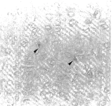
Figure 1. Apoptotic cells (arrow head) are characterized by loss of cell volume and condensation of chromatin. (H.E.
x200)
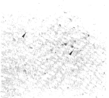
Figure 2. Pyknosis of tumor cells (arrow head) after irradiation. (H.E. x200)
Results
Histology
All fifteen of the cervical SCC specimens showed a proliferation of atypical squamous epithelium with confluent stromal invasion. The histological diagnosis of SCC, large cell non-keratinizing type was made in all cases.
Dead and dying carcinoma cells were recognized in both pre- and post-radiation biopsy specimens with their characteristic morphological features. The most distin- guishing features of the pre-radiation dead cells were loss of cell volume and condensation of chromatin material in the nuclei (Figure 1). The changes in tumor cells observed varied among the post-radiation biopsy specimens. The most pronounced change during irradiation was seen in the nuclei. Degenerated tumor cells showed cytoplasmic vesiculation and pyknosis (Figure 2).
Immunohistochemistry
Tables 2 summarize the results of immunohistoche- mistry. No specimens showed positive results for TNF- a , TNF- y or Fas expression prior to radiation. Positive reactions were observed in pre-radiated histologic sections for p 53 (Figure 3), c-fos (Figure 4) and bcl-2 (Figure 5) in 40%, 3 % and 12% of the specimens, respectively. In contrast, all of the histologic sections of irradiated cases showed a positive reaction for TNF- a (Figure 6). A positive result for c-fos was also observed in almost all post-radiation cases (Figure 7). About 50% of the irradi- ated specimens showed positive reactions for TNF-r (Figure 8) and p 53 (Figure 9). But only 12% of the irradiated specimens showed positive reactions for Fas and bcl-2.
The intensity of positive reaction was variable among the post-radiation specimens. TNF- a expressed weak to strong positive reactions, and c-fos and p 53 also showed similar results. TNF- y , Fas and bcl 2 showed only weak positive reactions.
Electron Microscopy
The tissue specimens for ultrastructural study were obtained from paraffin blocks. Therefore, the ultra- structural features of the cytoplasmic organellae of the tumor cells were not clear due to artifact. The tumor cells prior to radiotherapy showed intact nuclear membrane.
The chromatin materials were uniformly distributed and nucleoli were distinct. Desmosomes were also seen. The
Figure 3. Immunoreactivity for p 53 is seen in the cell nuclei (arrow head) prior to radiation. (LSAB x400)
Table 2 Result of Immunohistochemistry (n = 15) Pre-Radiation Post-Radiation
TNF-alpha 0/15 15/15
TNF-receptor 0/15 8/15
Fas * 0/15 2/15
p 53* 6/15 8/15
c-fos 5/15 13/15
bcl-2 * 1/15 1/15
* The result is not statistically significant according to Chi
Square test (p<0.05). Figure 4. Note a weak positive reaction for c-fos in pre- radiation tumor cells (arrow head). (LSAB x400)
Figure 5. Positive reaction for bcl-2 (arrow head) is seen in only one case prior to radiation. (LSAB x200)
Figure 7. Weak positive reaction for c-fos is noted in irradi- ated tumor cells (arrow head). (LSAB x400)
Figure 6. Post-radiation biopsy shows TNF-alpha-positive cells (arrow head). (LSAB x200)
Figure 8. TNF-receptor-positive cells are shown (arrow head) in half of the post-radiation cases. (LSAB x200)
post-radiation tumor cells showed marked ultrastructural changes in their nuclei. The cells had large and faintly stained nuclei. Unlike the pre-radiation tumor cells, the nuclei of the post-radiation tumor cells showed no distinct nucleoli. These cells had convoluted cytoplasmic mem-
branes and the cytoplasm showed a few number of vesicles and dense particles. Degenerated mitochondria with complete or partial loss of cristae were also seen. The presence of other organellae such as rough endoplasmic reticulum and ribosomes were decreased.
Figure 9. Note positive immunohistochemical reaction for p 53 in irradiated tumor cells (arrow head). (LSAB x400)
Discussion
Radiation can induce at least two distinct modes of cell death such as necrosis and apoptosis (14, 15). At the cellular level, necrosis is characterized by irreversible cell swelling and autolysis due to increased plasma membrane permeability, a decline in protein synthesis, swelling of the matrix of mitochondria, and dissolution of ribosomes and lysosomes (16). In contrast, apoptosis, a DNA-mediated cell death, is characterized by fragmentation of the nuclear chromatin, degradation of nuclear DNA, condensation of cell cytoplasm, alteration of the membrane permeability, and reduction in cell volume (17). Ultrastructural findings of the irradiated cervical SCC cells in the present study were compatible with necrotic change.
TNF- a , a cytokine generated from activated macro- phages, affects a wide range of biologic activities (18, 19).
TNF- a is reported to induce both apoptotic and necrotic forms of cell death in vitro (20). A cell culture study by Manchester et al. (21) showed that TNF- a was cytotoxic
for ME-180 human cervical carcinoma cells. In the present study, histologic sections of cervical SCC prior to radia- tion showed no positive result for TNF- a immunohisto- chemically. However, histologic sections of irradiated tumor biopsy specimens of the same patients immuno- histochemically showed positive reactions for TNF- a in
all cases, and for TNF- y in half of the cases. Herzog et al.
(22), in their cell culture study, indicated that cells derived from cervical malignancies were resistant to a direct cytolytic effect of TNF- a , but the cells became sensitive
to cytolysis after irradiation. Radiation inhibits the protein synthesis-dependent resistance mechanism and thus potentiates the cytolytic activity of TNF- a (24).
Therefore, the present results seem to support the hypothe- sis of Hallahan et al. (26) that the cytolytic activity of the TNF- a gene is induced by x-rays and regulated at the level of transcription. TNF- y belongs to the same family of tumor necrosis factor, showed almost similar reaction line TNF- a, but in a low intensity in the present study. So, we can surmise that TNF- y also have similar cytolytic effect like TNF- a in post-radiated cervical SCC. TNF- a and T NF- y were both expressed in pre and post-radiated speci- mens in other organs. But in the present study with cervical SCC, only the post radiated specimens expressed positive reaction for TNF- a and TNF- y .
Expression of c-fos was observed in the apoptotic cells in regressing rat prostate after castration (25). In the present study, c-fos showed a positive reaction in only 33%
of the pre-radiation specimens (5/15), and all but 2 post-radiation specimens (13/15) showed positive reaction for c-fos. Thus, the present results suggest that c-fos is responsible for cell death in post-radiation cervical SCC.
The oncogene bcl-2 has been reported to inhibit apoptosis (26). Overexpression of bcl-2 stimulates the development of follicular lymphoma by suppressing apoptosis in lymphoid tissue (27). In the present study, bcl-2 showed no significant positive reaction suggesting the inhibition of apoptosis. Tumor suppresser gene p 53 appears to have some role in radiation induced apoptosis in myeloid leukemic cells and mouse thymocytes (28, 29). But, p 53 almost similarly expressed in pre- and post-radiated specimens of cervical SCC in the present study suggesting cell death induced by radiation varies enormously from one tumor to another. Fas is a type of surface receptor mole- cule that can trigger apoptosis in some cell types including leukemic cell lines (30). However, Fas might have no significant inducing effect for cell death in SCC of the uterine cervix, since we observed positive result for Fas in only one post-radiation specimen in our study.
We observed that the cell death of uterine SCC induced by repeated radiation was ultrastructurally compatible with necrosis, but immunohistochemically compatible with apoptosis. We surmise that both necrotic and apoptotic cells might be present among the dead cells which were induced by repeated irradiation, since apoptosis has been reported to occur following low-dose radiation (2). Apoptotic necrosis, a type of cell death with features of both apoptosis and necrosis might also be present, because some parts of the biochemical process have been reported to be common between apoptosis and necrosis (18). Further study is required to elucidate the details of the process of cell death in irradiated cervical SCC, in particular using tissue specimens just after the initial mild dose of radiation.
Acknowledgments
We thank D. Morzek for critical reading of the manu- script.
References
1) Wyllie AH: The biology of cell death. Anticancer Res 5: 131-136, 1985 2) Kerr JFR, Winterford CM, Harmon BV : Apoptosis : its significance in
cancer and cancer therapy. Cancer 73: 2013-2023, 1994
3) Macklis RM, Lin JY, Beresford B, Atcher RW, Hines JJ, Humm JL : Cellular kinetics, dosimetry, and radiobiology of a-particle radioim-
munotherapy : induction of apoptosis. Radiat Res 130: 220-225, 1992 4) Cotter TG, Glynn JM, Echeverri F, Green DR: The induction of apoptosis by chemotherapeutic agents occurs in all phases of the cell
cycle. Anticancer Res 51 : 741-743, 1992
5) Kyprianou N, English HF, Davidson NE, Isaacs JT : Programmed cell death during regression of the MCF-7 human breast cancer following
estrogen ablation. Cancer Res 51: 162-166, 1991
6) Takano YS, Harmon BV, Kerr JFR : Apoptosis induced by mild hyperthermia in human and murine tumor cell lines : A study using
electron microscopy and DNA gel electrophoresis. J Pathol 163:
329-336, 1991
7) Munro TR : The relative radiosensitivity of the nucleus and the cytoplasm of the chinese hamster fibroblasts. Radiat Res 42: 451-457,
1970
8) Bradford JS : Sublethal damage, potentially lethal damage, and chromosomal aberrations in mammalian cells exposed to ionizing
radiation. Int J Radiat Oncol Biol Phys 21: 1457-1462, 1991 9) Sheehan JF, Schmitz HE : Histologic changes produced by radiation in
adenocarcinomas of uterus, comparison with changes produced in
squamous cell carcinoma of cervix. Am J Clin Pathol 20: 241-248, 1950 10) Wyllie AH : Apoptosis (The 1992 Frank Rose memorial lecture). Br J
Cancer 67: 205-208, 1993
11) Scully RE (ed) : Histological Typing of Female Genital Tract Tumors.
Ed. 2, Geneva, WHO, 1994
12) The general rules for clinical and pathological management of uterine corpus cancer. Japan Society of Obstetrics and Gynecology. Ed. 2,
Tokyo, Kinbara Shuppan, 1996
13) Hsu SM, Raine L, Fanger H : Use of avidin-biotin peroxidase complex (ABC) in immunoperoxidase techniques : A comparison between ABC
and unlabelled antibody (PAP) procedures. J Histochem Cytochem 29:
577-580, 1981
14) Okado S : Radiation-induced death. in : K. I. Altman, G. G. Gerber, and S. Okada (eds.), Radiation Biochemistry, Vol 1, pp. 247-307. New
York : Academic Press, Inc., 1970
15) Duvall E, Wyllie AH : Death and the cell. Immunol Today 7 : 115-119, 1986
16) Wylie AH : The significance of apoptosis. Int Rev Cytol 68: 251-306, 1980
17) Clarke PGH: Developmental cell death: morphological diversity and multiple mechanisms. Anat Embryol 181: 195-213, 1990
18) Old LJ : Tumor necrosis factor. Science 230: 630-632, 1985 19) Le J, Vilcek J : Biology of disease. Lab Invest 56: 234- 248, 1987 20) Lester SM, Wood JG, Gooding LR : Tumor necrosis factor can induce
both apoptotic and necrotic forms of cell death. J Immunol 141:
2629-2634, 1988
21) Manchester KM, Heston WDW, Donner DB: Tumor necrosis factor- induced cytotoxicity is accompanied by intracellular mitogenic signals
in ME-180 human cervical carcinoma cells. Biochem J 290: 185-190,
1993
22) Herzog TJ, Nelson PK, Mutch DG, Wright WD, Kao MS, Collins JL : Effects of radiation on TNF a-mediated cytolysis of cell lines derived
from cervical carcinomas. Gynecol Oncol 47: 196-202, 1992
23) Boothman DA, Wang M, Lee SW : Induction of tissue-type plasminogen activator by ionizing radiation in human malignant
melanoma cells. Cancer Res 51: 5587-5598, 1991
24) Hallahan DE, Spriggs DR, Beckett MA, Kufe DW, Weichselbaum RR:
Increased tumor necrosis factor alpha mRNA after cellular exposure to
ionizing radiation. Proc Natl Acad Sci 86: 10104-10108, 1989 25) Buttyan R, Zakeri Z, Lockshin R, Woglemuth D : Cascade induction of
c-fos, c-myc and heat shock protein 70k transcripts during regression
of rat ventral prostate gland. Mole Endocrinol 11: 650-656, 1988 26) Hockenberry D, Nunez G, Schreiber RD, Korsmeyer SJ : Bcl- 2 is an
inner mitochondrial membrane protein that blocks programmed cell
death. Nature 348: 334-336, 1990
27) Tsujimoto Y, Gorham J, Cossman J, Jaffe E, Croce CM: The t (14;
18) chromosome translocation involved in B-cell neoplasms result from
mistakes in VDJ joining. Science 229: 1390-1393, 1985
28) Trauth BC, Klas C, Peters AM, Matzku S, Moller P, Falk W, Debatin KM, Kramer PH : Monoclonal antibody-mediated tumor regression by
induction of apoptosis. Science 245: 301-305, 1989
29) Lowe SW, Schmitt EM, Smith SW, Osborne BA : p 53 is required for radiation-induced apoptosis in mouse thymocytes. Nature 362:
847-851, 1993
30) Yonish-Rouach E, Resnitsky D, Lotem J, Sachs K, Kimchi A, Oren M:
Wild type p 53 induces apoptosis of myeloid leukemic cells that is
inhibited by interleukin-6. Nature 352: 345-347, 1991