Acta med. Nagsaki . 12: 133 -141
Studies on the Unknown Amino Acids in Normal Human Urine
Ichiro YOSHIKAWA*
Department of Biochemistry Nagasaki University School of Medicine, Nagasaki, Japan
Received for publication, March 10, 1968
Many of the ninhydrin-positive substances excreted in human urine still remain unknown. The present author carried out the isolation of some un- known ninhydrin-positive substances from the neutral amino acid fraction of human adult male urine. In this paper, the isolation and characterization of four amino acids were described.
INTRODUCTION
Although a number of studies have been made on amino acids in urine, many ninhydrin-positive substances excreted in human urine remain unidentified and have not been studied enough. For several years, studies on unknown ninhydrin-positive substances in normal hu- man urine have been carried out in our laboratory and two of them have been isolated and identified as i9-hydroxyasparagine-15I and O-xylo- syl-serine'8. Recently, _other three amino acids were isolated and identified as O-mannosyl-serine, 0-mannosyl-threonine, and isoserine by us. 16) , 19) MAEKAWA6) investigated on the basic amino acid fraction of human urine from normal adult male subjects. The isolation and char- acterization of four unknown ninhydrin-positive substances in urine are the subjects of this paper.
EXPERIMENTS AND RESULTS
Collection and desalting of urine and fractionation of the desalted ampholytes were carried out by the same methods described by TOMI- NAGA et al. 15' ,18 )
1) Materials
Urine excreted between 10 a.m. and 3 p.m. was collected daily from normal male subjects. After desalted, the ampholytes were pooled
*.吉川 一・郎
and kept in a refrigerator. Every time when the stored ampholytes reached the amount obtained from 100f of urine, they were passed through columns of weak basic and weak acidic resins successively, and the resulting effluent was fractionated on the column of Amberlite CG- 120. This procedure was carried out once or twice a month all the year round, and the unknown ninhydrin-positive substances which appear through all seasons are the subjects of the present study.
2) Desalting
The urine was decolorized with charcoal (5g per liter) and filtered.
A portion of each filtrate (1 ~ ) was placed separately on the column (4.5 x 30cm) of Amberlite CG -120 (100 - 200 mesh) in the H+ form. Each column was washed with 2 ~ of water and eluted with 800 m ~ of 2N ammonia. The eluates were evaporated to dryness at 50°C under reduced pressure. The residue obtained from 10 Q of urine was dissolved in 1.5
Q of water and subjected to the column (4.5 x 30cm) of Amberlite CG -400 (100-200 mesh) in the OH- form . The column was washed with 2 ~ of water and eluted with 800 m 17 of 2N acetic acid. The eluate was evaporated to dryness at 50°C in vacuo.
3) Primary fractionation
The desalted ampholytes obtained from 50 ~ of urine were passed through columns (4.5 x 30cm) of Amberlite CG-45 (Type I) in the OH- form and CG-50 (Type I) in the H+ form, respectively. Most of the acidic and all of the basic amino acids present were adsorbed by these
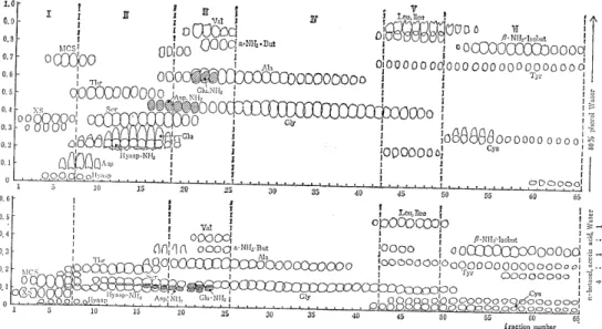
Fig. 1 The paper chromatograms of the neutral amino 'acid fractions eluted from Amberlite CG-120 column with 0.2N ammonia.
XS=O-xylosyl-serine
MCS=S-methylcysteine sulfoxide Hyasp = (3-hydroxyasparatic acid
Hyasp. NH2 = (3-hydroxyasparagine
resins and removed. The effluent containing the neutral amino acids which was obtained from 100 ~ of urine was next placed on the column (2.8 x 40cm) of Amberlite CG -120 (200 - 400 mesh) in the H+ form. The adsorbed amino acids were eluted with 0.2N ammonia and the effluent was collected into 20 m ~ -fractions. The amino acids in each fraction were examined by one-dimensional paper chromatography. Toyo Roshi No. 51 paper (40 x 40cm) and two solvent systems (I) n-butanol, acetic acid, water (4:1:1) and (II) 80% aqueous phenol were used for the paper chromatography.
Fig. 1 shows the result of the primary fractionation.
As shown in Fig. 1, the eluted amino acid fraction was divided into six subfractions by the order of elution , and the subfractions which contained little unknown ampholytes were discarded, except subfraction I and V.
4) Isolation and identification of sarcosine
The mother liquor prepared by removing O-xylosyl-serine (XS) and S-methylcysteine sulfoxide (MCS) from the subfraction I contains sev- eral unknown ninhydrin-positive substances, one of which moves to a position close to that occupied by methionine sulfoxide on the two-di- mensional paper chromatogram was isolated and identified as sarcosine.
Sarcosine-containing fractions were collected from more than 1000,C of urine, refractionated on the column (2.8 x 37cm) of Amberlite CG-
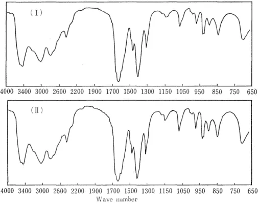
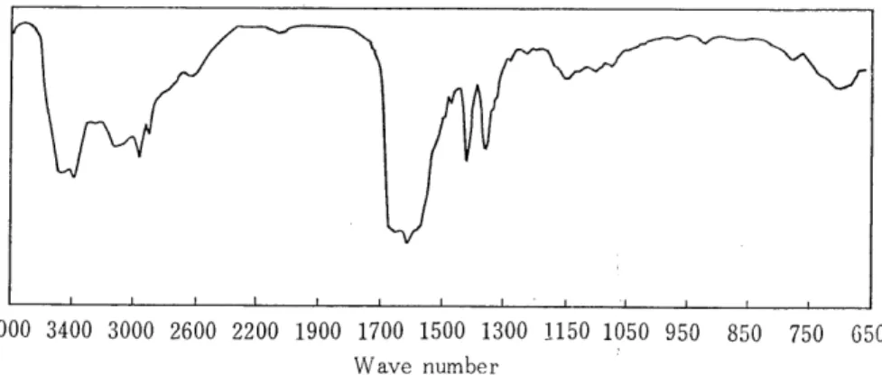
Fig. 2 The infrared absorption spectra of sarcosine (authentic sample) (I) and
ofithe substance isolated from human urine.
400 (200-400 mesh) in the OH- form, and eluted into 20 m ~ -fract- ions with 0.2N acetic acid. Sarcosine was contained in the first three tubes and the tube No. 1 was free from any other ninhydrin-positive substance.The solution in the tube No. 1 was evaporated in vacuo and the resulting residue (320 mg) was dissolved in 2 m ~ water, and to
this was added 18 m . of ethanol. A small amount of brown precipitate was yielded, but the crystallization was failed. After concentrated again, it was applied to 12 sheets of Toyo Roshi No. 51 paper and developed with the solvent (1). The position of the aiming substance
(Rf = 0.28) was cut out and extracted with water. 64 mg of powder was obtained. This material was crystallized twice from 80-90%
ethanol. The resulting hygroscopic crystals (20 mg) did not show the definite melting point (203-218°C with decomposition). The crystals were chromatographically identical with the authentic sarcosine (Rf values, 0.28 with solvent (I) and 0.73 with solvent (II) . The values of the elementary analysis were C; 39.96%, H; 7.82%, and N; 15.43%. The calculated values for C3 H, 0, N (sarcosine) were C; 40.44%, H; 7.92%, and N; 15.72%. Fig. 2 shows the infrared absorption spectra of the isolated and authentic sarcosines.
5) The unknown ampholytes in the subfraction V
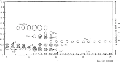
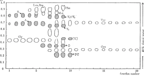
Subfraction V collected from 100 ~ of urine was applied to the column (1.3 x 2Ocm) of Amberlite CG-400 (200-400 mesh) and eluted into 8 m ~ of fractions with 0.2N acetic acid. Fig. 3 and Fig. 4 show the paper chromatograms of them. In these figures, the spot except the seven known amino acids (glycine isoleucine, leucine, methionine, phenylalanine, tyrosine, and histidine) indicate the unidentified subs- tances.
Fig. 3 The paper chromatogram of subfraction V eluted from Amberlite CG-400
column with-10.2N acetic acid.
Fig. 4 The paper chromatogram of subfraction V eluted from Amberlite CG-400 with 0.2N acetic acid.
(a) `A' and `B' Both substances were contained in the earliest portion of the eluate when the subfraction V was eluted from either Amberlites CG-120 or CG-400 column. But the substrnce `A' was eluted a little faster than `B' from the strong acidic resin (Amb. CG
-120) and it was in the reverse order from the strong basic resin (Amb . CG-400). They were not decomposed by boiling in 6N HCI for 20 hours. The Rf values of `A' and `B' with the solvents (I) and (II) were 0.15, 0.68 and 0.18', 0.81, respectively. Only `B' could be isola- ted.
(b) Isolation of `B' The pooled `B' fraction (80 m t ) was applied to a small column (1.3 x 11cm) of Amberlite CG - 400 (200 - 400 mesh).
A portion of ampholytes was flowed out into the effluent (10 m Q ).
The column was eluted with 0.2N acetic acid, and 10 m Q of fractions were collected. First three amino acid fractions mainly contained the substance 'B'. They were combined, concentrated, decolorized with charcoal, and filtered. The filtrate (2-3m k ) was added four volumes of ethanol, and amorphous precipitate was yielded. Further, 10m Q of ethanol was added to the solution, and the precipitate was filtered and dried (260mg). Into 2 m Q of water, 200mg of this powder was dissolved and. to this was added 8 m Q of ethanol. A small quantity of the amorphous precipitate yielded was removed by filtration, and the filtrate was again added 10 m t of ethanol. A large quantity of the amorphous precipitate (102 mg) was obtained. It decomposed with bub- bling at 190-191°C. The isolated `B' was free from other ninhydrin- reacting substance on the two-dimensional paper chromatogram. The
values of the elementary analysis were C; 42.69%, H; 8.00%, and
N; 20.04%. Fig. 5 shows the infrared spectrum of the isolated '13' ,
Fig'_ 9 The infrared absorption spectrum of `B'