Acta Med. Nagasaki 47: 155-160
Expressions of Vascular Endothelial Growth Factor (VEGF)-D and VEGF
Receptor-3 in Colorectal Cancer: Relationship to Lymph Node Metastasis
Masatoshi HASEBA1), Takashi Tsuji1), Hiroshi YANo1), Hideaki KOMATSU1), Shigekazu HIDAKA1), Terumitsu SAWAI1), Toru YASUTAKE1), Tohru NAKAGOE1), Yutaka TAGAWA2)
1) Division of Surgical Oncology, Department of Translational Medical Science, Nagasaki University Graduate School of Biomedical Sciences 2) Nagasaki University School of Health Sciences
Angiogenic factors play a major role in tumor growth and metastasis. Vascular endothelial growth factor (VEGF)- D is a ligand for VEGF receptor-3 (VEGFR-3/Flt-4), which mainly expressed on the lymphatic endothelium. Recent ex- perimental studies have shown that VEGF-D induces tumor lymphangiogenesis and promote metastatic spread of tumor cells via lymphatic vessels. However, the contribution of VEGF- D to lymph node metastasis in human colorectal cancer is less understood. We therefore examined VEGF-D and VEGFR-3 expression in patients with colorectal cancer. Sections of formalin-fixed and paraffin-embedded specimens from 76 colorectal cancers were immunohistochemically stained for VEGF-D and VEGFR-3. Staining for VEGF-D was positive in the cytoplasm of tumor cells in 43 of 76 examined tumors (56.6%). Staining for VEGFR-3 was positive in endothelial cells in 38 (50.0%) tumors. Univariate analysis showed that both VEGF-D and VEGFR-3 expressions correlated significantly with lymph node metastasis, histological type and depth of tumor invasion. However, logistic regression analysis indi- cated that VEGF-D expression, but not that of VEGFR-3, was an independent predictor for lymph node metastasis.
Our data suggest that VEGF-D plays an important role in lymph node metastasis in colorectal cancer.
ACTA MEDICA NAGASAKIENSIA 47: 155-160, 2002
Key Words: vascular endothelial growth factor (VEGF)-D, lymph node metastasis, colorectal cancer,
lymphangiogenesis
Address Correspondence: Masatoshi Haseba, M.D.
Division of Surgical Oncology, Department of Translational Medical Science, Nagasaki University Graduate School of Biomedical Sciences, Sakamoto 1-7-1, Nagasaki, 852-8501 Japan
TEL: +81-95-849-7304 FAX: +81-95-849-7306 E-mail: m-haseba@guitar.ocn.ne.jp
Introduction
The capacity of tumor cells to induce angiogenesis and lymphangiogenesis may regulate the probability of hematogenous or lymphatic metastasis. Lymph- angiogenesis is controlled, in part, by members of the vascular endothelial growth factor (VEGF) family, i.e., VEGF-C, and VEGF-D, and their receptor on lymphatic endothelium, VEGFR-31,2'. VEGF-C was initially identi- fied as a ligand for VEGFR-33'. Because expression of VEGFR-3 is mainly restricted to the lymphatic endo- thelium, the major function of VEGF-C appears to be the regulation of lymphatic vessel growth"'). VEGF-C has been shown to regulate the growth of lymphatic vessels in various experimental models' 6
VEGF-D is the most recently discovered member of the VEGF family. It was isolated as a fos-inducible factor from mouse skin fibroblasts and the mature form shares about 60% amino acid sequence identity with VEGF-C 2'' ) . In addition to their sequence identity, VEGF-C and VEGF-D are thought to have similar bio- logical functions because they can bond to common receptors. These secreted growth factors are synthe- sized as propeptides that are activated by proteolysis to form high-affinity ligands that activate VEGFR-3 and stimulate lymphangiogenesis 8'') . In experimental mouse tumor model, it is reported that VEGF-C and VEGF-D can induce tumor lymphangiogenesis and di- rect metastasis to the lymphatic vessels and lymph nodes'"'). In human, recent studies indicated that VEGF- D is upregulated in malignant melanoma"', inflamma- tory breast carcinoma"), and colorectal cancer"), sug- gesting that VEGF-D expression is associated with lymph node metastasis in various human malignan- cies.
In the present study, we examined the expression of VEGF-D and VEGFR-3 in colorectal cancer and inves- tigated their relationship to lymph node metastasis, one of the most important prognostic factors in colo- rectal cancer.
Masatoshi Haseba et al : VEGF-D and VEGFR-3 Expressions in Colorectal Cancer
Material and Methods
Patients
Seventy-six tissue samples were obtained from pa- tients with colorectal cancer who had been operated between 1997 and 1998 at the Division of Surgical Oncology, Department of Translational Medical Science, Nagasaki University Graduate School of Biomedical Sciences. The group consisted of 52 males and 24 fe- males, with a mean age at the time of surgery of 62.
5±12.0 years (± SD, range: 29-88 years). None had re- ceived chemotherapy or radiotherapy before surgery.
Forty tumors were localized in the colon and 36 tu- mors were in the rectum. Seventy-three patients un- derwent curative operation. The criteria of the American Joint Committee on Cancer Classification and stage grouping was used to classify tumors i61. The 76 pa- tients included 1 patient with stage 0, 12 with stage I, 30 with stage II, 22 with stage III, and 11 with stage IV cancer. Eight tumors were classified as 'well-differentiated adenocarcinoma', 61 tumors as 'moderately-differentiated adenocarcinoma', 5 tumors as 'poorly-differentiated adenocarcinoma', and 2 tumors as 'mucinous carci- noma'. A written informed consent was obtained from each patient for use in the present study.
Immunohistochemistry for VEGF-D and VEGFR-3
The mouse monoclonal anti-VEGF-D antibody pur- chased from R&D systems, Inc (Minneapolis, MN) was used for staining at a concentration of 10 u g/ml. The rabbit polyclonal anti-VEGFR-3 antibody purchased from Santa Cruz Biotechnology, Inc. (Santa Cruz, CA) was used at 2 u g/ml. Immunohistochemical staining was performed as follows. Serial sections (3-4-gm thick) from formalin-fixed, paraffin-embedded tissue blocks were deparaffinized and then heated in a mi- crowave oven in 10 mM citrate buffer, pH 6.0, for 10 min. The sections were then treated in methanol con- taining 3% hydrogen peroxide for 10 min at room temperature. They were subsequently washed in dis- tilled water, rinsed for phosphate buffered saline (PBS), and treated with nonspecific staining blocking reagent containing 0.25% casein (DAKO, Glostrup, Denmark) for 10 min at room temperature. The slides were then incubated overnight at 4°C in a humidified chamber with anti-VEGF-D or anti-VEGFR-3 antibody diluted in PBS with 1 % bovine serum albumin (BSA).
After washing the specimens with PBS, the slides were incubated with biotinylated anti-mouse or anti- rabbit IgG for 30 min at room temperature. After three washes in PBS, the sections were incubated with
streptavidin-peroxidase reagent for 30 min at room temperature. VEGF-D antigen and VEGFR-3 antigen were developed by incubating the slides in diaminobenzidine (DAKO) solution containing 0.06 mM diaminobenzidine and 2 mM hydrogen peroxide in 0.05% PBS (pH 7.6) for 5 min. Sections were counterstained with hematoxylin, dehydrated with alcohol and xylene, and mounted in a routine fashion. Negative controls were performed in all cases by omitting the first antibody.
Assessment of immunoreactivity
VEGF-D expression in the tumors was measured by assessing the percentage of positively stained cells.
Tumors were classified into three categories according to the extent of staining: (+), over 20% of tumor cells were stained; (± ), less than 20% of tumor cells were stained, and (- ), completely negative. (-) and (±) were classified as negative for VEGF-D, (+) was classi- fied as positive for VEGF-D. For VEGFR-3 expression in endothelial cells, we scored the tumors as positive for VEGFR-3 when VEGFR-3 positive vessels were found at the tumor periphery.
Statistical analysis
Patients were divided into two groups based on the median age (62 years). The statistical significance among VEGF-D, VEGFR-3 expressions, and clinicopathological features was evaluated using chi-square test. Backward stepwise logistic regression model was used for multi- variate adjustments for all covariates simultaneously.
All statistical. analyses -.described in this study were conducted using STATISTICATM software (StatSoft, Inc.
Tulsa, OK).
Results
VEGF-D expression in colorectal cancer
VEGF-D was detected in the cytoplasm of tumor cells, and no expression was observed in the nucleus (Figure 1). Expression of VEGF-D tended to be local- ized to the deep layer of the tumor. In specimens of normal colonic mucosa, no VEGF-D expression was observed. Among the 76 tumors, 43 (56.6%) were posi- tive for VEGF-D expression. The relationship between VEGF-D expression and clinicopathological features is summarized in Table 1. VEGF-D expression was sig- nificantly associated with histological type and depth of invasion (p=0.03 and <0.01, respectively). There was no correlation between VEGF-D expression and
Masatoshi Haseba et al : VEGF-D and VEGFR-3 Expressions in Colorectal Cancer
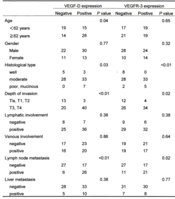
Table 1. VEGF-D and VEGFR-3 expressions, and clinicopathological features.
VEGF-D expression VEGFR-3 expression
Negative Positive P value Negative Positive P value
Age 0.04 0.65
<62 years 19 15 17 19
>_62 years 14 28 21 19
Gender 0.77 0.32
Male 22 30 28 24
Female 11 13 10 14
Histological type 0.03 <0.01
well 5 3 8 0
moderate 28 33 28 33
poor, mucinous 0 7 2 5
Depth of invasion <0.01 0.02
Tis, T1, T2 13 3 12 4
T3, T4 20 40 26 34
Lymphatic involvement 0.38 0.38
negative 8 7 9 6
positive 25 36 29 32
Venous involvement 0.86 0.64
negative 17 23 19 21
positive 16 20 19 17
Lymph node metastasis <0.01 0.02
negative 27 17 27 17
positive 6 26 11 21
Liver metastasis 0.38 0.77
negative 28 33 31 30
positive 5 10 7 8
well, well-differentiated adenocarcinoma; moderate, moderately-differentiated
adenocarcinomas; poor, poorly-differentiated adenocarcinoma; mucinous, mucinous
carcinoma.
Table 2. Logistic regression analysis of VEGF-D with respect to lymph node metastasis.
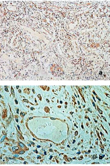
Figure 1. Immunohistochemical staining for VEGF-D in colon cancer.
Note the positive immunoreactivity for VEGF-D predomi- nantly in the cytoplasm of tumor cells. Magnification: (a), x100; (b), x400.
gender, lymphatic involvement, venous involvement, or liver metastasis. The positive expression of VEGF-D was significantly higher in tumors with lymph node metastasis than in those without lymph node metasta- sis (Table 1). Multivariate analysis revealed that VEGF- D expression in tumor cells is an independent factor influencing lymph node metastasis (Table 2).
VEGFR-3 expression in colorectal cancer
VEGFR-3 was detected in a subset of vessels, which were typically thin-walled and devoid of red blood cells (Figure 2). Thirty-eight of 76 tumors (50%) were positive for VEGFR-3 expression in endothelial cells.
Although VEGFR-3 was detected in the cytoplasm of tumor cells with a staining pattern similar to that of VEGF-D (49 of 76 tumors, 64.4%), in this study we evaluated VEGFR-3 only in endothelial cells. Like
Odds ratio (95% CI) P value Histological type
well 1
moderate 1.31 (0.09 -19.08) 0.84
poor, mucinous 8.81 (0.23 - 338.58) 0.23 Depth of invasion
Tis, T1, T2 1
T3, T4 1.90 (0.31 -11.71) 0.48
Venous involvement
negative 1
positive 0.64 (0.18 - 2.28) 0.48
Lymphatic involvement
negative 1
positive 24.31 (1.80 - 328.34) 0.015 VEGF-D expression
negative 1
positive 5.31 (1.56-18.09) <0.01 CI, confidence interval. Other abbreviations as in Table 1.
Masatoshi Haseba et al : VEGF-D and VEGFR-3 Expressions in Colorectal Cancer
Table 3. Logistic regression analysis of VEGFR-3 with re- spect to lymph node metastasis.
Odds ratio (95% CI) P value Histological type
well 1
moderate 0.89 (0.07 - 11.83) 0.92 poor, mucinous 9.28 (0.26 - 335.62) 0.22 Depth of invasion
Tis, T1, T2 1
T3, T4 2.37 (0.41 -13.39) 0.32
Venous involvement
negative 1
positive 0.64 (0.19 - 2.20) 0.48
Lymphatic involvement
negative 1
positive 22.01 (1.67 - 290.05) 0.02 VEGFR-3 expression
negative 1
positive 2.26 (0.70 - 7.34) 0.16
Abbreviations as in Tables 1 and 2.
Table 4. Relationship between VEGF-D expression and VEGFR-3 expression.
Figure 2. Immunohistochemical staining for VEGFR-3 in colon cancer.
Note the positive immunoreactivity for VEGFR-3 in the en- dothelium of vessels. Magnification: (a), x100; (b), x400.
VEGF-D, VEGFR-3 expression was significantly associ- ated with histological type and depth of invasion (p <
0.01 and p < 0.02, respectively). VEGFR-3 expression was significantly higher in tumors with lymph node me- tastasis than in those without lymph node metastasis (Table 1). However, multivariate analysis revealed that VEGFR-3 expression was not an independent factor influencing lymph node metastasis (Table 3). VEGFR-3 expression in endothelial cells was significantly associ- ated with VEGF-D expression in tumor cells (Table 4).
Discussion
VEGFR-3 expression
VEGF-D expression
Negative Positive P value
Negative 25 8 <0.01
Positive 13 30
The VEGF family consists of VEGF-A, -B, -C, -D, and -E as well as placenta growth factor"' . The founding member, VEGF-A, plays essential roles in vasculogenesis and angiogenesis'$'. Although its crucial role in tumor angiogenesis and hematogenous metastasis has been documented in a variety of cancers"', little is known
about the physiological and pathological roles of other family members. Recent experimental studies with VEGF-C and VEGF-D have shown that they can in- duce tumor lymphangiogenesis. However, few studies assessed the relevance of VEGF-D to lymph node me- tastasis in human malignancies. VEGF-D activates VEGFR-2 (KDR/Flk-1) and VEGFR-3220', both of which are essential for vascular development"-"'. VEGFR-2 is expressed on vascular endothelial cells"' and VEGFR-3 is mainly expressed in the endothelium of lymphatic vessels''. A recent study indicated that VEGF-D can stimulate both angiogenesis and lymphangiogenesis12'.
However, another study showed that VEGF-D binds only to VEGFR-3 and induces only lymphangiogenesis in mouse tumor models''.
In the present study, VEGF-D was highly expressed
Masatoshi Haseba et al : VEGF-D and VEGFR-3 Expressions in Colorectal Cancer
in primary tumors of colorectal cancer with lymph node metastasis. On the other hand, the expression of VEGF-D was not related to hematogenous metastasis, such as liver metastasis or venous involvement. Recent studies demonstrated that VEGF-D is proteolytically
processed in a manner similar to VEGF-C8'". It exists in numerous forms, as some molecules are completely
processed, whereas others are partially processed or remain unprocessed". There is evidence indicating that the unprocessed or partially processed VEGF-Ds are capable of binding VEGFR-3, albeit with low affin- ity, but fully proteolytic cleavage is necessary to gen- erate the mature form that binds VEGFR-2 as well.
These data suggest the possibility that the angiogenic versus lymphangiogenic responses to VEGF-D may de- pend on, at least in part, the degree of proteolytic processing of this growth factor in different tumor en- vironment, and that in colorectal cancer VEGF-D does not act as an angiogenic factor, but plays a role in the development of lymphatic vessels thereby promoting only lymphatic metastasis. Although the protease re- sponsible for the processing is yet to be determined, we speculate that in colorectal cancer the protease is not expressed enough to generate fully processed VEGF-D.
Our results also showed a significant correlation be- tween expression of VEGF-D and depth of tumor inva- sion. That VEGF-D tended to be expressed in the deep layers of tumors suggests the increased potential of lymphangiogenesis following growth of such tumors, or reflects the malignant potential of tumor invasion.
Several studies have examined the relationship be tween VEGF-D and human malignancies '3,14,23,24>, Although recent studies have examined the expression of VEGF- D in colorectal cancer, the role of this factor in lymph node metastasis is still controversial. White et al. 15) studied 84 patients with colorectal cancer and used univariate analysis to conclude that the expression of VEGF-D correlated with lymph node metastasis. On the other hand, George et al.25) reported that there was no correlation between VEGF-D expression and lymph node metastasis in 70 patients with colorectal cancer.
Our results were consistent with the former study. To our knowledge, the present study is the first report to indicate that VEGF-D expression is an independent predictor of lymph node metastasis in colorectal can- cer using multivariate analysis.
In the same population of patients, we examined the expression of VEGFR-3. VEGFR-3 expression was sig- nificantly associated with tumor histological type and with depth of invasion. Furthermore, VEGFR-3 expres- sion was significantly associated with VEGF-D expres- sion, suggesting that the co-expression of VEGF-D and
VEGFR-3 may play an important role in tumor lymph- angiogenesis. However, multivariate analysis revealed that VEGFR-3 expression was not an independent fac- tor influencing lymph node metastasis. In addition, VEGFR-3 expression was also detected in tumor cells (data not shown). Similar pattern of VEGFR-3 in colo- rectal cancer was also reported"'. Although the signifi- cance of such expression is still unclear, it is possible that VEGFR-3 plays a role in tumor growth in an autocrine manner. Further studies should be performed to elucidate the role of VEGFR-3 on tumor cells.
In conclusion, we have demonstrated in the present study a close relationship between VEGF-D expression and lymph node metastasis in colorectal cancer. Our study suggests that the detection of VEGF-D protein in the primary tumor may represent a potential risk
of lymph node metastasis in colorectal cancer.
References
1) Jeltsch M, Kaipainen A, Joukov V, et al: Hyperplasia of lymphatic vessels in VEGF-C transgenic mice. Science 276: 1423-1425, 1997 2) Achen MG, Jeltsch M, Kukk E, et al: Vascular endothelial growth
factor D (VEGF-D) is a ligand for the tyrosine kinases VEGF re-
ceptor 2 (Flkl) and VEGF receptor 3 (FIt4). Proc Natl Acad Sci
USA 95: 548-553, 1998
3) Joukov V, Pajusola K, Kaipainen A, et al: A novel vascular endo- thelial growth factor, VEGF-C, is a ligand for the Flt4 (VEGFR-3)
and KDR (VEGFR-2) receptor tyrosine kinases. EMBO J 15: 290-
298, 1996
4) Kaipainen A, Korhonen J, Mustonen T, et al: Expression of the fms-like tyrosine kinase 4 gene becomes restricted to lymphatic
endothelium during development. Proc Natl Acad Sci USA 92:
3566-3570, 1995
5) Kukk E, Lymboussaki A, Taira S, et al: VEGF-C receptor binding and pattern of expression with VEGFR-3 suggests a role in lym-
phatic vascular development. Development 122: 3829-3837, 1996 6) Veikkola T, Jussila L, Makinen T, et al: Signalling via vascular en
dothelial growth factor receptor-3 is sufficient for lymphangiogenesis
in transgenic mice. EMBO J 20: 1223-1231, 2001
7) Orlandini M, Marconcini L, Ferruzzi R, Oliviero S: Identification of a c-fos-induced gene that is related to the platelet- derived growth
factor/vascular endothelial growth factor family. Proc Natl Acad
Sci USA 93: 11675-11680, 1996
8) Joukov V, Sorsa T, Kumar V, et al: Proteolytic processing regu- lates receptor specificity and activity of VEGF-C. EMBO J 16:
3898-3911, 1997
9) Stacker SA, Stenvers K, Caesar C, et al: Biosynthesis of vascular endothelial growth factor-D involves proteolytic processing which
generates non-covalent homodimers. J Biol Chem 274: 32127-
32136, 1999
10) Mandriota SJ, Jussila L, Jeltsch M, et al: Vascular endothelial growth factor-C-mediated lymphangiogenesis promotes tumour
metastasis. EMBO J 20: 672-682, 2001
11) Skobe M, Hawighorst T, Jackson DG, et al: Induction of tumor lymphangiogenesis by VEGF-C promotes breast cancer metastasis.
Nat Med 7: 192-198, 2001
12) Stacker SA, Caesar C, Baldwin ME, et al: VEGF-D promotes the metastatic spread of tumor cells via the lymphatics. Nat Med 7:
186-191, 2001
13) Achen MG, Williams RA, Minekus MP, et al: Localization of vas- cular endothelial growth factor-D in malignant melanoma sug-
gests a role in tumour angiogenesis. J Pathol 193: 147-154, 2001 14) Kurebayashi J, Otsuki T, Kunisue H, et al: Expression of vascular
Masatoshi Haseba et al : VEGF-D and VEGFR-3 Expressions in Colorectal Cancer
endothelial growth factor (VEGF) family members in breast can- cer. Jpn j Cancer Res 90: 977-981, 1999
15) White JD, Hewett PW, Kosuge D, et al: Vascular endothelial growth factor-D expression is an independent prognostic marker
for survival in colorectal carcinoma. Cancer Res 62: 1669-1675,
2002
16) Fleming ID, Cooper JS, Henson DE, et al: Colon and Rectum. In:
AJCC Cancer Staging Manual, fifth ed. (Fleming ID, Cooper JS,
Henson DE, Hunter RVP, Kennedy BJ, Murphy GP, O'Sullivan B,
Sobin LH, Yarbro JW eds.; Lippincott-Raven, New York) pp. 83- 90, 1997
17) Veikkola T, Karkkainen M, Claesson-Welsh L, Alitalo K: Regulation of angiogenesis via vascular endothelial growth factor receptors.
Cancer Res 60: 203-212. 2000
18) Beck L Jr., D'Amore PA: Vascular development: cellular and mo- lecular regulation. FASEB J 11: 365-373, 1997
19) Ellis LM, Fidler IJ: Angiogenesis and metastasis. Eur J Cancer 32A: 2451-2460, 1996
20) Baldwin ME, Catimel B, Nice EC, et al: The specificity of receptor binding by vascular endothelial growth factor-d is different in
mouse and man. J Biol Chem 276: 19166-19171, 2001
21) Dumont DJ, Jussila L, Taipale J, et al: Cardiovascular failure in mouse embryos deficient in VEGF receptor-3. Science 282: 946- 949, 1998
22) Ferrara N, Davis-Smyth T: The biology of vascular endothelial growth factor. Endocr Rev 18: 4-25, 1997
23) Niki T, Iba S, Tokunou M, et al: Expression of vascular endothe- lial growth factors A, B, C, and D and their relationships to
lymph node status in lung adenocarcinoma. Clin Cancer Res 6:
2431-2439, 2000
24) O-Charoenrat P, Rhys-Evans P, Eccles SA: Expression of vascular endothelial growth factor family members in head and neck
squamous cell carcinoma correlates with lymph node metastasis.
Cancer 92: 556-568, 2001
25) George ML, Tutton MG, Janssen F, et al: VEGF-A, VEGF-C, and VEGF-D in colorectal cancer progression. Neoplasia 3: 420-427,
2001
26) Witte D, Thomas A, Ali N, Carlson N, Younes M: Expression of the vascular endothelial growth factor receptor-3 (VEGFR-3)
and its ligand VEGF-C in human colorectal adenocarcinoma.
Anticancer Res 22: 1463-1466, 2002