Prevention of hepatocellular carcinoma
development associated with chronic hepatitis by anti‑Fas ligand antibody therapy
著者 Nakamoto Yasunari, Kaneko Shuichi, Fan Hong, Momoi Takashi, Tsutsui Hiroko, Nakanishi Kenji, Kobayashi Kenichi, Suda Takashi journal or
publication title
Journal of Experimental Medicine
volume 96
number 8
page range 1105‑1111
year 2002‑10‑21
URL http://hdl.handle.net/2297/7453
doi: 10.1084/jem.20020633
J. Exp. Med. The Rockefeller University Press • 0022-1007/2002/10/1105/7 $5.00 Volume 196, Number 8, October 21, 2002 1105–1111
Brief Definitive Report
1105
Prevention of Hepatocellular Carcinoma Development Associated with Chronic Hepatitis by Anti-Fas Ligand Antibody Therapy
Yasunari Nakamoto, 1, 2 Shuichi Kaneko, 1 Hong Fan, 2 Takashi Momoi, 3 Hiroko Tsutsui, 4 Kenji Nakanishi, 4 Kenichi Kobayashi, 1 and Takashi Suda 2
1
Department of Gastroenterology, Graduate School of Medicine, and
2Center for the Development of Molecular Target Drugs, Cancer Research Institute, Kanazawa University, Kanazawa, Ishikawa 920-0934, Japan
3
Division of Development and Differentiation, National Institute of Neuroscience, National Center of Neurology and Psychiatry, Kodaira, Tokyo 187-8502, Japan
4
Department of Immunology and Medical Zoology, Hyogo College of Medicine, Nishinomiya, Hyogo 663-8501, Japan
Abstract
A persistent immune response to hepatitis viruses is a well-recognized risk factor for hepatocellu- lar carcinoma. However, the molecular and cellular basis for the procarcinogenic potential of the immune response is not well defined. Here, using a unique animal model of chronic hepa- titis that induces hepatocellular carcinogenesis, we demonstrate that neutralization of the activ- ity of Fas ligand prevented hepatocyte apoptosis, proliferation, liver inflammation, and the eventual development of hepatocellular carcinoma. The results indicate that Fas ligand is in- volved not only in direct hepatocyte killing but also in the process of inflammation and hepato- cellular carcinogenesis in chronic hepatitis. This is the first demonstration that amelioration of chronic inflammation by some treatment actually caused reduction of cancer development.
Key words: disease model • apoptosis • inflammation • cytotoxic T lymphocytes • cancer
Introduction
Hepatitis B virus (HBV) is one of the most common pathogens; more than 350 million people are estimated to be chronically infected with it, worldwide. Hepatitis C vi- rus is also a widespread pathogen, which has a worldwide seroprevalence of 1%. These two pathogens are the ma- jor cause of chronic liver inflammation, leading to hepato- cellular carcinoma (HCC).
CTLs have been implicated in both the eradication of viruses and liver injury in viral hepatitis patients (1, 2). It has been previously demonstrated that the transfusion of a highly active CD8
CTL clone that is specific for the hep- atitis B surface antigen (HBsAg) induces lethal fulminant hepatitis in transgenic mice that express the HBsAg specifi- cally in the liver (3). We further demonstrated that Fas ligand (FasL), one of the major cytocidal molecules pro- duced by CTLs (4, 5) plays an important role in the patho- genesis of this disease, and that the administration of soluble Fas (Fas–Fc fusion protein that can neutralize FasL) rescues
mice from the fatal disease (6, 7). On the other hand, it is generally believed that apoptosis is a mechanism to prevent carcinogenesis. Therefore, it was possible that treatment of hepatitis patients with a FasL-neutralizing agent might in- crease the risk of hepatic cancer. However, we have not been able to investigate this possibility, because the CTL clone induces neither chronic liver diseases nor HCC.
HCC occurs after many years of chronic hepatitis. The cycles of liver cell destruction and regeneration by repeti- tive inflammation are thought to set up the mitogenic and mutagenic environment leading to HCC development (8–
11). In an effort to clarify the carcinogenic potential of per- sistent inflammation, one of us (Y. Nakamoto) recently de- veloped a unique animal model of chronic hepatitis that leads to HCC (12). In this model, an investigator transfuses HBsAg-primed splenocytes from wild-type mice into the aforementioned HBsAg transgenic mice. Immune responses against HBsAg are essentially involved in the development of liver diseases including HCC, because the transgenic mice are healthy unless primed splenocytes are transfused.
Using this model, we have investigated how treatment by an anti-FasL neutralizing antibody affects in the progression of chronic hepatitis and the development of HCC.
Address correspondence to Takashi Suda, Center for the Development of
Molecular Target Drugs, Cancer Research Institute, Kanazawa University,
13-1 Takaramachi, Kanazawa, Ishikawa 920-0934, Japan. Phone: 81-76-
265-2736; Fax: 81-76-234-4525; E-mail: [email protected]
1106 Prevention of HCC by Anti-Fas Ligand Antibody
Materials and Methods
HBV Transgenic Mice. HBsAg transgenic mouse lineage 107–
5D (official designation Tg[Alb-1,HBV]Bri66; inbred B10D2, H-2
d) was provided by Dr. F.V. Chisari (The Scripps Research Institute, La Jolla, CA; reference 13). Lineage 107–5D contains the entire HBV envelope-coding region (subtype ayw) under the constitutive transcriptional control of the mouse albumin pro- moter (13). These mice express the HBV small, middle, and large envelope proteins in their hepatocytes (13). They are immuno- logically tolerant to HBsAg at the T cell level (14) and they dis- play no evidence of liver disease during their lifetime, without the adoptive transfer of HBsAg-specific CTLs (13, 15). There is no X-RNA or X-protein expression detectable in the livers of these animals (unpublished data).
Disease Model. The animal model of chronic hepatitis was generated as described previously (12). Briefly, male HBsAg transgenic mice were thymectomized, irradiated (900 cGy), and their hemopoietic system was reconstituted with bone marrow cells from syngeneic nontransgenic B10D2 (H-2
d) mice. 1 wk af- ter the bone marrow transfer, the animals received the indicated numbers of splenocytes from nontransgenic B10D2 (H-2
d) mice that were infected intraperitoneally with a recombinant vaccinia virus expressing HBsAg (HBs-vac) 3 wk before the splenocyte transfer (15).
Preparation and Administration of Anti-FasL mAb. The anti–
mouse FasL mAb (FLIM58) was produced and purified using a protein A column as described previously (16). 2 ng of anti-FasL mAb used in this study completely neutralized the cytotoxicity of 5 units of recombinant mouse FasL. 500 g of the anti-mouse FasL mAb or control hamster IgG (ICN Biomedicals), or PBS alone was administered intraperitoneally or subcutaneously daily for seven consecutive days (day 0 to 6), and 200 g anti–mouse FasL mAb, control IgG, or PBS alone was injected every other day between the second and fourth week (day 7 to 28) after adop- tive transfer of HBsAg-primed nontransgenic mouse splenocytes.
Measurement of Serum Alanine Aminotransferase Activity and IL- 18. Serum alanine aminotransferase (ALT) activity was deter- mined as described previously (11). Serum IL-18 was measured by ELISA kits provided by Hayashibara (Okayama, Japan), as re- ported previously (17).
Immunohistochemical Analysis. Tissue samples were fixed in buffered zinc formalin (Anatech Ltd.), embedded in paraffin, sec- tioned (at 3 m), and stained with hematoxylin and eosin as de- scribed previously (12). Some of the paraffin sections were treated with anti-proliferating cell nuclear antigen (PCNA) primary solu- tion (Dako) at a 1:10 dilution, followed by biotin-conjugated secondary antibody (Vector Laboratories). PCNA
cells were then visualized using a VECTASTAIN ABC Standard Kit (Vec- tor Laboratories), and the tissue sections were counterstained with hematoxylin before mounting. Liver tissues were also em- bedded in OCT compound (Sakura Finetek) and snap-frozen in liquid nitrogen. Cryostat sections of frozen tissues were fixed in 4% paraformaldehyde overnight at 4 C. After blocking biotin, the tissue sections were incubated with rabbit anti–mouse active caspase-3 antibodies (18) at a 1:400 dilution for 30 min at room temperature, followed by biotin-conjugated goat anti–rabbit IgG secondary antibodies (Vector Laboratories). The reaction was vi- sualized in the same way as the PCNA staining described above.
The TdT-mediated digoxigenin-dUTP nick-end labeling (TUNEL) analysis was performed on serial liver sections according to the manufacturer’s instructions (Roche).
Detection of HBV-specific CD8
T Lymphocytes. Intrahepatic lymphocytes (IHLs) were stained with Cy-Chrome–conjugated
anti–mouse CD8 mAb (53–6.7; BD Biosciences) in round-bot- tom 96-well plates. Otherwise, IHLs were cultured for 5 h at 37 C in complete RPMI medium containing 10% FBS in the presence or absence of 10
7M of the peptide representing resi- dues 28–39 of HBsAg (IPQSLDSWWTSL) or the lymphocytic choriomeningitis virus nucleoprotein (LCMV NP) peptide (PQASGVYML). Brefeldin A (Sigma-Aldrich), which inhibits exocytosis of the cytokines, was added at a final concentration 2 g/ml. Subsequently, the cells were stained with Cy-Chrome- anti–mouse CD8 mAb. They were then permeabilized using the Cytofix/Cytoperm kit (BD Biosciences) and stained with allo- phycocyanin (APC)-conjugated rat mAb specific for mouse IFN- (XMG1.2) or its isotype control Ab (rat IgG
1; BD Biosciences).
Cells were resuspended in PBS containing 2% formaldehyde, and analyzed on a FACSCalibur™ flow cytometer (50,000–300,000 gated events acquired per sample) using CELLQuest™ software (Becton Dickinson).
Results
Anti-FasL mAb Diminished Not Only Hepatocyte Apoptosis but Also Inflammation and Hepatocyte Proliferation. As we reported previously (12), transplantation of total spleno- cytes from HBsAg-primed mice into HBsAg-transgenic mice induced relatively slow and prolonged acute-phase liver injury, compared with CTL clone-induced hepatitis (3) or other animal models of acute hepatitis. The liver in- jury peaked 1 wk after the transplantation and gradually re- solved thereafter, as revealed by the change in the serum ALT level (Fig. 1 A). Consistent with this, on day 7 we ob- served strong activation of caspase-3 and DNA degradation (shown by TUNEL), indicating massive hepatocyte apop- tosis that occurred along the edge of inflammatory infiltra- tion (Fig. 1 B). Interestingly, the number of active caspase- 3
cells peaked in the second week and was much greater than the number of TUNEL
cells at the same time point (Fig. 1 C). This observation may suggest that some hepato- cytes became relatively resistant to caspase-3 activation after prolonged inflammation.
Because we previously demonstrated that the Fas–Fc fu- sion protein that can neutralize FasL has therapeutic poten- tial against the fulminant hepatitis induced by a CD8
CTL clone (6), we thought that treatment with an anti-FasL neutralizing antibody might suppress the activity of hepati- tis in this model. As we expected, administration of anti- FasL mAb intraperitoneally and subcutaneously decreased the mean ALT level at day 7 to 30% and 60%, respec- tively, of that in the mice that received PBS (Fig. 1 A).
Therefore, in the following experiments, we administered mAb intraperitoneally. In accordance with the reduction in serum ALT levels, both caspase-3 activation and DNA degradation in hepatocytes 1 to 4 wk after the splenocyte transfer were greatly diminished by the anti-FasL mAb treatment (Fig. 1, B and C). The anti-FasL mAb treatment not only inhibited hepatocyte apoptosis but also reduced the size and number of inflammatory foci in the liver (Fig.
1 B, and data not depicted). We investigated serum IL-18
levels, because we previously discovered that FasL induces
the activation of IL-1 and IL-18 in neutrophils and mac- rophages, respectively, and that IL-18 is at least partly in- volved in the FasL-induced liver injury (19, 20). Serum IL- 18 increased after the splenocyte transfer and anti-FasL mAb significantly reduced it (Fig. 1 D). These results indi- cate that anti-FasL mAb treatment inhibited inflammatory responses in this model. The anti-FasL mAb treatment also reduced the number of PCNA
hepatocytes, indicating suppression of the regenerative proliferation of hepatocytes (Fig. 1, B and C).
Anti-FasL mAb Did Not Deplete CTLs. CD8 CTL plays a primary role in the acute-phase liver injury in this animal model (reference 12, and unpublished data). There- fore, the reason that anti-FasL mAb protected the liver
could be because it eliminated CTLs. To test this possibil- ity, we compared the proportion of intrahepatic CD8
cells in control and anti-FasL mAb-treated mice at day 7.
Approximately 15% of the IHLs were CD8
in transgenic mice that had been transfused with primed splenocytes (Table I). The anti-FasL mAb treatment did not reduce the proportion of CD8
cells. Interestingly, we reproducibly detected low but significant numbers of CD8
IHLs that produced IFN- in response to stimulation by the peptide representing residues 28–39 of HBsAg in transgenic mice transfused with primed splenocytes. This peptide is the ma- jor epitope of the HBsAg-specific CTLs detected in this model (12). Therefore, these IFN- –producing CD8
cells are likely to be the HBsAg-specific CTLs. The proportion
Figure 1. Anti-FasL mAb reduced hepatocellular apoptosis, inflammation, and regeneration in the prolonged acute-phase liver injury. (A) Anti–mouse
FasL mAb or PBS was administered to HBsAg transgenic mice intraperitoneally or subcutaneously after the transfer of 5 10
7HBsAg-primed nontrans-
genic splenocytes. Liver injury was evaluated by monitoring serum ALT activity. Vertical lines indicate standard errors. Each group represents five ani-
mals. Data shown represent three similar experiments. (B) Immunohistochemical analyses for active caspase-3, TUNEL, and PCNA were performed on
liver sections from HBsAg transgenic mice that were treated with anti-FasL mAb or control hamster IgG, 14 d after the splenocyte transfer. TUNEL
and PCNA
hepatocyte nuclei are indicated with arrows. The bar represents 40 m. (C) The proportions (%) of active caspase-3
, TUNEL
, or
PCNA
hepatocytes were quantified in the transgenic mice that were treated with anti-FasL mAb (white bars) or control IgG (black bars) and killed at
the indicated time points after the splenocyte transfer. Each group represents three animals. (D) The serum IL-18 levels before or 7 d after the splenocyte
transfer were determined by ELISA.
1108 Prevention of HCC by Anti-Fas Ligand Antibody of the IFN- –producing cells in the total CD8
cells was
0.18%. This percentage is similar to the percentage of CD8
cells that stained with the phycoerythrin-labeled tet- ramer consisting of MHC class I molecule (H-2L
d) and the peptide epitope of HBsAg (data not depicted). In contrast, the percentage of IFN- –producing CD8
cells specific for LCMV NP peptide was less than 0.02%. The percentage of HBsAg peptide-induced IFN- –producing CD8
cells in unmanipulated transgenic mice was also less than 0.02%.
Importantly, the anti-FasL mAb treatment did not signifi- cantly affect the percentage of IFN- –producing CD8
cells. Therefore, the liver protective effect of anti-FasL mAb was not a result of CTL depletion, and thus it was likely due to its neutralizing effect against FasL.
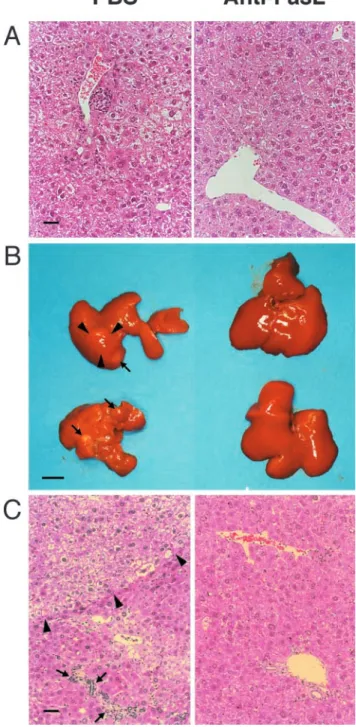
Prevention of Chronic Liver Disease Including Hepatocarcino- genesis by the Administration of Anti-FasL mAb. To evaluate the effect of anti-FasL mAb treatment on the development of chronic liver disease, including HCC, we histopathologi- cally examined the livers of mice that were treated with a control substance or with anti-FasL mAb and assessed tumor development by autopsy. 9 mo after splenocyte transfer, the control mice, which received PBS, displayed portal lympho- cytic infiltrates, lobular disarray, and marked variation in the size and shape of hepatocytes, reflecting long-term, persis- tent hepatitis (Fig. 2 A, left). In contrast, the mice treated with anti-FasL mAb showed minimal inflammatory infil- trates and no dysplastic or preneoplastic changes in the liver (Fig. 2 A, right). After one year or later, all the control ani- mals displayed marked hepatic atrophy and developed mul- tiple liver tumors, most of which were larger than 4 mm in diameter with the largest ranging up to 20 mm in diameter (Fig. 2 B, and Table II). The tumor specimens illustrated the classical histological features of HCC, consisting of relatively poorly differentiated hepatoma cells (Fig. 2 C, left). The sur- rounding hepatic parenchyma displayed focal lobular inflam- matory infiltrates associated with degenerating hepatocytes and marked lobular disarray. In contrast, most of the anti- FasL mAb-treated livers had an almost normal appearance Table I. Anti-FasL mAb Did Not Deplete HBsAg-specific CD8
T Lymphocytes
CD8
IFN-
stimulated with:
Treatment CD8
HBV S28–39 LCMV NP
Control IgG 15.4 0.5 0.18 0.02 0.02 Anti-FasL 16.1 0.8 0.13 0.02 0.02
Unmanipulated 9.1 1.1 0.02 0.02
IHLs were isolated from control IgG or anti-FasL mAb treated transgenic mice 7 d after the splenocyte transfer, or from unmanipulated transgenic mice. The numbers give the proportion of CD8
cells ([%] [number of CD8
cells]/[number of total cells] 100) or CD8
IFN-
cells ([%] [number of CD8
IFN-
cells]/[number of CD8
cells] 100) standard deviation.
Figure 2. Prevention of progressive liver dysplasia and HCC develop-
ment by anti-FasL mAb treatment. The transgenic mice described in the
legend to Fig. 1 were killed 9 (A) and 15 (B and C) mo after the spleno-
cyte transfer. (A) A 9-mo liver specimen with PBS (left) or anti-FasL
mAb treatment (right). (B and C) 15 mo after the splenocyte transfer, liv-
ers from PBS-injected animals displayed marked atrophy and multiple
liver tumors (arrows) up to 11 mm in diameter (arrowheads; B, left). A
representative specimen illustrates the classical histological features of
HCC (arrowheads), and the surrounding hepatic parenchyma displays fo-
cal lobular inflammatory infiltrates associated with degenerating hepato-
cytes (arrows; C, left). Most of livers from anti-FasL mAb-injected ani-
mals did not show apparent atrophy or liver tumors (B, right). A
representative specimen demonstrates minimal portal infiltrates and very
mild lobular disarray (C, right). Liver sections were stained with hema-
toxylin and eosin. The bars represent 40 m (A and C), and 10 mm (B).
both macro- and microscopically (Fig. 2, B and C, right).
Only two of the 15 animals developed solitary liver tumors, one of which was histologically classified as HCC (Table II).
Collectively, these results demonstrated that the hepatocar- cinogenesis was greatly suppressed by the administration of anti-FasL mAb in this chronic hepatitis model, suggesting that FasL expressed on liver-infiltrating CD8
T cells acti- vated the caspase cascade in the hepatocytes and induced hepatocellular apoptosis, liver inflammation, regeneration, and the eventual development of HCC.
Discussion
In this model, the major mechanism of hepatocyte in- jury seemed to be apoptosis, because we observed massive apoptotic hepatocytes associated with an elevation of the
serum ALT level. The anti-FasL mAb treatment markedly attenuated the hepatocyte apoptosis. However, because the proportion of HBsAg-reactive CD8
T cells to the to- tal population of infiltrates in the liver was so low (Table I), it is unlikely that FasL expressed on the surface of CTLs induced all the apoptosis. Recent studies have established that FasL has a proinflammatory activity (21–23). We have previously demonstrated that FasL induces the release of the activated form of proinflammatory cytokines such as IL-1 and IL-18 from neutrophils and/or macrophages (19, 20). Consistent with this, we found strong inflamma- tory infiltration in the liver and an elevated serum IL-18 level in this model, and anti-FasL mAb treatment reduced them (Fig. 1, B and D). Thus, it is more likely that the massive apoptosis was induced by inflammation that was exaggerated by FasL.
Table II. Prevention of Hepatocarcinogenesis by Anti-FasL mAb Treatment
Mouse ID
Mo after spl. transfer
Age (mo) at killing
No. of tumors
Largest tumor (mm)
aTumor histology
Treated with PBS or control hamster IgG (intraperitoneally)
110 9 16 1 2 Adenoma
113 8 15 2 2 Adenoma
189 9 15 1 4 Adenoma
266 13 17 1 11 HCC
276 15 18 1 5 HCC
323 15 18 2 11 HCC
341 15 18 4 3 HCC
343 14 17 11 3 HCC
391 15 17 3 7 HCC
6 17 19 1 10 HCC
55 17 19 3 10 HCC
59 20 22 2 20 HCC
Treated with anti-FasL mAb (intraperitoneally)
33 7 17 0 0
93 7 12 0 0
96 7 12 0 0
135 9 16 0 0
136 13 20 1 3 Adenoma
147 13 20 0 0
179 12 16 0 0
287 18 20 0 0
243 17 23 1 12 HCC
258 18 21 0 0
283 17 19 0 0
349 12 14 0 0
351 15 17 0 0
360 15 17 0 0
78 23 25 0 0
spl., splenocyte.
a