Acta Med. Nagasaki. 22: 110-115
Prosopagnosia with Acquired Colour Blindness
Isao FUJII* and Yoshibumi NAKANE**
Department of Neuropsychiatry, Nagasaki University School of Medicine,
Nagasaki, Japan
Received for publication, June 10, 1977
A 71-year-old male complained of a sudden attack of colour blindness and prosopagno- sia. The colour blindness was red-green defect. Left homonymous hemianopsia was confir- med by red-target perimetry. Brain scanning suggested the lesion was to be in the right in-
ferior occipital region. The symptoms disappeared spontaneously in three weeks. A 35-year- old male presented prosopagnosia and achromatopsia. These cases suggested that acquired colour blindness of central origin may exist and that the non-dominant hemisphere may play a specific role in the process of colour discrimination.
INTRODUCTION
Although prosopagnosia or a selective failure to recognize physiognomy or facial expression of persons previously known well was partly mentioned as a symptom in patients with cerebral lesions in the early leterature, this was for the first time systematically described by Bodamer3) in 1947 and proposed as one type of agnosia which is not directly connected with object agnosia. While prosopagnosia is often observed as an aspect of the clinical picture of severe object agnosia, he correctly emphasized that recognition of faces could be preserved in patients with object agnosia.
On the other hand, defective colour-vision can be caused by diseases or pathological changes of any part of the visual pathway before the chiasma opticus such as the eye media, the retina, and the optic nerve. However, there are very few reports of acquired colour blindness associated with cerebral focal lessions.
This paper reports two rare cases who showed prosopagnosia together with acquired colour blindness, probably caused by cerebrovascular disturbances. The findings of these cases are discussed with reference to cerebral localization of the lesions which appears to greatly contribute to the advancement of neuropsychology of perception or cognition.
*藤 井 薫 ,**中 根 允文
CASE REPORTS
Patient 1. -T. K. A right-handed man. A retired goverment official aged 71.
1) Present history
He was admitted on 31 October 1975 to our department, University Hospital. He was acutely ill, with symptoms of headache and trouble in recognizing colours and faces which started on 18 October 1975. Previously, his physician had pointed out that he had cardiomegalia with arrythmia when he was 45 years of age. His chief complaint during hospitalization was of inability to differentiate and recognize human faces he knew well.
He could not even recognize his own wife or his wife's sister by sight so that his wife seemed to be a woman he met for the first time, though he recognized them immediately by their voices. He could visualize in mind faces of persons long known to him but was unable to identify their faces from photographs. He found television disturbing, being unable to recognize properly a face which appeared in TV even though it was the face of a minister of the goverment whom he had been long acquainted with on business. He thought that the eyes of other people were changed in some way, a left-sided eye-ball appearing larger, and that their lips slanted upward, appearing in some way strangely darker.
2) Clinical and radiological examinations.
On neurological examination the only abnormalities suspected were in the visual fields. Uncorrected visual acuities were 0.2(r) and 0.4(l). Left homonymous hemianopsia was confirmed by only red-target perimetry. The colour blindness was red-green defect of a protan type, on the Ishihara's pseudoisochromatic plates.
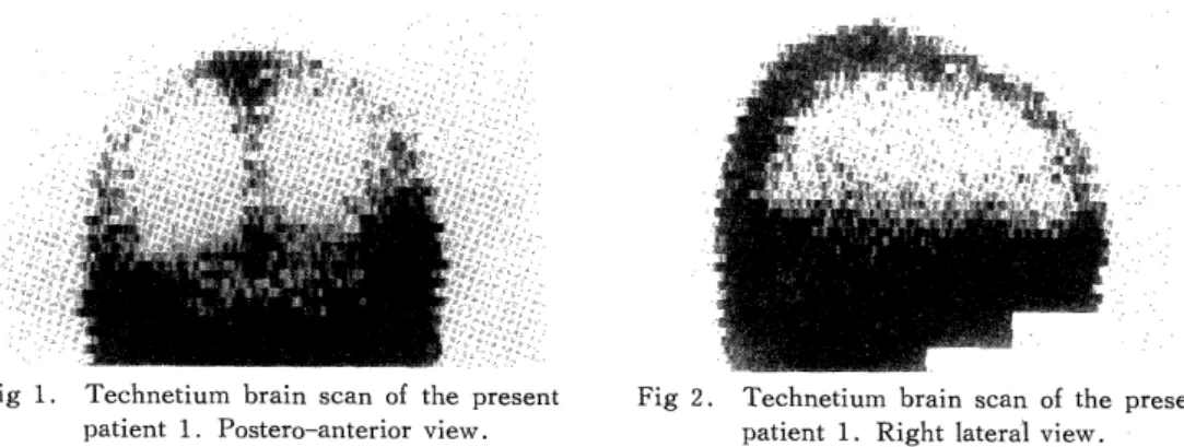
On Technetium brain scan, the hot spot was detected in the infero-posterior part of the right hemisphere. (Fig. 1 & 2)
On neuropsychological examination, he failed to recognize photographs of well-known public figures and also could not judge a person's age and sex from his face only. The patient showed slight difficulty in colour-matching, particularly failing with red. His WAIS verval IQ was 136, but he rejected the perfomance subtests. There were no symptoms nor signs of aphasia, apraxia, autotopagnosia, spatial agnosia and topographical
Fig 1. Technetium brain scan of the present patient 1. Postero-anterior view.
Fig 2. Technetium brain scan of the present patient 1. Right lateral view.
disorientation except for prosopagnosia. There was no change in the patient's condition until 5 November 1977. Thereafter his symptoms gradually improved and on 11 November he said that all his complaints disappeared. Uncorrected visual acuities were 0.5/0.7 and no colour blindness was detected. However, by the perimetry a slight hemianopic restriction in the left homonymous visual fields was detected by red-target only.
Patient 2. -H. K. A right-handed banker-businessman aged 35.
(1) Present history
On 11 April 1976, at a golf links, he complained of blurred vision of left eye when he saw the ball. On examination, uncorrected visual acuity was 0.3 for each eye and corrected one was 1.2/0.5. From 23 May 1976, he felt that other people had a more whitish complexion than usual. His chief complaint was of inability to differentiate and recognize human faces he knew well. He could not even recognize his own daughter by sight when she had changed her hair-style. Then he saw all faces as alike. He was referred to our department for thorough check-up on 2 July 1976.
(2) Clinical and radiological examination
On neurological examination, he showed no abnormal findings apart from the neuropsychological defects. On neuropsychological examination, he demonstrated prosop- agnosia, constructional apraxia and spatial dyslexia. However, autotopagnosia, spatial agnosia and topographical disorientation were not observed.
Uncorrected visual acuity was 0.2 for each eye and no visual field defect was found.
However, the Ishihara's test ended in a total failure. On the other hand, colour naming and colour matching were relatively good. His WAIS verbal IQ was 102, and he achieved total scaled score of 8 on the performance subtests.
Carotid and vertebral angiography revealed only a presence of persistent left primitive trigemineal artery. On Technetium scan and CT scan, there were no abnormal findings.
DISCUSSION
(1) Clinical picture
The main features of our unsusal 2 cases may be summarized as (1) selective failure to recognize faces previously known well, (2) acquired colour blindness, and (3) some evidence indicating right caudal cerebral lesions. (patient 1 : left visual field defect, patient 2 : right parietal syndromes). The faces of the nearest members of the family seem to be those of strangers. Patients are able to identify people by the help of addit- tional marks and signs not connected with the individual traits of the face. These can be
of different kinds, for example : voice, characteristic clothing, gait, peculiar hair style, etc. The phenomenon of prosopagnosia may appear as an isolated and pure manifestation, but the great majority of investigators8) are of the opinion that such pure cases are very rare. Our case 1 is such a rare case and also a reversible one. This phenomenon almost always appears in connection with other dysfunctions of gnosis, praxis, and topographic
orientation. True prosopagnosia is usually not connected with visual object agnosia and colour agnosia. (Table 1.)
2) The anatomical backgrouna
In the majority of published cases of prosopagnosia with autopsy findings, there is evidence of bilateral brain disease, although in some there is evidence only of right hemisphere involvement. Among the 9 cases of prosopagnosia caused by cerebrovascular lesions reported in the literature, 21. 9, 15, 16, 7, 12, 2, 4) some of the patients had multiple asymmetrical lesions, but all showed at least one occipital lobe lesion. (Table. 2) COHN et a14) pointed out that in all reported cases one or both fusiform gyri were destroyed, and there were variably sized lesions in the pericalcarine structures. Especially PEVZNER et al'6 presented a 54-year-old man who, together with failure to recognize faces previously he knew well, showed achromatopsia. At autopsy a narrow cavity was observed in the
Table 1. Associated symptoms in the present patients.
Case 1 Case 2
spatial dyslexia - +
constructional apraxia - +
apraxia for dressing - -
topographical disturbances - -
metamorphopsia of face + +
(vestibular disturbances) - +
unilateral spatial agnosia ± -
color agnosia - -
Table 2. Autopsy findings of cases with prosopagnosia.
Wilbrand (1892) bilateral occipital lobes
rt. calcarine fissure
Heidenhain (1927) bilateral calcarine fissures
rt. hypocampal gyrus
Nielsen (1946) bilateral occipital lobes (PCA)
Pevzner (1962) rt. calcarine fissure
It. angular gyrus
Gloning (1970) bilateral fusiform gyri
rt. lingual gyros
Lhermitte (1972) bilateral fusiform gyri
rt. lingual & hyppocampal gyri
Benson (1974) bilateral fusiform gyri
splenium
Cohn I (1977) bilateral fusiform & hypocampal gyri
It. lingual gyrus
II bilateral occipital lobes (PCA)
PCA : posterior cerebral artery
right hemisphere 3 cm anterior to the occipital pole and the lesion involved the lingual and fusiform gyri. The left cerebral hemisphere showed a lesion over the confluence between temporo-parietal lobes. Hence it is thought that prosopagnosia may correlate with right hemisphere lesions in the occipito-temporal region. Our patient 1 has the hot spot on brain scan in the right inferior occipital region.
3) Acquired colour blindness
In analysing the clinical signs of acquired colour defect of central origin, CRITCHLEYS) could not present definite evidence. LHERMITTE et ally studied 110 brain-damaged with homonymous hemianopsia and found disturbance of colour perception in 42 patients, of whom 32 had a left homonymous hemianopsia. SCOTTI and SPINNLER10) investigated the relationship between response to colour tasks and hemisphreric side of lesion in 164 unselected hemisphere-damaged patients, by means of Farnsworth-Munsell 100 Hue Test.
Their study showed that rate of occurrence of colour defect was higher in the right hemisphere - damaged patients than in the left hemisphere - damaged patients and that patients with visual field defects had more colour defects than those without visual field defects. MEADOWS 141 demonstrated that patients with prosopagnosia nearly always had a left homonymous upper quadrantic visual field defect, correlated clinically and pathologi- cally with a right occipito-temporal lesion. But he made no mention of colour blindness.
ALBERT et all) reported a case of hemianopic colour blindness and presumed that the right hemisphere might be greatly involved in the process of ciscrimination of sensory inputs, taking account into consideration the study in which DE RENZI et all) suggested a diffe- rential specialization of the two hemispheres for visual recognition. At present over the world 7 cases 10. 17, 18, 20, 13) including our 2 cases with prosopagnosia have been studied objectively on colour perception by means of the isochromatic colour plates or hues of colours. Among them 5 cases demonstrated some colour blindness. Our two cases with prosopagnosia suggested that acquired colour blindness of central origin may exist, and that the right (non-dominant) cerebral hemisphere may play a specific role in the process of colour discrimination and comparison.
A part of this work was presented at the 18th Annual Meeting of the Japanese Neurological Society, May 1977
REFERENCES
1) ALBERT, M.L., RECHES, A., & SILVERBERG, R.(1975) Hemianopic colour blindness.
.1. Neurol. Neurosurg. Psychiat. 38 : 546-549.
2) BENSON, D.F., SEGARRA, J., & ALBERT, M.L. (1974) Visual Agnosia-Prosopagnosia.
Arch. Neurol. 30 : 307-310.
3) BODAMER, J. (1947) Die Prosop-Agnosie. Arch. Psychiat. Nervenkr. 179 : 6-53.
4) COHN, R., N EUMANN, M. A . , & WOOD, D. H. (1977) Prosopagnosia : A clinicopa-
thological study. Annals of Neurology, 1 : 177-182.
5) CRITCHLEY, M. (1965) Acquired anomalies of colour perception of central origin.
Brain, 88: 711-724.
6) De RENZI, E., SCOTTI, G., & SPINNLER, H. (1969) Perceptual and associative disorders of visual recognlion. Neurology 19 : 634-642.
7) GLONING, I . , GLONING, K . , & JELLINGER, K. (1970) A case of "prosopagnosia"
with necropsy findings. Neuropsychologia 8 : 199-204
8) HECAEN, H. & ANGELERGUES (1962) Agnosia for faces (Prosopagnosia). Arch.
Neurol. 7 : 24-32.
9) HEIDENHAIN, A. (1927) Beitrag zur Kenntnis Seelenblindheit. Monatsschr. Psychiatr.
Neurol. 66 : 61-116.
10) KoRNER, F., REGLI, F., & HAYNAL, (1967) Ein durch Farbsinnstorung, Prosopagnosie
and Orientierungs-storung charakterisierte visuelle Agnosie. Arch. Psychiat. Nervenkr.
209 :1-20.
11) LHERMITTE, F., CHAIN, F., ARON, D., et al (1969) Les troubles de la vision des couleurs dans les lesions posterieures du cerveau. Rev. Neurol. 121 : 5-29.
12) LHERMITTE, F., CHAIN, F., ESCOUROLLE, B., et al (1972) Etude anatomo-clinique
d'un cas de prosopagnosie. Rev. Neurol. 126 : 329-346.
13) LHERMITTE, F. & PILLON, B. (1975) La prosopagnosie. Role de l'hemisphere droit dans la perception visuelle. Rev. Neurol. 131 : 719-812.
14) MEADOWS, J. C. (1974) The anatomical basis of prosopagnosia. J. Neurol. Neurosurg.
Psychiat. , 37 : 489-501.
15) NIELSEN, J.M. (1946) : Agnosia, Apraxia, Aphasia. New York, Hoeber.
16) PEVZNER, S., BORNSTEIN, B., & LOEWENTHAL, M. (1962) Prosopagnosia. J. Neuro.
Neurosurg. Psychiatry 25 : 336-338.
17) RONDOT, P., TZAVARAS, A., & GARCIN, R. (1967) Sur un cas de prosopagnosie
persistant depuis quinze ans. Rev. Neurol. 117: 424-428.
18) RTJBENS, A.B. & BENSON, D.F. (1971) Associative visual agnosia. Arch. Neurol. 24:
305-316.
19) SCOTTI, G. & SPINNLER, H. (1970) Colour imperception in unilateral hemisphere- damaged patients. J. Neurol. Neurosurg. Psychiat. 33:22-28.
20) TZAVARAS, A., MERIENNE, L., et MASURE, M. D. (1973) Prosopagnosie, amnesie et troubles du langage par lesion temporale gauche chez un sujet gaucher. Encephale
62 :382-394.
21) WILBRAND, H. (1892) Ein Fall von Seelenblindheit and Hemianopsie mit Sektionsbefund.
Dtsch. Zschr. Nervenheilk. 2 : 361-387.