ORIGINAL ARTICLE
1HXURQDOGL൵HUHQWLDWLRQSRWHQWLDORIPRXVHPHVHQFK\PDOVWHPFHOOVGHULYHGIURP ZKLWHYHUVXVEURZQDGLSRVHWLVVXH
Yohshiro Nitobe1),Gentaro Kumagai1),Ayako Sasaki1),Toshihide Nagaoki1),Kanichiro Wada1), Sunao Tanaka1),Toru Asari1),Ken-Ichi Furukawa2),and Yasuyuki Ishibashi1)
Abstract: Mesenchymal stem cells (MSCs) from white adipose tissue (WAT) have a higher osteogenic differentiation potential than those from brown adipose tissue (BAT). The purpose of this study was to compare the neuronal differentiation potentials of these cells. In addition, to examine the applicability of these cells for autologous neuronal regeneration, the mouse hind limbs were assessed for neurological function after adipose tissue was harvested from the inguinal and interscapular regions. Adipose-MSCs were isolated from WAT of the inguinal subcutaneous regions and BAT of the interscapular regions in 6-week-old female C57BL/6J mice. The expression of cell-surface markers on the adipose-MSCs was examined by flowcytometry. The markers examined were CD90, Sca1, CD34, and CD45.
Neuronal differentiation was assessed by immunocytochemistry using NeuN as a mature neuron marker. MSCs derived from WAT and BAT displayed similar immunophenotypes. Both cell populations differentiated into NeuN- positive cells (16.2% ± 1.0% of the WAT-MSCs and 14.7% ± 2.4% of the BAT-MSCs) in vitro. Harvesting the adipose tissue did not affect the neurological properties of the hind limbs. These findings indicate that both WAT-MSCs and BAT-MSCs may be attractive cells for autologous neuronal regeneration.
Hirosaki Med.J. 71:12―20,2020
Key words: Autologous; Brown adipose tissue; Mesenchymal stromal cells; Neuronal differentiation;
White adipose tissue.
1) Department of Orthopedic Surgery, Hirosaki University Graduate School of Medicine, Hirosaki, Aomori, Japan
2) Department of Pharmacology, Hirosaki University Graduate School of Medicine, Hirosaki, Aomori, Japan.
Correspondence: G. Kumagai
Received for publication, November 20, 2019 Accepted for publication, April 10, 2020
Introduction
Mesenchymal stem cells (MSCs) can be harvested from autologous donors and have the capacity to self-renew as well as the potential to differentiate into multiple lineages
1). These char- acteristics make MSCs very attractive for use in cell therapy and regenerative medicine
2). MSCs are found in various tissues, including bone marrow
3), umbilical cord blood
4), placenta
5), and adipose tissue
6). Adipose tissue is an accessible, abundant, and reliable source for the isolation of adult stem cells suitable for tissue engineering and regenerative medicine applications
7), and the treatment efficacy of adipose tissue-derived MSCs (AdMSCs) for neuronal regeneration in animal models has been reported
2, 8).
There are two types of adipose tissue in
mammals, white and brown. White adipose
tissue (WAT) contains lipids that act as ener-
getic fuel or as lipid reconstructive material, and
brown adipose tissue (BAT) can use fatty acids
for thermogenesis
9). WAT and BAT have differ-
ent antigenic features and cell differentiation
potentials, including osteoblasts, endothelial cells,
adipocytes, hematopoietic cells, and cardiomyo-
blasts. In these differentiation potentials, BAT-
derived cells display less plasticity
10). White
adipose-derived stem cells can differentiate into
neuronal and glial cells in vitro
11). Therefore,
WAT has been thought to be more plastic than
BAT and a more suitable source of stem cells
10).
The subcutaneous inguinal WAT is located
running ventrally across the femoral nerve
12-14).
Material and Methods
Animals
Six-week-old female C57BL/6J mice were used as the source of adipose tissue (CLEA Japan, Inc., Shizuoka, Japan) . All animal procedures were carried out in accordance with the guidelines of the institutional animal care and research advisory committee of Hirosaki University.
,VRODWLRQDQGFXOWXUHRIDGLSRVHWLVVXHGHULYHG 06&V$G06&VIURPLQJXLQDODQGLQWHUVFDSXODU
adipose tissuesAdMSCs were isolated from adult mouse adipose tissues (n=60) as previously described with minor modifications
19). WAT was dissected from the inguinal subcutaneous regions, while BAT was dissected from the interscapular region. The weight of the mice and resected adipose tissues was measured (n=10 each group). To confirm the identity of the WAT and BAT, each adipose tissue was assessed by HE staining. Adipose tissue samples were pooled in ice-cold DMEM, and then minced and digested with 10 ml of 0.2% collagenase type I at 37°C for 45 min. The resultant cell suspension was filtered through a 70-ȝm mesh to remove tissue debris, and then the collagenase was removed by centrifugation for 5 min. The cell pellet fraction was suspended in RBC lysis buffer (Becton Dickinson, Franklin Lakes, NJ, USA) to remove contaminating red blood cells.
The cells were resuspended in DMEM supple- mented with 10% fetal bovine serum (FBS), and then washed twice. The cells were then cultured with complete MesenCult
TM(MesenCult MSC Basal Medium [Mouse] supplemented with serum-containing MesenCult MSC Stimulatory Supplements [Mouse], both from Stem Cell Technologies, Vancouver, Canada) supplement- ed with 1% penicillin/streptomycin (Thermo Fisher Scientific, Waltham, MA, USA). After 24 h, the non-adherent cells were removed, and the So, harvesting inguinal WAT may affect the
motor and sensory function. BAT may become a suitable cell source for autologous regenera- tion research instead of WAT. However, reports on the neuronal differentiation potential of MSCs derived from BAT are limited, and there are no studies that have compared the neuronal differ- entiation potential between WAT-MSCs and BAT-MSCs. It is also unclear whether harvesting inguinal subcutaneous adipose tissue and inter- scapular adipose tissue affects the motor and sensory function in mice.
The repair effects of MSC transplants depend on the degree of their survival
15). Whether transplantations were autologous or allogeneic was important in transplantation experiments because cell survival rate was affected
16). In allogeneic transplantation studies, MSCs could not survive even 1 month
15, 17). However, MSCs could survive 3 months and could differentiate into neuronal and glial cells in an autologous transplantation study
18). Autologous transplanta- tion may promote the effect of MSCs thanks to long cell survival. Because adipose tissue is thought to be a suitable cell source for autolo- gous transplantation as the clinical trial showed
2), the potential effects of its harvesting and the neuronal differentiation potential should be carefully assessed before pre-clinical studies are performed.
We hypothesized that BAT should be used as
a cell source for autologous neuronal regenera-
tion in pre-clinical studies. The goal of this study
was to assess whether adipose tissues, WAT or
BAT, are suitable for autologous neuronal
regeneration. First, the neuronal differentiation
potential of MSCs derived from WAT and BAT
was compared. Second, whether the harvesting
of inguinal and interscapular adipose tissue
affected the neurological function of the hind
limbs was investigated.
adherent cells were expanded by serial passage.
When the cells reached 80~90% confluence, the adherent cells were detached with 0.125%
trypsin and 0.01% EDTA and replated at a density of 5000 ‒ 25,000 cells/cm
2. The live cell counts were performed with a TC20
TMAuto- mated Cell Counter (Bio-Rad Laboratories, Hercules, CA, USA). The second passage (P2)
adipose MSCs were used in the experiments
(n=10 in WAT-MSCs, n=8 in BAT-MSCs).
&HOOVXUIDFHPDUNHUVRI$G06&VIURP:$7DQG
BATConfluent mouse MSCs were analyzed using flowcytometry for the expression of cell-surface antigens CD90 (PE; Thermo Fisher Scientific), Sca1 (PE/Cy7; Becton Dickinson), CD34 (Alexa Fluor 647; Becton Dickinson), and CD45 (Alexa Fluor 700; Thermo Fisher Scientific). Two positive surface markers (CD90 and Sca1) and two negative surface markers (CD34 and CD45)
were used to identify mouse adipose MSCs.
Gating was set using rat monoclonal isotype antibodies which were used to detect any non- specific fluorescence, and in each analysis, at least 10000 events were collected. The cell- surface antigens of MSC markers were compared between WAT and BAT (n=3 in CD90, n=7 in Sca-1, CD34 and CD45).
&HOO6HSDUDWLRQRI$G06&VE\PDJQHWLFDFWLYDWHG
cell sorting (MACS)To avoid stem-cell heterogeneity, MSCs were sorted by MACS (Miltenyi Biotec, Bergisch Gladbach, Germany) before the assessment of their differentiation potential. AdMSCs were collected using Anti-Human/Mouse CD90-PE
(1:100; Thermo Fisher Scientific). Anti-PE microbeads (1:2, Miltenyi Biotec) were used for the MACS separation of the MSCs. The target cell-labeled microbeads were trapped in a magnetic field and later collected as a positive fraction. MSCs isolated from WAT and BAT are
hereafter referred to as WAT-MSCs and BAT- MSCs.
1HXURQDOGLIIHUHQWLDWLRQSRWHQWLDORI:$7DQG
%$706&V
Neuronal differentiation was performed as previously described
20). Experiments were repeated three times. Adipose-derived stem cells were harvested and transferred onto a sterile glass coverslip (1 × 10
4cells/cm
2) for immunos- taining. Cells were allowed to settle for at least 6 h and were then washed twice with serum-free DMEM. Neuronal differentiation was induced in Neurobasal medium (Thermo Fisher Scientific)
containing 1% FCS, 1 × B27 supplement, 0.5 mM 1-methyl-3-isobutylxanthine, 1
ȝM dexametha- sone, 50
ȝM 8CPT-cAMP, 10 mM valproic acid, and 10
ȝM forskolin. The neuronal differentia- tion medium was changed weekly. The cells were fixed with 4% paraformaldehyde in PBS for 30 min, and rinsed three times in PBS. The cells were blocked for 1 h with 10% (v/v) FBS, 3% (w/v) bovine serum albumin (BSA) in 0.1 M PBS (pH 7.4), 0.3 M sodium chloride, and 0.5% Triton X-100 (PBST), and incubated overnight at 4°C with primary antibodies in FBS+BSA+PBST. The cells were then washed three times with FBS+BSA+PBST, incubated for 1 h at room temperature with PBS/0.1%
BSA containing secondary immunofluorescent antibodies, and washed three times with PBS.
The cells were then incubated with murine- specific antibodies directed against NeuN
(Thermo Fisher Scientific; Alexa-555), a marker of neuronal cells. The samples were inspected under a digital microscope system (BZ-X700, Keyence, Osaka, Japan). The total cells and NeuN-positive cells were counted under a mi- croscope, and the ratio of NeuN-positive cells to total cells was determined.
1HXURORJLFDODVVHVVPHQWRIKLQGOLPEV
A resection group, in which both WAT and
BAT were resected, and a no resection (con- trol) group were tested for hind-limb motor function and mechanosensitivity every week from six weeks of age (the time of surgery)
until 12 weeks of age (n=8 at each time point).
Two non-biased observers analyzed hind-limb performance using the Basso Mouse Scale
(BMS) for locomotor scoring
21). Motor coordina- tion was also evaluated by a rotarod test at a speed of 10 rpm before resection and every 2 weeks after the adipose resection
22). Hind-paw withdrawal thresholds (measured at 5 grams)
in response to an innocuous mechanical stimulus were measured using a Dynamic Plantar Aesthesiometer set (Mechanical test)
23). In this test, mice were placed in Plexiglas containers resting on an elevated glass surface. A mobile infrared emitter below the glass was placed under the center of the mouseʼs plantar hind paw, and activating the emitter started a timer
24). The withdrawal latency was defined as the duration between the activation and termination
of the infrared stimulus (measured in seconds);
the final withdrawal latency was recorded as the mean of three measurements. A plantar test
(heat test) was used to assess reactions to heat.
Statistical analysis
All data are presented as the mean value ± standard deviation (S.D.). Differences between two groups were evaluated for statistical signifi- cance using Mann-Whitney U test. Differences were considered significant at p<0.05.
Results
,VRODWLRQDQGFXOWXUHRI$G06&VIURPLQJXLQDO
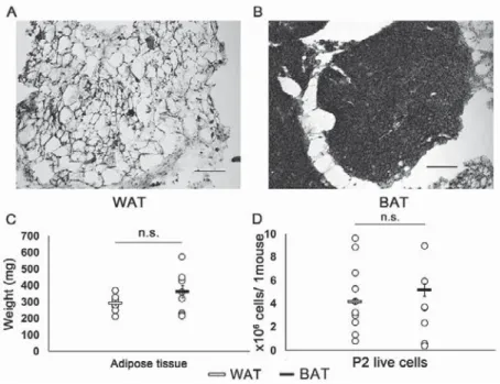
and interscapular adipose tissuesLarge fat droplets that were regarded as the WAT were seen more in inguinal subcutaneous regions than in interscapular regions (Fig. 1A).
In interscapular regions, there were small fat droplets that were regarded as the BAT (Fig.
1B). The average body weight of the 6-week-old
Fig. 1 Isolation and culture of AdMSCs from WAT and BAT
(A, B) HE staining of inguinal and interscapular adipose tissues. (A) Inguinal subcutaneous adipose tissue showed large fat droplets, which were defined as white adipose tissue (WAT). (B) Interscapular adipose tissue showed small fat droplets, which were defined as brown adipose tissue (BAT). Scale bars = 100 ȝm.
(C) Comparison of the weight of WAT and BAT that were dissected from one mouse (n = 10/group). (D)
Numbers of live cells in the passage 2 that were cultured per mouse (WAT n = 10, BAT n = 8).
mice was 17.6 g ± 0.6 g. The average weight of the WAT was 296 mg ± 45 mg, and that of the BAT was 366 mg ± 118 mg. There was no significant difference in the weight of adipose tissue per mouse between WAT and BAT (p = 0.165; Fig. 1C). In P2, the number of adherent live cells was 4.5 ± 3.0 × 10
6cells/mouse from WAT, and 3.2 ± 3.0 × 10
6cells/mouse from BAT. There was no significant difference in the number of harvested cells per mouse between WAT and BAT (p = 0.32; Fig. 1D).
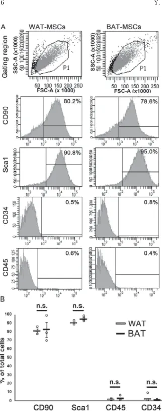
&HOOVXUIDFHPDUNHUVRI$G06&VIURP:$7DQG
BATFlow cytometric analysis showed positive expression for CD90 and Sca1, and low expres- sion of CD34 and CD45, indicating that cells derived from WAT and BAT had MSCs charac- teristics (Fig. 2A). Mean surface immunopheno- typed expression values and standard deviations of WAT-MSCs and BAT-MSCs were as follows:
CD90 (80.8 ± 2.6% and 82.3 ± 8.7%, respectively)
and Sca1 (90.1 ± 1.8% and 94.6 ± 2.2%)and negative for CD34 (0.9 ± 0.8%, 2.0 ± 2.6%) and CD45 (1.4 ± 3.1%, 0.5 ± 0.3%) (Fig. 2B). The percentage of CD90-positive, Sca1-positive, CD34-negative, and CD45-negative cells showed no significant difference between WAT-MSCs and BAT-MSCs (p = 1.00,
p = 0.089, p = 0.81, p = 0.32, respectively; Fig. 2B).1HXURQDOGLIIHUHQWLDWLRQSRWHQWLDORI:$7DQG
%$706&V
Both WAT-MSCs and BAT-MSCs differenti- ated into NeuN-positive cells (Fig. 3A). Cells positive for the mature neuron marker, NeuN, represented 16.2% ± 1.0% of the WAT-MSCs and 14.7% ± 2.4% of the BAT-MSCs (Fig. 3B).
There was no significant difference in the percentage of NeuN-positive cells between WAT and BAT (p = 0.45; Fig. 3B).
Fig. 2 Cell-surface markers of AdMSCs
(A) Typical flowcytometry profiles of white adipose tissue (WAT)-mesenchymal stem cells (MSCs) and brown adipose tissue (BAT)-MSCs. Rat isotype antibodies served as control and at least 10000 labeled cells were acquired. (B) Comparison of the cell-surface marker expression in MSCs derived from WAT and BAT (n=3 in CD90, n=7 in Sca-1, CD34 and CD45).
1HXURORJLFDODVVHVVPHQWRIKLQGOLPEV
The BMS score was 9 in both the resection and control groups (Table 1) . The rotarod score was 119 ± 2.5 ‒ 120 ± 0 seconds throughout the experimental period in the resection group, and 117 ± 4.7 ‒ 120 ± 0 second in the control group
Table 1. Neurological assessment of hind limbs
Week after resection pre 2 weeks 4 weeks 6 weeks
BMS (score) resection 9±0 9±0 9±0 9±0
control 9±0 9±0 9±0 9±0
Rotarod test (sec) resection 120±0 119±1.5 120±0 119±2.5
control 117±4.7 120±0 117±7.8 119±2.0
p-value 0.107 0.334 0.334 0.855
Dynamic plantar test (g) resection 5.7±1.1 6.0±0.8 5.3±0.5 6.1±0.9
control 5.9±0.9 5.4±0.8 4.8±0.8 5.9±0.7
p-value 0.625 0.154 0.135 0.591
Plantar test (sec) resection 6.6±1.7 6.8±1.2 6.4±1.7 5.8±1.9
control 6.4±1.7 7.6±1.3 6.8±1.5 6.8±2.4
p-value 0.776 0.213 0.553 0.365
The BMS (Basso Mouse Scale), Rotarod test, Dynamic plantar test, and Plantar test were analyzed 6 weeks after both white adipose tissue (WAT) and brown adipose tissue (BAT) resection. The averages were compared with the no resection (control) group. n=8 for each group.
Fig. 3 Neuronal differentiation potential of WAT-MSCs and BAT-MSCs
(A) After neuronal induction, white adipose tissue (WAT)- and brown adipose tissue (BAT)- mesenchymal stem cells (MSCs) exhibited morphology similar to neuronal cells and were positive for NeuN. Scale bars = 100 ȝm. (B) Percentage of total cells that were NeuN-positive (n = 3/group).
(Table 1) . In the mechanical test, the withdrawal
thresholds throughout the experimental period
were 5.3 ± 0.5 ‒ 6.1 ± 0.9 g and 4.8 ± 0.8 ‒ 5.9 ± 0.9
g in the resection group and control group,
respectively (Table 1). In the heat test, the
withdrawal latencies throughout the experimen-
tal period were 5.8 ± 1.9 ‒ 6.8 ± 1.2 seconds in the resection group and 6.4 ± 1.7 ‒ 7.6 ± 1.3 seconds in the control group (Table 1) . Studentʼs t-test showed no significant difference in any of the behavioral tests between the resection group and the control group. These findings indicated that the mice did not display any hind limb paralysis after undergoing the resection of both WAT and BAT.
Discussion
AdMSCs were isolated at around 4.0 × 10
6cells/mouse from WAT in the inguinal subcuta- neous regions, and 3.0 × 10
6cells/mouse from BAT in the interscapular region in P2. The WAT- and BAT-MSCs differentiated similarly into mature neurons (16.2% in WAT-MSCs and 14.7% in BAT-MSCs). The adipose resection did not affect the neurological functions of the hind limbs.
In this study, WAT- and BAT-MSCs could be isolated from one mouse (Fig. 1D). Only a few studies have assessed the number of live cells isolated from one mouse, so it has been unclear whether enough MSCs could be isolated from one mouse for autologous transplantation.
In previous studies on neuronal regeneration in mice, the number of transplanted no-induced- MSCs was 2.0 × 10
5cells/mouse
25), and neuronal induced-MSCs was 2.5 × 10
5cells/mouse
26). Thus, our findings indicate that sufficient amount of AdMSCs is possible to be isolated for autologous neuronal regeneration in mice.
In a previous study, only a minority of the CD45-negative fraction was positive for CD90 and Sca1 in the BAT-stroma vascular fraction
10). In the present study, MSCs derived from WAT and BAT displayed similar immunophenotypes:
CD90(+), Sca1(+), CD34(‑), CD45(‑). Our method for preparing MSCs has several differ- ences from those of the previous study, including the cell culture material, the number of passages,
and the cell-surface markers used for flowcy- tometry. The Mesencult
TMmedium, which was used in this study, is reported to be a good choice for AdMSCs, which exhibit a stable morphology and surface marker expression in this medium
27). Using our methods, we were able to isolate and culture BAT-MSCs successfully.
This study demonstrated that there were no significant differences in the neuronal differenti- ation potential between WAT and BAT. A previous in vitro study showed that the osteo- genic differentiation potential is lower in BAT than in inguinal WAT
10). Adipocytes in the neck were generated from the neuroectoderm rather than mesoderm
28). The difference between neu- ronal and osteogenic differentiation potentials may be caused by the origin (neuroectoderm or mesoderm) of the adipose tissue. WAT is also reported to be a more suitable source of stem cells than BAT
10). Nevertheless, the present study revealed that BAT might be a useful source of MSCs that have a similar neuronal differentiation potential as those from WAT.
In many studies, inguinal adipose tissue, which is near the femoral nerve, is harvested after sacrifice of the mouse
13, 19, 26). Because few studies have performed behavioral tests after adipose resection, it was unclear whether harvesting the inguinal adipose tissue affects hind limb function. Here we found that adipose resection did not affect the motor or sensory function in mice. Notably, motor and sensory behavioral tests are often performed to assess the effect of MSCs transplantation after spinal cord injury
25, 29). Therefore, it was important to confirm that the resection of inguinal and interscapular adipose tissue itself does not affect the neurological assessment of the hind limbs of mice.
There were some limitations in this study.
First, the MSC differentiation potentials were
assessed only in vitro, and the findings still need
to be confirmed by in vivo studies. Second, we
did not compare the glial differentiation potential between WAT-MSCs and BAT-MSCs. Finally, other behavioral and electrophysiological tests were not performed in this study. Because the WAT from inguinal adipose tissue is located near the femoral nerve, its dissection may affect some other tests of hind limbs.
In conclusion, we isolated AdMSCs from BAT, and found that BAT-MSCs and WAT-MSCs differentiated similarly into mature neurons in vitro. The harvesting of adipose tissue did not affect the neurological properties of the mouse hind limbs. These results suggest that both WAT and BAT are suitable as a cell source for autologous transplantation research.
'HFODUDWLRQRI&RQÀLFWLQJ,QWHUHVWV
The authors declare that there is no conflict of interest.
$FNQRZOHGJHPHQWV
We thank members of the Department of Orthopedic Surgery at Hirosaki University for helpful discussions. We also thank Dr. Chikara Ohyama, Mr. Tohru Yoneyama, and Mr. Kenji Kabasawa of the Department of Urology, Hirosaki University Graduate School of Medicine, for providing technical assistance with the flow cytometric analysis. This study was funded by the Karoji Memorial Fund for Medical Research at Hirosaki University, Grants-in-Aid for Scientific Research from the Japan Society for the Promotion of Science (17K10917), and a Hirosaki University Grant for Distinguished Researchers
(FY2017-2018).
5HIHUHQFHV
1)Azari MF, Mathias L, Ozturk E, Cram DS, Boyd RL, Petratos S. Mesenchymal stem cells for treatment of CNS injury. Curr Neuropharmacol.
2010;8:316-23.
2)Ra JC, Shin IS, Kim SH, Kang SK, Kang BC, Lee HY, Kim YJ, et al. Safety of intravenous infusion of human adipose tissue-derived mesenchymal stem cells in animals and humans. Stem Cells Dev. 2011;20:1297-308.
3)Friedenstein AJ, Chailakhjan RK, Lalykina KS.
The development of fibroblast colonies in mono- layer cultures of guinea-pig bone marrow and spleen cells. Cell Tissue Kinet. 1970;3:393-403.
4)Erices A, Conget P, Minguell JJ. Mesenchymal progenitor cells in human umbilical cord blood. Br J Haematol. 2000;109:235-42.
5)Zhang Y, Li CD, Jiang XX, Li HL, Tang PH, Mao N. Comparison of mesenchymal stem cells from human placenta and bone marrow. Chin Med J.
(Engl) 2004;117:882-7.
6)Zuk PA, Zhu M, Mizuno H, Huang J, Futrell JW, Katz AJ, Benhaim P, et al. Multilineage cells from human adipose tissue: implications for cell-based therapies. Tissue Eng. 2001;7:211-28.
7)Kokai LE, Marra K, Rubin JP. Adipose stem cells:
biology and clinical applications for tissue repair and regeneration. Transl Res. 2014;163:399-408.
8)Aras Y, Sabanci PA, Kabatas S, Duruksu G, Subasi C, Erguven M, Karaoz E. The effects of adipose tissue-derived mesenchymal stem cell transplanta- tion during the acute and subacute phases follow- ing spinal cord injury. Turk Neurosurg. 2016;26:
127-39.
9)Giralt M, Martin I, Iglesias R, Viñas O, Villarroya F, Mampel T. Ontogeny and perinatal modulation of gene expression in rat brown adipose tissue.
Unaltered iodothyronine 5'-deiodinase activity is necessary for the response to environmental temperature at birth. Eur J Biochem. 1990;193:297- 302.
10)Prunet-Marcassus B, Cousin B, Caton D, André M, Pénicaud L, Casteilla L. From heterogeneity to plasticity in adipose tissues: site-specific differences.
Exp Cell Res. 2006;312:727-36.
11)Safford KM, Safford SD, Gimble JM, Shetty AK, Rice HE. Characterization of neuronal/glial differentiation of murine adipose-derived adult
stromal cells. Exp Neurol. 2004;187:319-28.
12)Kochi T, Imai Y, Takeda A, Watanabe Y, Mori S, Tachi M, Kodama T. Characterization of the arterial anatomy of the murine hindlimb: functional role in the design and understanding of ischemia models. PLoS One. 2013;8:e84047.
13)de Jong JM, Larsson O, Cannon B, Nedergaard J.
A stringent validation of mouse adipose tissue identity markers. Am J Physiol Endocrinol Metab.
2015;308:E1085-105.
14)Pratt SJP, Lovering RM. A stepwise procedure to test contractility and susceptibility to injury for the rodent quadriceps muscle. J Biol Methods.
2014;1:e8.
15)Ritfeld GJ, Rauck BM, Novosat TL, Park D, Patel P, Roos RA, Wang Y, et al. The effect of a polyurethane-based reverse thermal gel on bone marrow stromal cell transplant survival and spinal cord repair. Biomaterials. 2014;35:1924-31.
16)Muraoka K, Shingo T, Yasuhara T, Kameda M, Yuan W, Hayase H, Matsui T, et al. The high in- tegration and differentiation potential of autolo- gous neural stem cell transplantation compared with allogeneic transplantation in adult rat hippo- campus. Exp Neurol. 2006;199:311-27.
17)Ritfeld GJ, Patel A, Chou A, Novosat TL, Castillo DG, Roos RA, Oudega M. The role of brain-derived neurotrophic factor in bone marrow stromal cell- mediated spinal cord repair. Cell Transplant. 2015;
24:2209-20.
18)Zurita M, Vaquero J, Bonilla C, Santos M, De Haro J, Oya S, Aguayo C. Functional recovery of chronic paraplegic pigs after autologous trans- plantation of bone marrow stromal cells. Trans- plantation. 2008;86:845-53.
19)Yamamoto N, Akamatsu H, Hasegawa S, Yamada T, Nakata S, Ohkuma M, Miyachi E, et al. Isolation of multipotent stem cells from mouse adipose tissue. J Dermatol Sci. 2007;48:43-52.
20)Boulland JL, Mastrangelopoulou M, Boquest AC, Jakobsen R, Noer A, Glover JC, Collas P. Epigenetic regulation of nestin expression during neurogenic differentiation of adipose tissue stem cells. Stem
Cells Dev. 2013;22:1042-52.
21)Basso DM, Fisher LC, Anderson AJ, Jakeman LB, McTigue DM, Popovich PG. Basso Mouse Scale for locomotion detects differences in recovery after spinal cord injury in five common mouse strains. J Neurotrauma. 2006;23:635-59.
22)Brooks SP, Trueman RC, Dunnett SB. Assessment of Motor Coordination and Balance in Mice Using the Rotarod, Elevated Bridge, and Footprint Tests. Curr Protoc Mouse Biol. 2012;2:37-53.
23)Chaplan SR, Bach FW, Pogrel JW, Chung JM, Yaksh TL. Quantitative assessment of tactile allodynia in the rat paw. J Neurosci Methods. 1994;
53:55-63.
24)Hargreaves K, Dubner R, Brown F, Flores C, Joris J. A new and sensitive method for measur- ing thermal nociception in cutaneous hyperalge- sia. Pain. 1988;32:77-88.
25)Watanabe S, Uchida K, Nakajima H, Matsuo H, Sugita D, Yoshida A, Honjoh K, et al. Early transplantation of mesenchymal stem cells after spinal cord injury relieves pain hypersensitivity through suppression of pain-related signaling cas- cades and reduced inflammatory cell recruitment.
Stem Cells. 2015;33:1902-14.
26)Zhao Y, Jiang H, Liu XW, Chen JT, Xiang LB, Zhou DP. Neurogenic differentiation from adipose- derived stem cells and application for autologous transplantation in spinal cord injury. Cell Tissue Bank. 2015;16:335-42.
27)Al-Saqi SH, Saliem M, Asikainen S, Quezada HC, Ekblad A, Hovatta O, Le Blanc K, et al. Defined serum-free media for in vitro expansion of adipose- derived mesenchymal stem cells. Cytotherapy.
2014;16:915-26.
28)Lee YH, Mottillo EP, Granneman JG. Adipose tissue plasticity from WAT to BAT and in between. Biochim Biophys Acta. 2014;1842:358-69.
29)Kumagai G, Tsoulfas P, Toh S, McNiece I, Bramlett HM, Dietrich WD. Genetically modified mesenchy- mal stem cells (MSCs) promote axonal regenera- tion and prevent hypersensitivity after spinal cord injury. Exp Neurol. 2013;248:369-80.