1
Anatomical features of the aortic root in aortic stenosis and a novel approach for transcatheter aortic valve implantation
Norio Tada
1, 2• Teruo Inoue
2• Takashi Matsumoto
1,2• Mie Sakurai
1•
Yukiko Mizutani
1• Yusuke Enta
1• Kazunori Ishii
1• Hiroshi Inoue
3• Masataka Taguri
4• Masaki Hata
5• Masashi Sakuma
2• Shigeru Toyoda
2• Tatsushi Ootomo
11
Department of Cardiology, Sendai Kousei Hospital, Sendai, Japan
2
Department of Cardiovascular Medicine, Dokkyo Medical University, Mibu, Japan
3
Department of Anesthesiology, Sendai Kousei Hospital, Sendai, Japan
4
Department of Biostatistics and Epidemiology, Yokohama City University, Yokohama, Japan
5
Department of Cardiovascular Surgery, Sendai Kousei Hospital, Sendai, Japan
For correspondence: Norio Tada, MD
Department of Cardiology, Sendai Kousei Hospital 4-15 Hirosemachi Aoba Sendai, Miyagi 980-0873, Japan E-mail: noriotada@hotmail.com
Tel: +81-22-222-6181 Fax: +81-22-222-6189
1 2 3 4 5 6 7 8 9 10 11 12 13 14 15 16 17 18 19 20 21 22 23 24 25 26 27 28 29 30 31 32 33 34 35 36 37 38 39 40 41 42 43 44 45 46 47 48 49 50 51 52 53 54 55 56 57 58 59
Abstract
A narrow and calcified sinotubular junction (STJ) represents a risk for ascending aortic dissection after balloon-expandable transcatheter aortic valve implantation (TAVI). The aim of this study was to assess computed tomography (CT)-based aortic root morphology in patients with aortic stenosis (AS), and to evaluate the feasibility of a two-step inflation technique that we devised for TAVI using the SAPIEN 3 in patients with a narrow and calcified STJ. We retrospectively analyzed the STJ diameter (STJD) as well the as aortic annulus diameter (AAD) and STJ calcification using CT imaging in 412 patients undergoing TAVI. We defined a "narrow STJ" as a minimum STJD that was smaller than the diameter corresponding to a 10% oversized annulus area, and a "calcified STJ" as an STJ calcification angle>90°. A "narrow and calcified STJ" was identified in 54 patients (13.1%) of patients.
Among them, we performed TAVI using the two-step inflation technique with SAPIEN 3 in 20 patients and compared with 11 patients that underwent the conventional inflation
procedure. Two-step inflation was successfully performed without ascending aortic
dissection in all 20 patients. The effective orifice area index at discharge in these 20 patients was similar to that in 11 patients who underwent the conventional inflation procedure for a
“narrow and calcified STJ” [1.40 (1.20-1.51) vs. 1.33 (1.18-1.41) cm
2/m
2, p=0.23]. Although further assessment is required, the two-step inflation technique with the SAPIEN 3 is feasible for a narrow and calcified STJ.
Keywords: transcatheter aortic valve implantation, aortic stenosis, aortic root anatomy, sinotubular junction, calcification
1 2 3 4 5 6 7 8 9 10 11 12 13 14 15 16 17 18 19 20 21 22 23 24 25 26 27 28 29 30 31 32 33 34 35 36 37 38 39 40 41 42 43 44 45 46 47 48 49 50 51 52 53 54 55 56 57 58 59
Introduction
Transcatheter aortic valve implantation (TAVI) is an effective and safe therapeutic option for patients with symptomatic severe aortic stenosis [1]. When TAVI was introduced for the first time, its indication was limited to high-risk surgical patients because of
procedural complications, such as aortic annulus rupture [2, 3] and coronary obstruction [4].
However, the clinical outcomes have improved owing to various advances in the procedural technique, imaging modality and device technology. The SAPIEN 3 (Edwards Lifesciences Inc., Irvine, CA, USA), a newly designed balloon-expandable transcatheter heart valve (THV), has achieved low mortality and less complications such as stroke and aortic
regurgitation in intermediate-risk patients [5]. However, balloon-expandable THV still has several problems because of the nature of its design. Especially in patients with a narrow and calcified sinotubular junction (STJ), balloon-expandable TAVI may cause ascending aortic dissection [2, 6-8]. Therefore, it is important to assess the anatomical features of the STJ such as its diameter and the degree of calcification prior to TAVI. However, there is little
information of how precise STJ anatomical assessment should be used to establish the TAVI strategy.
We recently devised a two-step inflation technique for TAVI using the
balloon-expandable SAPIEN 3 valve in patients with a narrow and calcified STJ. This
technique can avoid stressful contact with the STJ and allows valve expansion at the annulus.
In the present study, we assessed the anatomical features of the aortic root using computed tomography (CT) in patients with aortic stenosis who underwent TAVI, especially focusing on the size and degree of STJ calcification. In addition, we assessed the feasibility of the novel two-step inflation technique in patients who had a narrow and calcified STJ.
1 2 3 4 5 6 7 8 9 10 11 12 13 14 15 16 17 18 19 20 21 22 23 24 25 26 27 28 29 30 31 32 33 34 35 36 37 38 39 40 41 42 43 44 45 46 47 48 49 50 51 52 53 54 55 56 57 58 59
Methods
CT assessment of aortic root morphology
In a total of 412 patients who underwent TAVI between January 2014 and June 2017 at Sendai Kousei Hospital, preprocedural CT was performed using a 320-detector row CT system (Aquilion ONE, VISION edition, Toshiba Medical Systems, Tokyo, Japan) to assess the anatomic features of the aortic root. This study complies with the Declaration of Helsinki and was approved by our institutional ethics and committee. Written informed consent was obtained from all patients. Each morphological parameter was measured using ZIO
STATION 2 PLUS, version 2.4.0.4 (Ziosoft, Tokyo, Japan), and OsiriX v.4.1.2 32-bit (OsiriX Foundation, Geneva, Switzerland). The CT measurement of the aortic root was performed based on a previous report [9]. The mean aortic annulus diameter was calculated as (2 × √ [basal ring area in mm
2/π]), as proposed previously [10,11]. STJ diameter was measured involving the thickness of its calcification (Figure 1A). The mean STJ diameter was calculated as the mean of the maximum and minimum diameters. The STJ area was calculated as maximum STJ diameter × minimum STJ diameter ×π /4. The mean STJ height was derived from the mean of the STJ heights measured in the slices at the right and left coronary ostium level. The STJ calcification angle was measured using a transverse STJ image and was summed if there were multiple calcifications (Figure 1B). The aortic valve Agatston calcium score was measured by standard Agatston methodology, with a threshold for calcium detection of 130 Hounsfield units [12]. The annular area sizing was determined as follows: (nominal THV area/annulus area–1)×100. Nominal areas of the 20-, 23-, 26-, and
1 2 3 4 5 6 7 8 9 10 11 12 13 14 15 16 17 18 19 20 21 22 23 24 25 26 27 28 29 30 31 32 33 34 35 36 37 38 39 40 41 42 43 44 45 46 47 48 49 50 51 52 53 54 55 56 57 58 59
29-mm SAPIEN 3 THVs were 309.2, 407.2, 519.2, and 649.2 mm
2, respectively, as provided by the manufacturer [13].
Echocardiographic measurement
All subjects underwent standard two-dimensional B mode and Doppler transthoracic echocardiography prior to the procedure and the conventional parameters were measured according to the American Society of Echocardiography (ASE) guidelines [14,15].
Definition of “narrow STJ” and “calcified STJ”
A “narrow and calcified STJ” was defined as the presence of both a "narrow STJ" and a "calcified STJ”. A "narrow STJ” was defined as a minimum STJ diameter smaller than the diameter derived from a 10% oversized annulus area. This criterion was based on the concept that the THV or balloon may contact the STJ when a 10% oversized SAPIEN 3 is selected, according to a previous report [16]. A "calcified STJ” was defined as an STJ calcification angle >90°.
The two-step inflation technique
With "the two-step inflation technique", the SAPIEN 3 is first deployed with an underfilled volume, and then post-dilation is performed for the lower half of SAPIEN 3. We devised this technique as a novel TAVI strategy using SAPIEN 3 for patients with a "narrow and calcified STJ". The choice of the inflation method, SAPIEN 3 sizing and the inflation volume were at the operator’s discretion, taking into account the preprocedural CT findings.
Figure 2 shows a representative case with a “narrow and calcified STJ” in which the SAPIEN 3 was deployed using the two-step inflation technique. Preprocedural CT demonstrated a
1 2 3 4 5 6 7 8 9 10 11 12 13 14 15 16 17 18 19 20 21 22 23 24 25 26 27 28 29 30 31 32 33 34 35 36 37 38 39 40 41 42 43 44 45 46 47 48 49 50 51 52 53 54 55 56 57 58 59
“narrow and calcified STJ”, where the minimum diameter was 16.7 mm and the calcification angle was 212.7° (Figure 2A, B). Since the aortic annulus area was 351 mm
2, a 23-mm SAPIEN 3 was selected. The THV balloon was slowly inflated to avoid stressful contact with the STJ calcification, and then the SAPIEN 3 was deployed at an underfilled volume (-3 mL from nominal inflation volume) (Figure 2D). Then, the THV balloon was shifted downward and post-dilated up to a nominal inflation volume (0 mL), where the central marker was at the left ventricular edge of the SAPIEN 3 (Figure 2E). Final aortography and transesophageal echocardiography demonstrated that the SAPIEN 3 had a skirt-like shape, and neither aortic dissection nor paravalvular leakage occurred (Figure 2F, G). Transthoracic echocardiography at discharge revealed that the effective orifice area (EOA) index was 1.33 cm
2/m
2, and the mean pressure gradient (MPG) was 7 mmHg (Figure 2H). The deployment position was approximately 2 mm between the SAPIEN 3 marker and the annulus plane to minimize the need for pacemaker implantation [17].
Definition of ascending aortic dissection
The definition of ascending aortic dissection in this study is Stanford type A aortic dissection originating notified during or after procedure.
Statistical analysis
Continuous variables were assessed for normality of distribution using the
Shapiro-Wilk test; those that followed a normal distribution were reported as the mean ± SD and those that did not were reported as the median (interquartile range). For intergroup comparisons, a Student’s t-test was used for data with a parametric distribution, and a Mann-Whitney U test was used for data with a non-parametric distribution. Categorical
1 2 3 4 5 6 7 8 9 10 11 12 13 14 15 16 17 18 19 20 21 22 23 24 25 26 27 28 29 30 31 32 33 34 35 36 37 38 39 40 41 42 43 44 45 46 47 48 49 50 51 52 53 54 55 56 57 58 59
variables were reported as a number (percentage) and compared using Pearson’s chi-square test or Fisher’s exact test. Correlations between two variables were assessed using simple linear regression. A p-value <0.05 was considered statistically significant. JMP
®11 for Mac
®(SAS Institute, Cary, NC, USA) was used for all statistical analyses.
Results
Morphological characteristics of STJ
In all 412 patients, the mean STJ diameter was larger than the mean aortic annulus diameter (25.8 ± 2.8 vs. 22.8 ± 2.0 mm, p<0.0001) (Figure 3A). However, the mean STJ diameter was smaller than the mean aortic annulus diameter in 22 patients (5.4%). A “narrow STJ”, as defined in the Methods section, was found in 125 patients (30.3%). There was a strong correlation between the mean aortic annulus diameter and the mean STJ diameter (r=0.61, p<0.0001) (Figure 3B). The mean STJ calcification angle was 66.0 ± 73.5° (Figure 3C). In 121 patients (29.3%), STJ calcification was absent. The STJ calcification angle was 0–90° in 177 patients (43.0%), 90–180° in 80 (19.4%), 180–270° in 21 (5.1%), and 270–360° in 13 (3.2%). Thus, a “calcified STJ”, as defined in the Methods section, was seen in 114 patients (27.7%). A “narrow and calcified STJ” was seen in 54 patients (13.1%). There was a negative correlation between the STJ calcification angle and the ratio of the mean STJ diameter to the mean aortic annulus diameter (r=-0.24, p<0.0001) (Figure 3D).
Comparison between patient groups with and without a “narrow and calcified STJ”
1 2 3 4 5 6 7 8 9 10 11 12 13 14 15 16 17 18 19 20 21 22 23 24 25 26 27 28 29 30 31 32 33 34 35 36 37 38 39 40 41 42 43 44 45 46 47 48 49 50 51 52 53 54 55 56 57 58 59
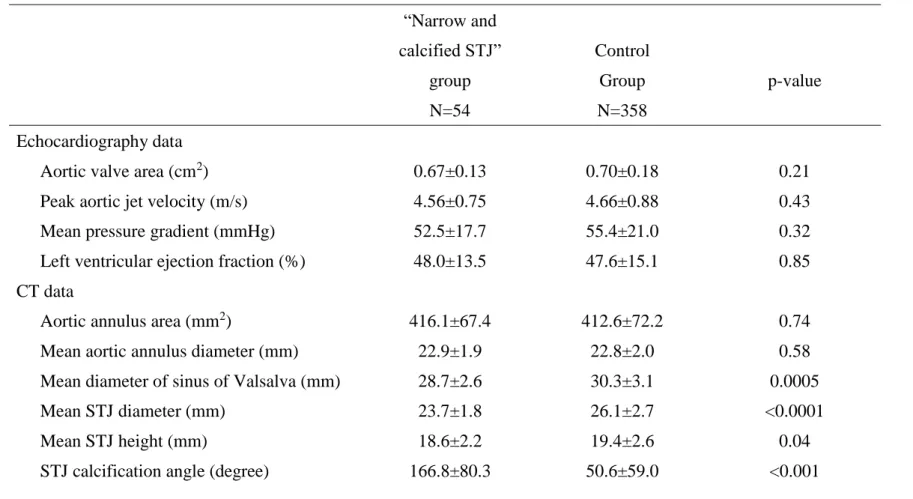
We compared patient characteristics, transthoracic echocardiographic parameters and CT data between patients with and without “narrow and calcified STJ”. The “narrow and calcified STJ” group showed a trend of smaller body size as compared to the control group in height (148.8±8.6 vs 151.2±8.9 cm, p=0.06), weight (50.4±11.2 vs 52.9±10.9 kg, p=0.11), and body surface area (1.4±0.2 vs 1.5±0.2 m
2, p=0.06). The frequency of prior open heart surgery was higher in the “calcified STJ” group than in the control group (14.9 vs 3.6 %, p<0.001). The other patient characteristics did not differ between the two groups (Table 1).
Preoperative echocardiographic data showed that aortic stenosis severity and left ventricular function were similar between the two groups (Table 2). Based on the preprocedural CT data, aortic annulus area, mean aortic annulus diameter were similar between the two groups (Table 2). The Mean diameter of sinus of Valsalva was smaller in the “narrow and calcified STJ” group than in the control group (28.7±2.6 vs 30.3±3.1 mm, p=0.0005). The mean STJ diameter was smaller in the “narrow and calcified STJ” group than in the control group (23.7±1.8 vs. 26.1±2.7 mm, p<0.0001). The mean STJ height was smaller in the “narrow and calcified STJ” group than in the control group (18.6±2.2 vs. 19.4±2.6 mm, p=0.04).
The STJ calcification angle in the “narrow and calcified STJ” group was 166.8±80.3°, whereas that in the control group was 50.6±59.0° (p<0.001). The aortic valve Agatston score was similar between the two groups.
Feasibility of the two-step inflation technique
Although the period of the CT assessment study with 412 patients was between January 2014 and June 2017, we started using the SAPIEN 3 in June 2016. After SAPIEN 3 was available, all 31 cases with “narrow and calcified STJ” were treated with SAPIEN 3. We performed the conventional inflation procedure for TAVI in the first 11 patients with a
1 2 3 4 5 6 7 8 9 10 11 12 13 14 15 16 17 18 19 20 21 22 23 24 25 26 27 28 29 30 31 32 33 34 35 36 37 38 39 40 41 42 43 44 45 46 47 48 49 50 51 52 53 54 55 56 57 58 59
“narrow and calcified STJ”. Thereafter, we used the two-step inflation technique in the next 20 patients with a “narrow and calcified STJ”. We compared preoperative echocardiographic parameters, CT data, procedural data, and the clinical and echocardiographic data at
discharge between the two-step inflation group (n=20) and the conventional inflation group (n=11). All preoperative echocardiographic and CT data were similar between the two groups (Table 3). The SAPIEN 3 size was similar between the two groups (Table 4). Annular area sizing [11.3 (4.5-15.3) vs 10.4 (-0.7-21.6)%, p=0.84] did not differ. Pre-dilation was
performed in 2 patients (10.0%) in the two-step inflation group and 2 patients (18.2%) in the conventional inflation group (p=0.60). The inflation volume at the time of THV deployment was significantly lower in the two-step inflation group than in the conventional inflation group (difference from nominal volume: -1.78±0.95 vs. -0.91±1.04 ml, p=0.02). Post-dilation was performed in all patients in the two-step inflation group and in 7 patients (65.4%) in the conventional inflation group (p=0.004). The final inflation volume was equivalent between the two groups (difference from nominal volume: -0.08±0.77 vs. -0.05±1.01 ml, p=0.93).
These was no postoperative mortality in the two-step inflation group, but one patient in the conventional inflation group died because of congestive heart failure (0 vs 9.1 %, p=0.35).
Ascending aortic dissection did not occur in any patient in either group (Table 4). A new pacemaker implantation was required in 3 (15.0%) and 2 patients (18.2%) in the two-step inflation and conventional inflation groups, respectively (p=1.0). Echocardiographic data at discharge showed that the EOA index was similar between the two groups [(1.40 (1.20-1.51) vs. 1.33 (1.18-1.41) cm
2/m
2, p=0.23]. In addition, the THV peak velocity (2.33±0.48 vs.
2.34±0.31 m/s, p=1.00) and MPG (10.9±4.4 vs. 10.8±2.9 mmHg, p=0.95) was also similar between the groups. Aortic regurgitation of > grade 2 was present in one patient (5.0%) in the two-step inflation group and no patient (0%) in the conventional inflation group (p=1.0).
1 2 3 4 5 6 7 8 9 10 11 12 13 14 15 16 17 18 19 20 21 22 23 24 25 26 27 28 29 30 31 32 33 34 35 36 37 38 39 40 41 42 43 44 45 46 47 48 49 50 51 52 53 54 55 56 57 58 59
Discussion
In the present study, we first elucidated the anatomical features of the aortic root in patients with aortic stenosis, especially focusing on size and calcification at the STJ. As a result, a “narrow STJ”, defined as a minimum STJ diameter smaller than the diameter derived from a 10% oversized annulus area, was found in 30.3% of the patients in this study. A
“calcified STJ”, defined as a STJ calcification angle > 90°, was seen in 27.7% of patients. A
“narrow and calcified STJ” defined as the presence of both a "narrow STJ" and a "calcified STJ” was seen in 13.1% of patients. Next, we demonstrated the feasibility of a novel TAVI strategy, the two-step inflation technique, for patients with a “narrow and calcified STJ”.
Anatomical features of the STJ in aortic stenosis
The STJ is recognized as the interface between the aortic root and the ascending aorta.
STJ calcification is predominant in the elderly and is associated with atherosclerosis [18.19].
Finkelhor et al. [20] reported that STJ calcification was observed in 18% of elderly individuals without any cardiac disorders. Colli et al. [7] used transesophageal echocardiography and reported that 78% of 103 aortic stenosis patients undergoing
transapical TAVI had localized STJ calcification. In addition, complete circumferential STJ calcification was found in 18% of the patients. In contrast, our CT assessment showed that an STJ calcification angle ≤ 360° but > than 270° was present in only 3.2%. These discrepant data may be due to the difference in the imaging approaches. To our knowledge, our study is the first to assess the distribution of STJ calcification using CT.
1 2 3 4 5 6 7 8 9 10 11 12 13 14 15 16 17 18 19 20 21 22 23 24 25 26 27 28 29 30 31 32 33 34 35 36 37 38 39 40 41 42 43 44 45 46 47 48 49 50 51 52 53 54 55 56 57 58 59
The size relationship between the aortic annulus and the STJ has been controversial.
Some studies reported that the STJ and aortic annulus showed a similar size in both healthy individuals and aortic stenosis patients [20,21], whereas other studies reported that the STJ was smaller than the annulus [22]. The discrepancy may be due to the difference in the imaging modalities and the methods of measurement. In our CT study, the aortic annulus diameter, which was calculated from the traced annulus area [10,11], was smaller than that of the STJ in all of the aortic stenosis patients. However, in 5.4% of patients, the mean STJ diameter was smaller than that of the aortic annulus. Additionally, we found that STJ calcification showed a negative correlation with the ratio of the mean STJ diameter to the mean aortic annulus diameter. This result suggests that STJ size may depend on STJ calcification, and that negative remodeling due to atherosclerosis may have an important influence on STJ morphology. However, further investigations are required to clarify these assumptions.
Iatrogenic ascending aortic dissection
Aortic dissection is a rare but serious complication in TAVI. A meta-analysis of 9251 patients from 46 studies reported that emergent cardiac surgery due to aortic dissection during TAVI was very rare, as it occurred in only 14 patients (0.15%) [23]. However, Barbanti et al. [2] reported two cases of STJ injury after TAVI, although they provided no details. On the other hand, Yashima et al. [7] reported clear details about iatrogenic ascending aortic dissection for a narrow and calcified STJ using SAPIEN XT. An important finding in their study was that even though SAPIEN XT itself did not reach the STJ, STJ injury
occurred due to the overfilling of the delivery balloon. The potential risk of interference with the STJ may be higher with the SAPIEN 3 than with the SAPIEN XT, because SAPIEN 3
1 2 3 4 5 6 7 8 9 10 11 12 13 14 15 16 17 18 19 20 21 22 23 24 25 26 27 28 29 30 31 32 33 34 35 36 37 38 39 40 41 42 43 44 45 46 47 48 49 50 51 52 53 54 55 56 57 58 59
tends to reach the STJ for the following two reasons. First, the height of the SAPIEN 3 is taller than that of the SAPIEN XT. Second, the SAPIEN 3 is recommended to be deployed at a higher position to minimize the risk of atrioventricular block [17]. Therefore, we pay meticulous attention to this issue when we perform TAVI using the SAPIEN 3 in patients with a narrow and calcified STJ.
A self-expanding THV may be preferred for a narrow and calcified STJ because it does not require excessive force to expand. In addition, even though iatrogenic dissection occurs at the ascending aorta, a self-expanding THV can seal it [24]. However, the use of a
self-expanding THV is limited by the device eligibility in institutions or countries and other morphological characteristics like horizontal aorta, ascending aorta dilatation, and device access.
The two-step inflation technique
We devised the two-step inflation technique utilizing the conformability of the SAPIEN3 platform as a novel approach for patients with a narrow and calcified STJ. Using this technique, the outflow portion is relatively under expanded to accommodate a narrow and calcified STJ, and was flared at the aortic annulus. The two-step inflation technique seemed to be safe and might contribute to the prevention of STJ injury induced by direct SAPIEN 3 THV contact as well as balloon delivery at the time of THV deployment. With the two-step inflation technique, the pigtail catheter should be fully pulled back from the sinus of Valsalva before full dilation, because the catheter as well as the balloon might cause aortic dissection [25, 26].
The inflation volume at the first inflation is a key factor in the two-step inflation technique. We inflate the delivery balloon very slowly so that we can stop the inflation when
1 2 3 4 5 6 7 8 9 10 11 12 13 14 15 16 17 18 19 20 21 22 23 24 25 26 27 28 29 30 31 32 33 34 35 36 37 38 39 40 41 42 43 44 45 46 47 48 49 50 51 52 53 54 55 56 57 58 59
the delivery balloon or THV itself pushes on the STJ calcification, which can be seen on fluoroscopy. In our experience, when STJ calcification is mild to moderate, the balloon should be underfilled by -1 to -2 ml compared with the nominal inflation volume for the annulus. However, when the STJ calcification is severe and thick, -3 ml may be beneficial. At the time of post-dilation, the center marker of the delivery balloon is positioned at the inflow edge of SAPIEN 3 so that it dilates at the annulus and does not interfere with the outflow edge (Figure 2E). The stiff wire should be positioned deep inside the left ventricle and trapping in the papillary muscle should be avoided. Also, blood pressure should be lowered by rapid pacing during inflation. These efforts are important for the prevention of injury of the left ventricle and papillary muscle, because the delivery balloon is deep inside the left ventricle at the time of post-dilation.
In this study, we deployed the SAPIEN 3 in patients with a “narrow and calcified STJ”
and compared various parameters between 20 patients who underwent two-step inflation and 11 patients who underwent conventional inflation. Our study showed that there was no significant difference in echocardiographic parameters, EOA and paravalvular leak at discharge, and pacemaker implantation between two groups, and this suggests that the two-step inflation technique is safe. Although the postoperative course of the two groups patient was not significantly different, it is important to note that that there was neither ascending aortic dissection nor postoperative death in the two-step inflation group. Although this pilot study showed the feasibility of the two-step inflation technique, evaluation using a larger sample of patients is required to establish the safety and efficacy of this technique.
Study limitations
1 2 3 4 5 6 7 8 9 10 11 12 13 14 15 16 17 18 19 20 21 22 23 24 25 26 27 28 29 30 31 32 33 34 35 36 37 38 39 40 41 42 43 44 45 46 47 48 49 50 51 52 53 54 55 56 57 58 59
There are several potential limitations in this study. First, the study sample size was too small to discuss the novel methods to prevent a rare complication of balloon-expandable TAVI such as ascending aortic dissection. Comparison between the two-step inflation
technique and the conventional inflation technique was based on retrospective analysis rather than a prospective randomized trial. In this study, we used an empirical formula to define a
“narrow and calcified STJ” as an indication for two-step inflation; thus, the optimal
indication for this technique will require further refinement in the future. In addition, a study with a larger number of patients is warranted to estimate the benefit of this technique. Next, this study included only single-center data from Japan, and there might be racial differences in the anatomical features of the STJ. Another limitation is that post-procedural CT study was not performed in all cases, therefore subclinical aortic dissection was possibly missed. Finally, this study evaluated only short-term valve function, and a long-term outcome study is
required.
Conclusion
Our data in the present study suggested the two-step inflation technique with the SAPIEN 3 would be a feasible TAVI approach for a narrow and calcified STJ. Studies with a larger number of patients and a longer follow-up time are needed to confirm its efficacy and safety.
Conflict of Interest
1 2 3 4 5 6 7 8 9 10 11 12 13 14 15 16 17 18 19 20 21 22 23 24 25 26 27 28 29 30 31 32 33 34 35 36 37 38 39 40 41 42 43 44 45 46 47 48 49 50 51 52 53 54 55 56 57 58 59
Norio Tada is a clinical proctor for Edwards Lifesciences. All the other authors have no conflict of interest to disclose.
1 2 3 4 5 6 7 8 9 10 11 12 13 14 15 16 17 18 19 20 21 22 23 24 25 26 27 28 29 30 31 32 33 34 35 36 37 38 39 40 41 42 43 44 45 46 47 48 49 50 51 52 53 54 55 56 57 58 59
References
1. Smith CR, Leon MB, Mack MJ, Miller DC, Moses JW, Svensson LG, Tuzcu EM, Webb JG, Fontana GP, Makkar RR, Williams M, Dewey T, Kapadia S, Babaliaros V,
Thourani VH, Corso P, Pichard AD, Bavaria JE, Herrmann HC, Akin JJ, Anderson WN, Wang D, Pocock SJ; PARTNER Trial Investigators (2011) Transcatheter versus surgical aortic-valve replacement in high-risk patients. N Engl J Med 364 :2187-2198
2. Barbanti M, Yang TH, Rodes Cabau J, Tamburino C, Wood DA, Jilaihawi H, Blanke P, Makkar RR, Latib A, Colombo A, Tarantini G, Raju R, Binder RK, Nguyen G, Freeman M, Ribeiro HB, Kapadia S, Min J, Feuchtner G, Gurtvich R, Alqoofi F, Pelletier M, Ussia GP, Napodano M, de Brito FS Jr, Kodali S, Norgaard BL, Hansson NC, Pache G, Canovas SJ, Zhang H, Leon MB, Webb JG, Leipsic J (2013) Anatomical and procedural features associated with aortic root rupture during balloon-expandable transcatheter aortic valve replacement. Circulation 128: 244-253
3. Hayashida K, Bouvier E, Lefevre T, Hovasse T, Morice MC, Chevalier B, Romano M, Garot P, Farge A, Donzeau-Gouge P, Cormier B (2013) Potential mechanism of annulus rupture during transcatheter aortic valve implantation. Catheter Cardiovasc Interv 82:
E742-E746
4. Yamamoto M, Shimura T, Kano S, Kagase A, Kodama A, Koyama Y, Watanabe Y, Tada N, Takagi K, Araki M, Shirai S, Hayashida K (2016) Impact of preparatory coronary protection in patients at high anatomical risk of acute coronary obstruction during transcatheter aortic valve implantation. Int J Cardiol 217: 58-63
5. Thourani VH, Kodali S, Makkar RR, Herrmann HC, Williams M, Babaliaros V, Smalling R, Lim S, Malaisrie SC, Kapadia S, Szeto WY, Greason KL, Kereiakes D,
1 2 3 4 5 6 7 8 9 10 11 12 13 14 15 16 17 18 19 20 21 22 23 24 25 26 27 28 29 30 31 32 33 34 35 36 37 38 39 40 41 42 43 44 45 46 47 48 49 50 51 52 53 54 55 56 57 58 59
Ailawadi G, Whisenant BK, Devireddy C, Leipsic J, Hahn RT, Pibarot P, Weissman NJ, Jaber WA, Cohen DJ, Suri R, Tuzcu EM, Svensson LG, Webb JG, Moses JW, Mack MJ, Miller DC, Smith CR, Alu MC, Parvataneni R, D'Agostino RB Jr, Leon MB (2016) Transcatheter aortic valve replacement versus surgical valve replacement in
intermediate-risk patients: a propensity score analysis. Lancet 387: 2218-2225
6. Langer NB, Hamid NB, Nazif TM, Khalique OK, Vahl TP, White J, Terre J, Hastings R, Leung D, Hahn RT, Leon M, Kodali S, George I. Injuries to the Aorta, Aortic Annulus, and Left Ventricle During Transcatheter Aortic Valve Replacement: Management and Outcomes. Circ Cardiovasc Interv. 2017 Jan;10(1).
7. Yashima F, Hayashida K, Fukuda K (2016) Delivery balloon-induced ascending aortic dissection: An unusual complication during transcatheter aortic valve implantation.
Catheter Cardiovasc Interv 87: 1338-1341
8. Colli A, D'Amico R, Kempfert J, Borger MA, Mohr FW, Walther T (2011)
Transesophageal echocardiographic scoring for transcatheter aortic valve implantation:
impact of aortic cusp calcification on postoperative aortic regurgitation. J Thorac Cardiovasc Surg 142: 1229-1235
9. Leipsic J, Gurvitch R, Labounty TM, Min JK, Wood D, Johnson M, Ajlan AM, Wijesinghe N, Webb JG (2011) Multidetector computed tomography in transcatheter aortic valve implantation. JACC Cardiovasc Imaging. 4: 416-429
10. Jilaihawi H, Kashif M, Fontana G, Furugen A, Shiota T, Friede G, Makhija R, Doctor N, Leon MB, Makkar RR (2012) Cross-sectional computed tomographic assessment
improves accuracy of aortic annular sizing for transcatheter aortic valve replacement and reduces the incidence of paravalvular aortic regurgitation. J Am Coll Cardiol 59:
1275-1286
1 2 3 4 5 6 7 8 9 10 11 12 13 14 15 16 17 18 19 20 21 22 23 24 25 26 27 28 29 30 31 32 33 34 35 36 37 38 39 40 41 42 43 44 45 46 47 48 49 50 51 52 53 54 55 56 57 58 59
11. Schultz CJ, Moelker A, Piazza N, Tzikas A, Otten A, Nuis RJ, Neefjes LA, van Geuns RJ, de Feyter P, Krestin G, Serruys PW, de Jaegere PP (2010) Three dimensional evaluation of the aortic annulus using multislice computer tomography: are
manufacturer’s guidelines for sizing for percutaneous aortic valve replacement helpful?
Eur Heart J 31: 849-856
12. Agatston AS, Janowitz WR, Hildner FJ, Zusmer NR, Viamonte M Jr, Detrano R.
Quantification of coronary artery calcium using ultrafast computed tomography (1990) J Am Coll Cardiol 15: 827-832
13. Blanke P, Pibarot P, Hahn R, Weissman N, Kodali S, Thourani V, Parvataneni R, Dvir D, Naoum C, Nørgaard BL, Douglas P, Jaber W, Khalique OK, Jilaihawi H, Mack M, Smith C, Leon M. Webb J, Leipsic J (2017) Computed tomography-based oversizing degree and incidence of paravalvular regurgitation of a new generation transcatheter heart valve. JACC Cardiovasc Interv 10: 810-820
14. Lang RM, Badano LP, Mor-Avi V, Afilalo J, Armstrong A, Ernande L, Flachskampf FA, Foster E, Goldstein SA, Kuznetsova T, Lancellotti P, Muraru D, Picard
MH, Rietzschel ER, Rudski L, Spencer KT, Tsang W, Voigt JU (2015)
Recommendations for cardiac chamber quantification by echocardiography in adults: an update from the American Society of Echocardiography and the European Association of Cardiovascular Imaging. J Am Soc Echocardiogr. 28:1-39 e14.
15. Baumgartner H, Hung J, Bermejo J, Chambers JB, Evangelista A, Griffin BP, Iung B, Otto CM, Pellikka PA, Quiñones M (2009) American Society of
Echocardiography; European Association of Echocardiography.: Echocardiographic assessment of valve stenosis: EAE/ASE recommendations for clinical practice. J Am Soc Echocardiogr 22:1-23; quiz 101-102.
16. Maeno Y, Abramowitz Y, Jilaihawi H, Israr S, Yoon S, Sharma RP, Kazuno
1 2 3 4 5 6 7 8 9 10 11 12 13 14 15 16 17 18 19 20 21 22 23 24 25 26 27 28 29 30 31 32 33 34 35 36 37 38 39 40 41 42 43 44 45 46 47 48 49 50 51 52 53 54 55 56 57 58 59
M, Chakravarty T, Nakamura M, Cheng W, Makkar RR (2017) Optimal sizing for SAPIEN 3 transcatheter valve replacement in patients with or without left ventricular outflow tract calcification. EuroIntervention 12: e2177-e2185
17. Schwerg M, Fulde F, Dreger H, Poller WC, Stangl K, Laule M (2106) Optimized implantation height of the Edwards SAPIEN 3 valve to minimize pacemaker implantation after TAVI. J Interv Cardiol 29: 370-374
18. Tveter KJ, Edwards JE (1988) Calcified aortic sinotubular ridge: a source of coronary ostial stenosis or embolism. J Am Coll Cardiol 12: 1510-1514
19. Finkelhor RS, Youssefi ME, Mohan SK, Bahler RC (1998) Aortic sinotubular junction calcium as a marker of severe aortic atherosclerosis. Am J Cardiol 82: 1549-1552 20. Akhtar M, Tuzcu EM, Kapadia SR, Svensson LG, Greenberg RK, Roselli EE,
Halliburton S, Kurra V, Schoenhagen P, Sola S (2009) Aortic root morphology in patients undergoing percutaneous aortic valve replacement: Evidence of aortic root remodeling, J Thorac Cardiovasc Surg 137: 950-956
21. Davis AE, Lewandowski AJ, Holloway CJ, Ntusi NA, Banerjee R, Nethononda R, Pitcher A, Francis JM, Myerson SG, Leeson P, Donovan T, Neubauer S, Rider OJ, Observational study of regional aortic size referenced to body size: production of a cardiovascular magnetic resonance nomogram (2014) J Cardiovasc Magn Reson 16: 9.
22. Kunzelman KS, Grande KJ, David TE, Cochran RP, Verrier ED (1994) Aortic root and valve relationships. Impact on surgical repair. J Thorac Cardiovasc Surg 107: 162-170 23. Eggebrecht H, Schmermund A, Kahlert P, Erbel R, Voigtländer T, Mehta RH (2013)
Emergent cardiac surgery during transcatheter aortic valve implantation (TAVI): a weighted meta-analysis of 9251 patients from 46 studies. Eurointervention 8: 1072-1080
1 2 3 4 5 6 7 8 9 10 11 12 13 14 15 16 17 18 19 20 21 22 23 24 25 26 27 28 29 30 31 32 33 34 35 36 37 38 39 40 41 42 43 44 45 46 47 48 49 50 51 52 53 54 55 56 57 58 59
24. Ong SH, Mueller R, Gerckens U (2011) Iatrogenic dissection of the ascending aorta during TAVI sealed with the CoreValve revalving prosthesis. Catheter Cardiovasc Interv 77: 910-914
25. Solar RJ, Ischinger TA (2003) Focused force angioplasty: theory and application.
Cardiovasc Radiat Med 4: 47-50
26. Tada N, Ootomo T, Meguro T (2012) Percutaneous Focused Force Aortic Valvuloplasty Using the Buddy-Catheter Technique, J Invasive Cardiol 24: 287-289
1 2 3 4 5 6 7 8 9 10 11 12 13 14 15 16 17 18 19 20 21 22 23 24 25 26 27 28 29 30 31 32 33 34 35 36 37 38 39 40 41 42 43 44 45 46 47 48 49 50 51 52 53 54 55 56 57 58 59
Figure legends
Figure 1 A computed tomography (CT) image of sinotubular junction (STJ) calcification and measurement of the maximum and minimum diameter (A) and STJ calcification angle (B).
STJ diameter was measured involving the thickness of its calcification. Maximum and minimum STJ diameter is measured as 22.5 mm and 16.7 mm, respectively (A). STJ calcification angle is summed if there are multiple (45.6°, 46.2°, and 120.9°, total 212.7°)(B). The same case as Figure 2.
Figure 2 A representative case of the two-step inflation technique for a “narrow and calcified STJ”.
A narrow and heavily calcified STJ is shown (A.B). There is bulky calcification above the non-coronary cusp (white arrow). Aortography shows heavy calcification at the STJ on the non-coronary cusp (white arrow) (C). A 23-mm SAPIEN 3 is dilated and stopped at an underfilled volume of -3 mL to avoid pushing on the bulky calcification (D). The
transcatheter heart valve (THV) balloon is shifted downward and post-dilation is performed up to nominal volume, where the central marker is at the left ventricular edge of the
SAPIEN 3 (E). Final aortography (F) and transesophageal echocardiography (G) shows that the THV has a skirt-like shape. There is neither aortic dissection nor paravalvular leakage.
Transthoracic echocardiography at discharge revealed that the effective orifice area (EOA) index was 1.33 cm
2/m
2, and the mean pressure gradient (MPG) was 7 mmHg (H).
Figure 3 Morphological characteristics of the sinotubular junction (STJ).
1 2 3 4 5 6 7 8 9 10 11 12 13 14 15 16 17 18 19 20 21 22 23 24 25 26 27 28 29 30 31 32 33 34 35 36 37 38 39 40 41 42 43 44 45 46 47 48 49 50 51 52 53 54 55 56 57 58 59
The mean STJ diameter was larger than the mean aortic annulus diameter (A).
Relationship between the mean STJ diameter and the mean aortic annulus diameter showing a strong correlation (B). Distribution and box plot of the STJ calcification angle (C). Relationship between the STJ calcification angle and the ratio of the mean STJ diameter to the mean aortic annulus diameter showing a negative correlation (D).
1 2 3 4 5 6 7 8 9 10 11 12 13 14 15 16 17 18 19 20 21 22 23 24 25 26 27 28 29 30 31 32 33 34 35 36 37 38 39 40 41 42 43 44 45 46 47 48 49 50 51 52 53 54 55 56 57 58 59