• 検索結果がありません。

Protective Effects of Olmesartan and Azelnidipine against Cardiovascular Organ Injuries in Spontaneously Hypertensive Rats

N/A
N/A
Protected

Academic year: 2021

シェア "Protective Effects of Olmesartan and Azelnidipine against Cardiovascular Organ Injuries in Spontaneously Hypertensive Rats"

Copied!
9
0
0

読み込み中.... (全文を見る)

全文

(1)

INTRODUCTION

The ultimate goal of antihypertensive therapy is not only to normalize the blood pressure level but also to prevent end-organ damage, such as cardiac hypertro- phy and renal dysfunction, and to prevent the cardio- vascular disease, such as stroke and myocardial infarc- Received June 30, 2015;accepted July 17, 2015

Reprint requests to:Toshihiko Ishimitsu, M.D.

Department of Cardiology and Nephrology Dokkyo Medical University Mibu, Tochigi 321-0293, Japan

Protective Effects of Olmesartan and Azelnidipine against Cardiovascular Organ Injuries in Spontaneously

Hypertensive Rats

Takeaki Honda, Masahito Furuichi, Yoshiki Murayama, Hiroshi Satonaka, Toshihiko Ishimitsu

Department of Cardiology and Nephrology, Dokkyo Medical University, Mibu, Tochigi 321-0293, Japan

SUMMARY

In the treatment of hypertension, care should be taken for preventing of hypertensive organ injuries as well as lowering blood pressure to the adequate level in order to reduce the risk of cardiovascular diseases.

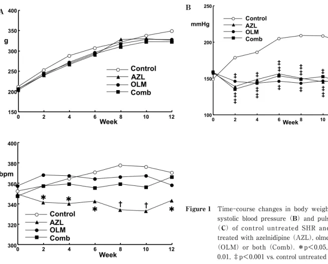
The purpose of this study is to examine the effects of angiotensin II receptor blockers (ARB), calcium chan- nel blockers (CCB) and their combination on the development of cardiovascular organ injuries in spontane- ously hypertensive rats (SHR). Four groups of male 8-week-old SHR (n=9 each) were given vehicle(con- trol), 10 mg/kg azelnidipine (AZL), 10 mg/kg olmesartan (OLM, n=9), or the combination of AZL and OLM(5 mg/kg each)for 12 weeks, and their effects on cardiovascular organ injuries were evaluated. Tail- cuff blood at 12 weeks was similarly lowered by AML, OLM and the combination therapy(148, 143 and 143 mmHg, respectively)as compared with the control SHR (198 mmHg). Pulse rate was significantly less in the AZL group but not in the OLM group or the combination therapy group than in the untreated con- trol group (−27, −12, +6 bpm, respectively). The cardiac ventricular weight (AZL −12%, OLM −15%, combination −18% vs. control) and aortic thickness (AZL −17%, OLM −16%, combination −19% vs. con- trol) were reduced by similar extents in the three groups given antihypertensive treatments. Regarding the myocardial fibrosis, left ventricular hydroxyproline content was reduced in the OLM and the combination groups but the change was not significant in the AZL group (AZL −14%,OLM −30%, combination −27

% vs. control). In the echocardiographic evaluation of cardiac function, the index of left ventricular diastolic function is significantly improved in the OLM and the combination groups but not in the AZL group, while the index of systolic function was not different between the four groups. It is suggested that the antihyper- tensive therapy including ARB is superior to the monotherapy by CCB in preventing the myocardial fibro- sis and preserving the left ventricular diastolic function.

Key Words: hypertension, spontaneously hypertensive rat, angiotensin II receptor antagonist, calcium channel blocker, left ventricular hypertrophy, arteriosclerosis

Original

(2)

tion. Therefore, the efficacy of antihypertensive drugs on inhibition of hypertensive tissue injuries and preser- vation of cardiovascular organ function has to be taken into consideration in order to achieve maximal im- provement of long-term prognosis of hypertensive pa- tients. In the real world clinical practice, angiotensin II receptor antagonists (ARB) and long-acting calcium channel blockers (CCB) are widely used for the treat- ment of hypertensive patients because these classes of antihypertensive drugs have few chances to cause ad- verse effects. In addition, CCB exerts consistently cer- tain hypotensive effects by dilating arteries directly and ARB is expected to have protective effects against the cardiac and the renal injuries partially by the mechanism independent of blood pressure lowing

1,2)

. Thus, it is obviously important to collect the informa- tion as to the effects of CCB and ARB on the develop- ment and the progression of hypertensive injuries in cardiovascular organs such as the heart, the kidneys and the arteries.

It should be also kept in mind that generally only one third of hypertensive patients achieve the blood pressure level lower than 140/90 mmHg with a single antihypertensive agent irrespective of the class of drug used and two thirds of patients require a combination of two or more drugs in order to obtain adequate blood pressure reduction

3,4)

. Therefore, it is also a matter of concern to delineate the therapeutic efficacy of combi- nation therapy especially with ARB and CCB in terms of protecting cardiovascular organs from hypertension.

In this experimental study, the efficacies of CCB, ARB and their combination therapy were examined and compared focusing on the inhibition of cardiovascular organ injuries in spontaneously hypertensive rat

(SHR)which is assumed to be an appropriate animal model of human essential hypertension.

METHODS Treatment of rats

Male 8-week-old SHR (n=36) were purchased from Charles River Japan (Atsugi, Kanagawa, Japan). They were fed standard chow and tap water, and were housed in a temperature- and light-controlled room throughout the study period. They were divided into 4 groups of 9 SHR each. As a dihydropyridine deriva- tive CCB, 10 mg/kg azelnidipine (AZL) suspended in

0.5 ml of 0.5% methyldellulose was given once daily by gastric tube in morning hours to 9 rats. As an ARB, 10 mg/kg olmesartan (OLM) was given in the similar way to another 9 rats. In addition, 5 mg/kg AZL and 5 mg/kg OLM were simultaneously given in 9 rats of the combination therapy group, and 9 rats of the con- trol group were given vehicle.

The treatments were continued for 12 weeks and the systolic blood pressure was measured biweekly during the study period using the tail-cuff method. At the end of 12-week period, the rats were evaluated for the physiological, biochemical and histological evalua- tions described below. The experiments were per- formed in accordance with the institutional guide for care and use of laboratory animals, and the study pro- tocol was approved by the institutional animal research committee.

Echocardiography

After 12 weeks, transthoracic echocardiographic studies were performed under light anesthesia with in- traperitoneal injection of ketamine HCl(10 mg/kg)and xylazine (10 mg/kg). Two-dimensional echocardiogra- phy and M-mode tracing were recorded at the level of the papillary muscles using a Toshiba(Tokyo, Japan)

SSH-260A unit with a 7.0 MHz transducer placed on the shaved left hemithorax of the rats in the left decu- bitus position. M-mode measurements included left ventricular end-systolic and end-diastolic diameters

(LVDs, LVDd), end-diastolic left ventricular posterior wall thickness ( PWT), and interventricular septal thickness ( IVST). Midwall fractional shortening

(mFS) was calculated as follows

5,6)

:mFS=[(LVDd+

IVST/2+PWT/2)−{(LVDd+IVST/2+PWT/2)

3

− LVDd

3

+LVDs

3

1/3

]/(LVDd+IVST/2+PWT/2). The values of mFS were multiplied by 100 and expressed as % values.

Pulsed-wave Doppler spectra of mitral inflow veloci-

ties were recorded from the apical 4-chamber view

with the sample volume placed near the tips of the mi-

tral leaflets and adjusted to the position where the ve-

locity was maximal and the flow patterns were lami-

nar. The Doppler spectra were recorded on paper at

100 mm/s and analyzed off-line to determine peak ear-

ly diastolic filling velocity(E)and peak filling velocity

at atrial contraction(A). The heart rate was main-

(3)

tained around 300 bpm with the anesthesia during the recording, which enabled the separation of E and A wave peaks in each rat. Measurements represent the mean of at least 3 consecutive cardiac cycles, and the E/A ratio was used for the evaluation of left ventricu- lar diastolic function

7)

.

Biochemical Assay

After performing the echocardiogram, blood samples were drawn from the inferior vena cava, transferred into ice-cooled tube containing 1 mg/ml EDTA and centrifuged at 4℃ to obtain plasma. Plasma renin activ- ity and concentrations of angiotenisin II and aldoste- rone were measured by respective radioimmunoassays using the commercial kits(SRL, Inc., Tokyo, Japan).

A portion of the left ventricular free wall tissue was homogenized in ten equivalent volumes of saline. A 0.5 mL aliquot of the homogenate was then mixed with 36% hydrochloride and heated to 100℃ for 20 hours.

Next, the mixture was centrifuged at 1 , 5 0 0 g for 30 minutes, and a 0.1 mL aliquot of the supernatant was

mixed with 1.5 mL of 0.3 N hydroxylithium. Hydroxy- proline content in the reaction product was determined by high-performance liquid chromatography, and the value was expressed relative to tissue weight

8)

.

Histological Examination

The cardiac ventricles and descending thoracic aorta were excised and weighed. The weight of cardiac ven- tricles was corrected with the body weight and the weight of aorta was expressed as weight per unit area.

The upper half of cardiac ventricles were fixed in neu- tral-buffered 8% formaldehyde solution and embedded in paraffin. The 2-mm sections were cut for histologi- cal examination which was conducted in a blind man- ner. The sections were stained with Masson trichrome and the fibrosis of the left ventricular wall was evalu- ated. The area stained in blue with aniline was quanti- fied in ten randomly-selected high-power fields

(x200)using a computer system(Image Quest, Hama- matsu Photonics;Hamamatsu and MacScope, Mitani Co., Fukui, Japan), and the average percent value was Control

OLM AZL Comb

12 10

8 6

4 2

150 0 200 250 300 350 400

Week g

A Control

AZL OLM Comb

12 10 8 6 4 2 1000 150 200 250

Week mmHg

B

Control AZL OLM Comb

*

* *

12 10

8 6

4 2

300 0 320 340 360 380 400

Week bpm

*

C

Figure 1  Time-course changes in body weight (A), systolic blood pressure (B) and pulse rate

(C) of control untreated SHR and SHR treated with azelnidipine (AZL), olmesartan

(OLM) or both (Comb). *p<0.05, †p<

0.01, ‡p<0.001 vs. control untreated SHR.

(4)

used for comparison.

Statistical Analysis

Values are expressed as means±SE. Comparison of the 4 groups was performed by one-way ANOVA and the post-hoc analysis by Dunnett’s multiple-range test.

Time-course changes in parameters were analyzed by two-way ANOVA with post-hoc multiple comparisons using the Bonferroni-Dunn test. Non-parametric data were analyzed by the Kruskal-Wallis H-test followed by Tukey’s method for post-hoc between-group com- parisons. A p value less than 0.05 was considered to indicate statistical significance.

RESULTS

Physical Measurements and Organ Weights

All the rats have survived and completed the 12- week study period. Figure 1 shows the time-course changes in body weight(panel A), blood pressure

(panel B)and pulse rate(panel C)during the study period. The values of body weight, blood pressure and pulse rate at 12 weeks were listed in Table 1. The body weight increases were similar among the control, the AZL, the OLM and the combination groups. The blood pressures were comparably lowered in the AZL, the OLM and the combination groups as compared with the control group throughout the study period.

The pulse rate was significantly reduced in the AZL group than in the control group, while it was not sig- nificantly changed in the OLM or the combination group as compared with the control group.

Figure 2 depicts the weights of cardiovascular or- gans such as the heart and the aorta. The weights of cardiac ventricles were significantly reduced in the AZL group, the OLM group and the combination group as compared with the control group. The reductions in cardiac weights were not significantly different be- tween the 3 antihypertensive treatments. The weight Table 1 Physical measurements at the end of 12-week study period.

Parameter Control

(n=9)

Azelnidipine

(n=9)

Olmesartan

(n=9)

Combination

(n=9)

Body weight, g 337±7 327±7 326±7 322±8

Systolic blood pressure, mmHg 198±5 148±3‡ 143±3‡ 143±3‡

Pulse rate, bpm 370±10 343±7

358±9 376±10

Values are means±SE.

p<0.05, ‡ p<0.001 vs. control untreated SHR.

Figure 2  Cardiovascular organ weights in control untreated SHR and SHR treated

with azelnidipine (AZL), olmesartan (OLM) or both (Comb) for 12

weeks. *p<0.05, ‡p<0.001 vs. control untreated SHR.

(5)

of descending thoracic aorta per unit area was also sig- nificantly reduced in the 3 treatment groups to similar extents as compared with the control group.

Circulation Levels of renin-Angiotensin-Aldosterone System

Figure 3 presents the plasma levels of renin, angio- tensin II and aldosterone in the 4 experimental groups.

Plasma renin activity and plasma angiotensin concen- tration were significantly increased in the SHR groups treated with antihypertensive drugs as compared with

the control untreated SHR. Especially, the increases were prominent in the groups given OLM. Regarding the aldosterone in plasma, the level was significantly increased in the AZL group while it was significantly decreased in the OLM and the combination groups as compared with the control group.

Evaluation of Cardiac Injury

Echocardiographic data obtained at the end of the 12-week study period are shown in Figure 4. The mid- wall fractional shortening, an index of left ventricular Figure 3  Circulating levels of renin-angiotensin-aldosterone system in control

untreated SHR and SHR treated with azelnidipine (AZL), olmesartan (OLM)

or both (Comb) for 12 weeks. *p<0.05, †p<0.01, ‡p<0.001 vs. control untreated SHR.

Figure 4  Parameters of systolic and diastolic function of left ventricle in control untreated SHR and SHR treated with azelnidipine (AZL), olmesartan (OLM) or both (Comb) for 12 weeks. *p<0.05, †p<

0.01 vs. control untreated SHR.

(6)

systolic function, was not significantly different be- tween the control untreated SHR and the 3 groups of SHR given AZL, OLM or the combination therapy.

With regard to the index of left ventricular diastolic function, the E/A ratio of transmitral flow velocity was improved in the OLM and the combination groups as compared with the control group. However, this pa- rameter of left ventricular diastolic function did not dif- fer in the AZL group as compared with the control group.

The bar graphs of Figure 5 compare the extents of left ventricular wall tissue fibrosis between the 4 groups of rats. As shown in the left panel, the fibrosis area stained in the Masson trichrome sections was sig- nificantly reduced in the OLM and the combination groups as compared with the control group. However, the fibrosis area observed in the AZL group was not significantly different with the control group. The mea- surements of hydroxyproline in the left ventricular tis- sue showed the results similar to the histological find- ings(right panel, Figure 5). The content of this amino acid composing collagen was lower in the OLM and the combination groups than in the control group, however, the content was not significantly different between the AZL and the control groups.

DISCUSSION

Several studies have investigated the protective ef- fects of antihypertensive drugs against cardiovascular

organ injuries using various animal models of hyper- tension, including mineralocorticoid-salt administra- tion

9,10)

, renovascular hypertension

11〜13)

and renal abla- tion

14,15)

, in which hypertensive organ injuries progress rapidly over a period of several weeks to months. How- ever, these are more likely the experimental models of secondary hypertension and the rapid temporal course of organ damage does not accurately reflect the organ damage that occurs over a span of decades in humans with essential hypertension. In this context, the patho- physiology of organ damage in SHR may more closely parallel to that which occurs in humans with essential hypertension

16〜18)

. Thus, it seems more appropriate to evaluate the effects of antihypertensive therapy using SHR in order to apply the findings to clinical medicine, considering that the majority of hypertensive patients are essential hypertension. SHR exhibit cardiac hyper- trophy and arterial wall thickening with the develop- ment of hypertension although the organ injury, such as renal dysfunction does not occur until a later stage of life. Therefore, SHR is thought to be a suitable model to evaluate the therapeutic effects against hyperten- sive injuries in the heart and the arteries.

It has been suggested that the enhancement of re- nin-angiotensin-aldosterone system ( RAAS) is in- volved in the progression process of cardiovascular tis- sue and organ injuries

19,20)

. Particularly, angiotensin II promotes hypertrophy of cardiovascular cells, and aldo- sterone induces fibrosis of the cardiovascular tis- Figure 5  Parameters of left ventricular fibrosis in control untreated SHR and SHR

treated with azelnidipine (AZL), olmesartan (OLM) or both (Comb) for

12 weeks. *p<0.05 vs. control untreated SHR.

(7)

sues

19,20)

. Fibrosis and deposition of intercellular matri- ces, such as collagen, in the cardiac tissue cause reduction in left ventricular distensibility

21)

. Therefore, antihypertensive drugs suppressing the RAAS, such as ACE inhibitors and ARBs, may be expected to protect cardiovascular organs from hypertensive injury inde- pendent of their effects on blood pressure

1,2)

. It has been indicated that the growth of cardiovascular cells is enhanced in SHR and hypertrophy of cardiovascular tissues and organs develops before blood pressure ele- vation

22〜25)

. In the present study, the antihypertensive therapy including ARB mitigated the increase in left ventricular mass and fibrosis of left ventricular tissue.

This mitigation of cardiac fibrosis, combined with inhi- bition of cardiac hypertrophy, may account for the ob- served preservation of myocardial contractility and distensibilty by ARB as compared with CCB alone.

Considerable amount of clinical evidence indicates that inhibitors of the RAAS improve the prognosis of patients with myocardial infarction or heart fail- ure

26〜29)

. However, with regard to the primary preven- tion, it is not necessarily clear if there is an advantage to RAAS inhibitors over other classes of antihyperten- sive drugs in the prevention of coronary artery disease and heart failure in hypertensive patients. In CASE-J

(Candesartan Antihypertensive Survival Evaluation in Japan)trial

30)

, Japanese hypertensive patients with high risks of cardiovascular diseases were treated with an ARB(candesartan)or a CCB(amlodipine)for an average of 3.2 years, which produced no significant dif- ferences in cardiovascular morbidity or mortality. ALL- HAT(Antihypertensive and Lipid-Lowering treat- ment to Prevent Heart Attack Trial)

31)

is a large-scale clinical study in which the effects of a diuretic

(chlorthalidone), a CCB (amlodipine), and an ACE in- hibitor (lisinopril) were compared in hypertensive pa- tients with high risks of cardiovascular diseases. The trial investigators reported that the incidence of heart failure showed a nonsignificant trend towards lower incidence in the ACE inhibitor group than in the CCB group. The VALUE ( Valsartan Antihypertensive Long-term Use Evaluation) trial

32)

, which compared the effects of an ARB, valsartan, and a CCB, amlodip- ine, in high-risk hypertensive patients, also reported a nonsignificant trend towards lower incidence of heart failure in the ARB group than in the CCB group. Fur-

ther, the meta-analyses of large-scale clinical trials of hypertension treatment by the Blood Pressure Lower- ing Treatment Trialists’ Collaboration

33,34)

suggested that ACE inhibitors were more effective than CCBs in preventing heart failure and ARBs have comparable effects to ACE inhibitors in this respect. Thus, it is sug- gested that inhibitors of RAAS are advantageous for the primary prevention of cardiac injury in hyperten- sion.

Epidemiological studies have indicated that an in- creased heart rate is associated with a higher incidence of cardiovascular diseases and a worse prognosis

35〜37)

. Tachycardia due to reflex activation of the sympathet- ic nervous system is one of the major adverse effects of CCB therapy. In this respect, unlike other dihydropyri- dine CCB, azelnidipine has been shown to inhibit sym- pathetic nerve activity

38)

. In the present study, the pulse rate of SHR was reduced in the AZL group. This property of azelnidipine may be beneficial for the inhi- bition of cardiovascular events in hypertensive pa- tients. However, the pulse rate was not significantly changed in SHR given lower dose of AZL in combina- tion with OLM. Therefore, the inhibitory effect of AZL on sympathetic nerve activity seems dose-dependent.

Considering the physiological actions of angiotensin II, it is quite natural that the plasma renin and angio- tensin II levels were increased and plasma aldosterone is decreased by ARB such as OLM in the present study. In addition, OLM has been reported to suppress plasma aldosterone more prominently than other ARB

39)

. The reduction in blood pressure and renal per- fusion pressure by CCB is also expected to enhance plasma renin and angiotensin II as observed in this study. Regarding the effect on aldosterone secretion, the in vitro study has shown that AZL rather inhibits aldosterone synthesis in cultured adrenocortical cells

40)

. However, this effect does not seem potent enough to counteract the stimulation of RAAS by blood pressure reduction in vivo because plasma aldosterone was in- creased in SHR given AZL in the present study.

The present study demonstrated that the antihyper-

tensive drug therapy including ARB such as OLM is

superior to the monotherapy using CCB in inhibiting

the myocardial fibrosis of left ventricle and preserving

its diastolic function. Thus, long-term antihypertensive

therapy using the inhibitors of RAAS may be advanta-

(8)

geous in preventing the occurrence of heart failure in hypertensive patients.

Acknowledgments. The authors thank Ms. Yasuko Mamada, Ms. Masako Minato, Ms. Machiko Sakata and Ms. Noriko Suzuki for technological assistance.

REFERENCES

1) Maggioni AP:Efficacy of Angiotensin receptor block- ers in cardiovascular disease. Cardiovasc Drugs Ther 20:295-308, 2006.

2) Ibrahim MM:RAS inhibition in hypertension. J Hum Hypertens 20:101-108, 2006.

3) Elliott WJ:Is fixed combination therapy appropriate for initial hypertension treatment? Curr Hypertens Rep 4:278-285, 2002.

4) Mori H, Ukai H, Yamamoto H, et al:Current status of antihypertensive prescription and associated blood pressure control in Japan. Hypertens Res 29:143- 151, 2006.

5) Shimizu G, Hirota Y, Kita Y, et al:Left ventricular midwall mechanics in systemic arterial hypertension.

Myocardial function is depressed in pressure-overload hypertrophy. Circulation 83:1676-1684, 1991.

6) de Simone G, Devereux RB, Roman MJ, et al:Assess- ment of left ventricular function by the midwall frac- tional shortening/end-systolic stress relation in hu- man hypertension. J Am Coll Cardiol 23:1444-1451, 1994.

7) Litwin SE, Katz SE, Morgan JP, et al:Serial echocar- diographic assessment of left ventricular geometry and function after large myocardial infarction in the rat. Circulation 89:345-354, 1994.

8) Brilla CG, Matsubara LS, Weber KT:Anti-aldoste- rone treatment and the prevention of myocardial fi- brosis in primary and secondary hyperaldosteronism.

J Mol Cell Cardiol 25:563-575, 1993.

9) Ishimitsu T, Ono H, Ogawa Y, et al:Renoprotective effect of nisoldipine in rats with severe hypertension.

J Hypertens 12:751-759, 1994.

10) Pu Q, Amiri F, Gannon P, et al:Dual angiotensin- converting enzyme/neutral endopeptidase inhibition on cardiac and renal fibrosis and inflammation in DOCA-salt hypertensive rats. J Hypertens 23:401- 409, 2005.

11) Brilla CG:Regression of myocardial fibrosis in hyper-

tensive heart disease:diverse effects of various anti- hypertensive drugs. Cardiovasc Res 46:324-331, 2000.

12) Thone-Reineke C, Olivier J, Godes M, et al:Effects of angiotensin-converting enzyme inhibition and calcium channel blockade on cardiac apoptosis in rats with 2K1C(two-kidney/one-clip)renovascular hyperten- sion. Clin Sci 104:79-85, 2003.

13) Wenzel UO, Helmchen U, Schoeppe W, et al:Combi- nation treatment of enalapril with nitrendipine in rats with renovascular hypertension. Hypertension 23:

114-122, 1994.

14) Anderson S, Rennke HG, Brenner BM:Therapeutic advantage of converting enzyme inhibitors in arrest- ing progressive renal disease associated with systemic hypertension in the rat. J Clin Invest 77:1993-2000, 1986.

15) Tornig J, Amann K, Ritz E, et al:Arteriolar wall thickening, capillary rarefaction and interstitial fibro- sis in the heart of rats with renal failure:the effects of ramipril, nifedipine and moxonidine. J Am Soc Nephrol 7:667-675, 1996.

16) Yamori Y:Pathogenesis of spontaneous hypertension as a model for essential hypertension. Jpn Circ J 41:

259-266, 1977.

17) Frohlich ED:Left ventricular hypertrophy:dissocia- tion of structural and functional effects by therapy.

Adv Exp Med Biol 308:175-190, 1991.

18) Frohlich ED:Arthus C. Corcoran Memorial Lecture.

Influence of nitric oxide and angiotensin II on renal involvement in hypertension. Hypertension 29:188- 193, 1997.

19) Brewster UC, Setaro JF, Perazella MA:The renin- angiotensin-aldosterone system:cardiorenal effects and implications for renal and cardiovascular disease states. Am J Med Sci 326:15-24, 2003.

20) Struthers AD, MacDonald TM:Review of aldoste- rone- and angiotensin II-induced target organ dam- age and prevention. Cardiovasc Res 61:663-670, 2004.

21) Burlew BS, Weber KT:Cardiac fibrosis as a cause of diastolic dysfunction. Herz 27:92-98, 2002.

22) Sen S, Tarazi RC, Khairallah PA, et al:Cardiac hy- pertrophy in spontaneously hypertensive rats. Circ Res 35:775-781, 1974.

23) Cutilletta AF, Benjamin M, Culpepper WS, et al:

(9)

Myocardial hypertrophy and ventricular performance in the absence of hypertension in spontaneously hy- pertensive rats. J Mol Cell Cardiol 10:689-693, 1978.

24) Olivetti G, Melissari M, Marchetti G, et al:Quantita- tive structural changes of the rat thoracic aorta in early spontaneous hypertension. Tissue composition, and hypertrophy and hyperplasia of smooth muscle cells. Circ Res 51:19-26, 1982.

25) Ishimitsu T, Uehara Y, Ishii M, et al:Thromboxane and vascular smooth muscle cell growth in genetically hypertensive rats. Hypertension 12:46-51, 1988.

26) GISSI Study Group. GISSI-3:effects of lisinopril and transdermal glyceryl trinitrate singly and together on 6-week mortality and ventricular function after acute myocardial infarction. Lancet 343:1115-1122, 1994.

27) Pfeffer MA, McMurray JJ, Velazquez EJ, et al:Val- sartan, captopril, or both in myocardial infarction com- plicated by heart failure, left ventricular dysfunction, or both. N Engl J Med 349:1893-1906, 2003.

28) The CONSENSUS Trial Study Group. Effects of enala- pril on mortality in severe congestive heart failure.

Results of the Cooperative North Scandinavian Enala- pril Survival Study(CONSENSUS). N Engl J Med 316:1429-1435, 1987.

29) Pfeffer MA, Swedberg K, Granger CB, et al:Effects of candesartan on mortality and morbidity in patients with chronic heart failure:the CHARM-Overall pro- gramme. Lancet 362:759-766, 2003.

30) Ogihara T, Nakao K, Fukui T, et al:Effects of cande- sartan compared with amlodipine in hypertensive pa- tients with high cardiovascular risks:candesartan antihypertensive survival evaluation in Japan trial.

Hypertension 51:393-398, 2008.

31) The Antihypertensive and Lipid-Lowering Treat- ment to Prevent Heart Attack Trial Collaborative Re- search Group. Major outcomes in high-risk hyperten- sive patients randomized to angiotensin-converting enzyme inhibitor or calcium channel blocker vs di- uretic:The Antihypertensive and Lipid-Lowering Treatment to Prevent Heart Attack Trial(ALLHAT).

JAMA 288:2981-2897, 2002.

32) Julius S, Kjeldsen SE, Weber M, et al:Outcomes in hypertensive patients at high cardiovascular risk treated with regimens based on valsartan or amlodip- ine:the VALUE randomised trial. Lancet 363:2022- 2031, 2004.

33) Blood Pressure Lowering Treatment Trialists’ Collab- oration. Effects of different blood-pressure-lowering regimens on major cardiovascular events:results of prospectively-designed overviews of randomised tri- als. Lancet 362:1527-1535, 2003.

34) Blood Pressure Lowering Treatment Trialists’ Collab- oration. Blood pressure-dependent and independent effects of agents that inhibit the renin-angiotensin system. J Hypertens 25:951-958, 2007.

35) Gillman MW, Kannel WB, Belanger A, et al:Influ- ence of heart rate on mortality among persons with hypertension:the Framingham Study. Am Heart J 125:1148-1154, 1993.

36) Seccareccia F, Pannozzo F, Dima F, et al:Heart rate as a predictor of mortality:the MATISS project. Am J Public Health 91:1258-1263, 2001.

37) Hozawa A, Ohkubo T, Kikuya M, et al:Prognostic value of home heart rate for cardiovascular mortality in the general population:the Ohasama study. Am J Hypertens 17:1005-1010, 2004.

38) Konno S, Hirooka Y, Araki S, et al:Azelnidipine de- creases sympathetic nerve activity via antioxidant ef- fect in the rostral ventrolateral medulla of stroke- prone spontaneously hypertensive rats. J Cardiovasc Pharmacol 52:555-560, 2008.

39) Ichikawa S, Takayama Y:Long-term effects of olmesartan, an Ang II receptor antagonist, on blood pressure and the renin-angiotensin-aldosterone sys- tem in hypertensive patients. Hypertens Res 24:641- 646, 2001.

40) Isaka T, Ikeda K, Takada Y, et al:Azelnidipine inhib-

its aldosterone synthesis and secretion in human ad-

renocortical cell line NCI-H295R. Eur J Pharmacol

605:49-52, 2009.

Figure 1  Time-course  changes  in  body  weight (A),  systolic  blood  pressure (B) and  pulse  rate
Figure 2 depicts the weights of cardiovascular or- or-gans such as the heart and the aorta
Figure 3 presents the plasma levels of renin, angio- angio-tensin II and aldosterone in the 4 experimental groups

参照

関連したドキュメント

Kilbas; Conditions of the existence of a classical solution of a Cauchy type problem for the diffusion equation with the Riemann-Liouville partial derivative, Differential Equations,

– Solvability of the initial boundary value problem with time derivative in the conjugation condition for a second order parabolic equation in a weighted H¨older function space,

In Section 13, we discuss flagged Schur polynomials, vexillary and dominant permutations, and give a simple formula for the polynomials D w , for 312-avoiding permutations.. In

Analogs of this theorem were proved by Roitberg for nonregular elliptic boundary- value problems and for general elliptic systems of differential equations, the mod- ified scale of

Then it follows immediately from a suitable version of “Hensel’s Lemma” [cf., e.g., the argument of [4], Lemma 2.1] that S may be obtained, as the notation suggests, as the m A

Definition An embeddable tiled surface is a tiled surface which is actually achieved as the graph of singular leaves of some embedded orientable surface with closed braid

Correspondingly, the limiting sequence of metric spaces has a surpris- ingly simple description as a collection of random real trees (given below) in which certain pairs of

[Mag3] , Painlev´ e-type differential equations for the recurrence coefficients of semi- classical orthogonal polynomials, J. Zaslavsky , Asymptotic expansions of ratios of