Histopathological and Immunohistochemical Studies of the Distribution of Elastic Fibers in Oral Fibrous Hyperplasia
㸦ཱྀ⭍⥺⥔㐣ᙧᡂኚ࠾ࡅࡿᙎᛶ⥺⥔ࡢศᕸ㛵ࡍࡿ
⌮⤌⧊Ꮫⓗ࠾ࡼࡧච⤌⧊Ꮫⓗ◊✲㸧
᪥ᮏᏛᏛ㝔ᯇᡞṑᏛ◊✲⛉ṑᏛᑓᨷ
ᓥ 㯞⪨
㸦ᣦᑟ㸸⛅ඖ ⰾ᫂ ᩍᤵ㸧
㸦ᣦᑟ㸸㇂ 㚽 ᩍᤵ㸧
1 Abstract
Oral fibrous hyperplasias (OFH) are thought to result from hyperplasia of collagen
fibers. However, details regarding the presence of elastic fibers and reticular fibers
other than collagen fibers in OFH are unclear.
Therefore, this study focused on elastic fibers in the connective tissue with regard
to OFH, and assessed the histopathological, histochemical, and immunohistochemical
distribution of the elastic fibers.
All cases of OFH (120 cases) were performed Elastica van Gieson (EvG) staining,
and the distribution of elastic fibers was assessed using image analysis (binarization).
Cases were classified into 2 groups; one group with elastic fibers (EF+ group) and one
group without elastic fibers (EFí group).
Elastic fibers were observed in 20 cases of fibroma of the buccal mucosa, 20 cases
of fibroma of the labial mucosa, 19 cases of fibroma of the dorsal surface of tongue, 8
cases of fibroma of the gingiva, 1 case of fibrous epulis, and 1 case of fibromatous
epulis. Histopathologically, elastic fibers were observed with mingled hyperplastic
collagen fiber bundles and extended in the lesion. The distribution quantity of elastic
fibers was fibroma of labial mucosa and buccal mucosa, and there were fewer elastic
2
fibers in fibrous epulis and fibromatous epulis. Immunohistochemically, spindle cells
in all cases of OFH were diffusely positive for Vimentin and negative for Actin, and
CD34-positive spindle cells were interspersed into the connective tissue in EF+ group.
In conclusion, elastic fibers were observed in 57.5% of OFH cases. The
distribution of these fibers was site-specific, and differed from the collagen fibers that
constituted OFH. CD34 positivity was observed in the spindle cells constituting OFH
accompanied by elastic fibers, and undifferentiated mesenchymal cells around
myogenic blood vessels near the lesion were related to the formation of a part of elastic
fibers that constituted OFH.
Keywords
oral fibrous hyperplasia, elastic fibers, binarization, CD34, site-specific localization
3 Introduction
Connective tissue lesions of the oral region, particularly oral fibrous hyperplasia
(OFH), which includes traumatic fibroma, irritation fibroma, denture hyperplasia, and
fibrous epulis, are thought to result from hyperplasia of collagen fibers (1). However,
details regarding the presence of elastic fibers and reticular fibers other than collagen
fibers in OFH are unclear.
On the other hand, elastofibroma, which is a lesion caused by hyperplasia of
elastic fibers, occurs in the subscapular region (2-8). Furthermore, several cases
presenting elastofibromatous changes in the oral mucosa have been reported in recent
years (9-14). As mentioned above, elastic fibers are also distributed around the
vascular wall in the oral mucosal tissues and are speculated to be involved in the
pathogenesis of OFH.
Therefore, this study focused on elastic fibers in the connective tissue which
constituted OFH, and performed histopathological, histochemical, and image analyses
of the distribution of elastic fibers in OFH. Furthermore, we performed an
immunohistochemical study to explore the origins of elastic fibers in OFH.
4 Materials and Methods
1. Materials
Subjects comprised 120 cases of OFH including fibroma (sites were buccal
mucosa, labial mucosa, dorsal surface of tongue, and gingiva), fibrous epulis, and
fibromatous epulis. Samples were surgically excised at the Nihon University Hospital
at Matsudo, Japan, from 1995 to 2013, and were histopathologically diagnosed by
hematoxylin-eosin (HE) staining (Fig. 1). Normal tissues of the same sites as subjects
were used as controls to determine the distribution quantity of existing elastic fibers.
Exclusion criteria for subjects were inflammatory reaction, ulcer formation, and cases
containing hard tissue.
This study was approved by the ethics committee of Nihon University School of
Dentistry at Matsudo (approval number: EC 11-029).
2. Methods
2.1. Histopathological and histochemical staining
Specimens were immediately fixed in 10% neutral formalin solution for 24-48 h
at room temperature, and paraffin blocks were prepared according to conventional
5
methods. Paraffin blocks sliced at a thickness of 4 ȝm were performed HE and Elastica
van Gieson (EvG) staining.
In addition, all cases were classified into 2 groups based on EvG staining results;
one group with elastic fibers (EF+ group) and one group without elastic fibers (EFí
group).
2.2. Image analysis (binarization) of elastic fibers
The lamina propria of OFH was classified into 2 layers according to collagen fiber
orientation in specimens stained by EvG under an optical microscope (×400) (Fig. 2).
The collagen fibers run parallel to the free surface of the epithelium at the layer of
100μm below the basal lamina, but intertwined with each other at the deeper layer.
For image analysis, the lamina propria of OFH was divided into 3 areas. The
superficial area was defined as the epithelial side ( 100μm below the basal lamina).
The deepest area was defined as the base (clinical base of the mass), and the remaining
area was defined as the central area. As the control cases have no clinical base, the
lamina propria was divided into 2 areas.
For each area (epithelial side, central area, and base), the hot spot (15) was
6
defined as the 100 μm × 100 μm area with the greatest distribution of elastic fibers (Fig.
2). The images of 3 spots in each area were recorded to accurately determine the hot
spots. However, the elastic fibers that constituted the elastic lamina of myogenic blood
vessels were excluded. After images were binarized using Image J (ver. 1.46r; National
Institutes of Health; Bethesda, MD) (16), elastic fibers were extracted, and the largest
pixel counts in each area were recorded (Fig. 2).
2.3. Statistical tests for quantity of elastic fibers
“Quantity” express the value of elastic fibers of the hot spot in this study. Hot spot
pixel counts were used to perform the following nonparametric tests in each area using
IBM SPSS statistical software (ver. 22.0; IBM, Chicago, IL).
1) Sex differences in quantity of elastic fibers in OFH: Mann-Whitney U test
(two-independent samples test)
2) Correlations between quantity of elastic fibers and age, and macrosize in OFH:
Spearman’s rank correlation
3) Comparison of quantity of elastic fibers between control and OFH: Mann-Whitney
7 U test (two-independent samples test)
4) Comparison of quantity of elastic fibers among OFH in the same area:
Kruskal-Wallis H test (several-independent samples test)
5) Comparison of quantity of elastic fibers among 3 areas in the same case: Friedman
test (several-related samples test)
2.4. Immunohistochemical staining
Subjects for immunohistochemical staining were the EF+ and EFí groups.
Specimens were prepared using standard techniques. After deparaffinization in a
xylene-alcohol series, specimens were treated with 3% hydrogen peroxide to block
endogenous peroxidase activity. The primary antibodies used in the present study were
monoclonal mouse anti-Vimentin (Vimentin) (Clone V9, IgG1, dilution 1: 100; Dako
Cytomation, Glostrup, Denmark), monoclonal mouse anti-human Actin (Actin) (clone
HHF35, IgG1, dilution 1: 100; Dako Cytomation), and monoclonal mouse anti-human
CD34 Class II (CD34) (clone QBEnd10, IgG1, dilution 1: 100; Dako Cytomation). For
antigen activation, Vimentin, Actin, and CD34 were treated by using 10 mM/l citrate
buffer solution (pH 6.0) in a microwave for 13 min. Secondary antibody was
8
ChemMate Envision (Dako Cytomation). The chromogenic substrate was liquid DAB+
(Dako Cytomation), and counterstaining was performed by using Mayer’s
hematoxylin.
Mouse IgG1-negative control (Dako Cytomation) was used as a negative control
and tissue containing healthy oral mucosa was used as a positive control.
Results
1. Histopathological and histochemical findings
1) HE findings
Specimens showed relatively dense hyperplasia with irregular bundles of
eosinophilic collagen fibers in the lamina propria under stratified squamous epithelium
with hyperkeratosis and acanthosis.
2) EvG findings
In all cases of OFH, collagen fibers in the lesion that were stained red with acid
fuchsin showed irregular bundles.
The EF+ group, which included elastic fibers stained black-purple with resorcin
9
and fuchsin in the lesion, consisted of 69 OFH cases; 20 cases of fibroma of the buccal
mucosa, 20 cases of fibroma of the labial mucosa, 19 cases of fibroma of the dorsal
surface of tongue, 8 cases of fibroma of the gingiva, 1 case of fibrous epulis, and 1 case
of fibromatous epulis (Table 1) (Fig. 4).
Elastic fibers were observed in the EF+ group and either paralleled or intertwined
collagen fibers (Fig. 2). Elastic fibers showed a fine granular appearance (i.e.,
cross-sections of elastic fibers were frequently observed) on the epithelial side, and
elongated filamentous morphology in the central area and base. Elastic fibers were
observed into the elastic lamina, which constitutes the vascular wall of myogenic blood
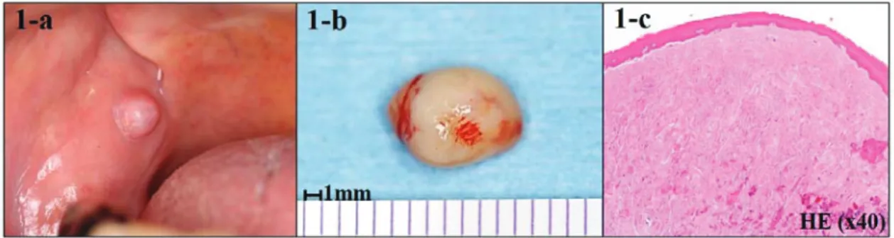
vessels stained yellow with picric acid, in the submucosal tissues (Fig. 3). These elastic
fibers were comparatively isolated, intermingled with hyperplastic collagen fibers
bundles, and extended toward the epithelial side in the lesion.
Two cases of fibroma of the labial mucosa showed different patterns of elastic
fibers (Fig. 2). On the epithelial side in these 2 cases, conspicuous quantities of elastic
fibers showed thick irregular fascicular bundles or globules.
2. Statistical tests for quantity of elastic fibers
10
1) Sex differences in quantity of elastic fibers in OFH: Mann-Whitney U test
(two-independent samples test)
Clinical data for 120 cases are summarized in Table 2. Significant differences
were observed in the quantity of elastic fibers in the central area of fibroma of the
labial mucosa between males and females (p < 0.05) (Table 3). However, sex
differences were not considered for further statistical tests because there were very few
males with fibroma of the labial mucosa (male to female ratio, 4: 16; Table 2).
2) Correlations between quantity of elastic fibers and age, and macrosize in OFH:
Spearman’s rank correlation
No significant correlations were observed in quantity of elastic fibers and age, and
macrosize (í0.39 r 0.43). Therefore, further comparisons were performed without
considering sex, age, or macrosize.
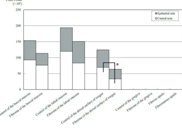
3) Comparison of quantity of elastic fibers between control and OFH: Mann-Whitney
U test (two-independent samples test)
There were significantly greater quantities of elastic fibers in the central areas of
11
the dorsal surface of tongue in control than in fibroma (p < 0.05) (Table 3) (Fig. 6).
4) Comparison of quantity of elastic fibers among OFH in the same area:
Kruskal-Wallis H test (several-independent samples test)
Significant differences were observed in each area, and quantities of elastic fibers
were particularly marked in fibroma of the labial mucosa (Table 4) (Fig. 7). Two cases
of fibroma of the labial mucosa showed particularly high quantities of elastic fibers on
the epithelial side (Fig. 2). Moreover, the lesion with the second highest quantity of
elastic fibers was fibroma of the buccal mucosa, while fewer elastic fibers were
observed in fibrous epulis and fibromatous epulis.
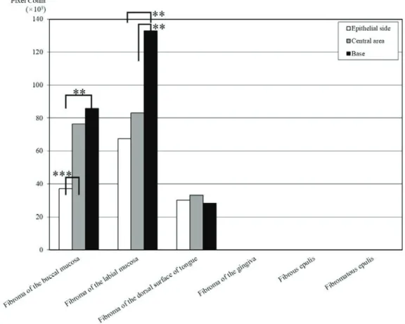
5) Comparison of quantity of elastic fibers among 3 areas in the same case: Friedman
test (several-related samples test)
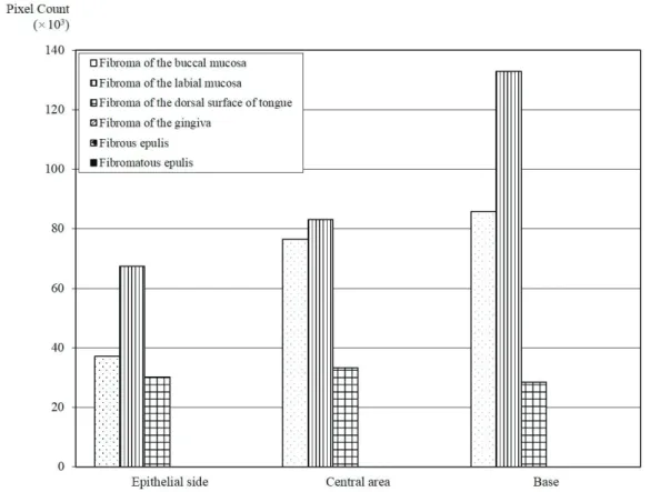
Significant differences were observed in fibroma of buccal mucosa (p < 0.001)
and fibroma of labial mucosa (p < 0.01). Thereafter, each area between fibroma of
buccal mucosa and fibroma of labial mucosa was compared, elastic fibers were mostly
distributed in the base (p < 0.01) (Fig. 8).
12 3. Immunohistochemical findings
Immunohistochemically, cytoplasm of spindle cells consisted in all OFH cases
showed diffusely positive reactions for Vimentin, and negative for Actin except for
cells composed of the vascular wall, respectively (Table 1) (Figs. 4, 5). In addition,
positive reactions for CD34 were observed in spindle cells and undifferentiated
mesenchymal cells around myogenic blood vessels, which were interspersed in the
connective tissue in the EF+ group.
Discussion
This study focused on elastic fibers in the connective tissue with regard to OFH,
and performed histopathological and histochemical studies of the distribution of elastic
fibers. In addition, the quantity of elastic fibers in OFH was investigated using image
analysis. To clarify the role of elastic fibers in OFH, lesions were classified into 3 areas
and the quantity of elastic fibers was measured and compared among these.
Significant differences were observed only in the tongue, as compared to the
quantity of elastic fibers in control and OFH (Fig. 6). This indicated that the quantity of
13
elastic fibers in OFH was reflected the quantity of elastic fibers in each existing tissue.
The quantity of elastic fibers was particularly high in fibroma of the labial mucosa
(Fig. 7), and 2 OFH cases showed markedly higher quantities of elastic fibers on the
epithelial side (Fig. 2). The second highest quantities of elastic fibers were seen in
fibroma of the buccal mucosa, and there were fewer elastic fibers in fibrous epulis and
fibromatous epulis.
On functional classification, the oral mucosa can be divided into 3 main types:
lining; specialized; and masticatory. For example, the buccal and labial mucosa are
lining mucosa, the dorsal surface of tongue is specialized mucosa, and the gingiva is
masticatory mucosa. Elastic fibers are more abundant in flexible lining mucosa, and
can be seen in most regions of the oral mucosal tissue. Therefore, the quantity of elastic
fibers is higher in the buccal and labial mucosa than on the dorsal surface of tongue
and gingiva (17). This indicated that existing elastic fibers, which have site-specific
localization, in contrast to collagen fibers in each tissue, were influenced by the
distribution of elastic fibers in OFH.
Elastic fibers were commonly concentrated at the base of lesions, near the
submucosal tissue in both fibroma of the buccal and labial mucosa wherein elastic
14
fibers were significantly abundant (Fig. 8). The buccal and labial mucosa accompany
submucosal tissue. The capillary plexus in normal buccal and labial mucosa is derived
from well-developed myogenic blood vessels present in the submucosal tissue, forming
loops while entering the lamina propria, and elastic lamina comprising elastic fibers is
present in these vascular walls.
On the other hand, the dorsal surface of tongue and gingiva lack submucosal
tissue. However, the deep layer of lamina propria on the dorsal surface of tongue has
myogenic blood vessels, similarly to the buccal and labial mucosa. Blood vessels
distributed in the gingiva are thought to pass through the surrounding periodontal
ligament, alveolar bone, and alveolar mucosa (17). In other words, myogenic tissue is
absent near the gingival mucosa, while myogenic tissues such as the buccinator,
orbicularis oris muscle, and tongue muscle are present near the mucosal tissue of the
buccal mucosa, labial mucosa, and dorsal surface of tongue. This suggested that the
presence of myogenic blood vessels with elastic lamina greatly influenced site-specific
localization.
We also performed an immunohistochemical study to explore the origin of elastic
fibers in OFH. Based on the results of immunohistochemical staining (Table 1) (Figs. 4,
15
5), spindle cells in all cases of OFH were positive for Vimentin and negative for Actin,
respectively. This suggested that OFH was of mesenchymal cell origin, not
myofibroblastic origin (5-8). With regard to CD34, spindle cells were positive in all
cases in the EF+ group and negative in all cases in the EF– group. This suggested that
no correlations existed between collagen fibers and CD34, but that correlations existed
between elastic fibers and CD34 (6-8). Therefore, it was a possible that CD34 was
involved in the elastic fibers observed in OFH.
It has been reported that CD34-positive spindle cells participate in the formation
of elastofibroma (6-8). CD34 is a single-chain transmembrane protein of approximately
116 kDa and is expressed on immature haematopoietic stem/progenitor cells, capillary
endothelial cells, and embryonic fibroblasts (18). Consequently, it has been suggested
that undifferentiated mesenchymal cells around myogenic blood vessels are related to
the formation of the elastic fibers that constitute OFH. In addition, histochemical
findings suggested that elastic fibers were derived from the elastic lamina, constituting
the vascular wall, and entered the lesion in a continuous or transitional pattern in the
upper parts of the lesion (Fig. 3). These results suggested that elastic fibers in OFH
were derived from the elastic lamina of myogenic blood vessels in the submucosa or
16 deep layer of the lamina propria.
CD34-positive cases in the EF+ group were mainly fibroma of the buccal mucosa,
labial mucosa, and dorsal surface of tongue (Table 1) (Fig. 4). However, there were
also cases with no CD34 expression in the gingiva (Table 1) (Figs. 4, 5). We compared
OFH cases occurring in the gingiva, and found 8 CD34-positive cases in the EF+ group
with fibroma of the gingiva, and 2 cases with epulis, including fibrous and fibromatous
(Table 1). The difference in the number of positive cases may be attributed to the
differences in pathogenesis of fibroma and epulis.
There is much discussion over whether fibroma is a real tumor, but it is classified
as a nonepithelial benign tumor, whereas epulis is classified as a lesion that synthesizes
localized masses in the gingiva, and that originates from connective tissues such as
gingiva, periodontal ligament, and alveolar bone periosteum (1). No detailed
information is available on the pathogenesis of fibroma of gingiva, but it is commonly
accepted that both gingiva and periodontal ligament are involved in epulis.
The elastic system fibers are constituted by fibers of three different types;
oxytalan, elaunin, and elastic fibers. The microfibrils consist of the fundamental
framework of the elastic system fibers, and elastogenesis is the process of deposition of
17
elastin. Oxytalan fibers are the bundles of microfibrils that first appear during
elastogenesis. The elaunin fibers are considered to be a transitional pattern between
oxytalan fibers and elastic fibers (19, 20). All elastic system fibers are observed in the
gingival lamina propria, but only oxytalan fibers are observed in the periodontal
ligament (21-25). In other words, periodontal ligament fibroblasts with the possibility
of the pathogenesis of epulis are not related to elastin secretion. Elastin and
microfibrils are produced as well as collagen proteins by fibroblasts (19-25). Therefore,
variations in the proteins secreted by the fibroblast in OFH lead to site-specific
localization.
In conclusion, elastic fibers were observed in 57.5% of OFH cases. The
distribution of these fibers was site-specific, and differed from the collagen fibers that
constituted OFH. CD34 positivity was observed in the spindle cells constituting OFH
accompanied by elastic fibers, and undifferentiated mesenchymal cells around
myogenic blood vessels near the lesion were related to the formation of a part of elastic
fibers that constituted OFH.
18 References
1. Regezi JA, Sciubba JJ, Jordan RC: Oral pathology: clinical pathologic
correlations. 6th ed. 2012. 162-167. Elsevier. St. Louis. USA.
2. Weiss SW, Goldblum JR: Soft Tissue Tumors. 4th ed. 2001. 286-290. Mosby. St.
Lo2uis. USA.
3. Järvi OH, Saxén AE, Hopsu-Havu VK, Wartiovaara JJ, Vaissalo VT:
Elastofibroma--a degenerative pseudotumor. Cancer, 23 (1): 42-63, 1969.
4. Daum O, Ferda J, Curik R, Choc M, Mukensnabl P, Michal M: Elastofibromatous
changes in tissues from spinal biopsies. A degenerative process afflicting a small
but important subset of patients operated for spinal canal compression: report of
18 cases. Int J Surg Pathol, 18 (6): 508-15, 2010.
5. Kayaselçuk F, Demirhan B, Kayaselçuk U, Ozerdem OR, Tuncer I: Vimentin,
Smooth Muscle Actin, Desmin, S-100 Protein, p53, and Estrogen Receptor
Expression in Elastofibroma and Nodular Fasciitis. Ann Diagn Pathol. 6 (2): 94-9,
2002.
19
6. Gun BD, Bahadir B, Behzatoglu K, Gun MO, Ozdamar SO: Elastofibroma: a
clinicopathologic and immunohistochemical study of seven cases and literature
review. APMIS. 115: 115-9, 2007.
7. Hisaoka M, Hashimoto H: Elastofibroma: clonal fibrous proliferation with
predominant CD34-positive cells. Virchows Arch. 448: 195-9, 2006.
8. Kuroda N, Hamaguchi N, Ohara M, Hirouchi T, Mizuno K, Hayashi Y, Lee GH:
Elastofibroma: a histochemical, immunohistochemical, and ultrastructural study
of two patients. Med Mol Morphol. 41: 179-82, 2008.
9. Daley T, Darling M: Elastofibroma Oralis. Head Neck Pathol, 5: 259-60, 2011.
10. Nonaka CF, Rêgo DM, Miguel MC, de Souza LB, Pinto LP: Elastofibromatous
change of the oral mucosa: case report and literature review. J Cutan Pathol, 37:
1067-71, 2010.
11. Potter TJ, Summerlin DJ, Rodgers SF: Elastofibroma: the initial report in the oral
mucosa. Oral Surg Oral Med Oral Pathol Oral Radiol Endod, 97: 64-7, 2004.
12. Manchandu R, Foot J, Alawi F: Elastofibroma presenting as an oral soft tissue
mass. J Oral Pathol Med, 37: 125-6, 2008.
20
13. Tosios KI, Economou I, Vasilopoulos NN, Koutlas IG: Elastofibromatous
changes and hyperelastosis of the oral mucosa. Head Neck Pathol, 4: 31-6, 2010.
14. Darling MR, Kutalowski M, Macpherson DG, Jackson-Boeters L, Wysocki GP:
Oral elastofibromatous lesion: a review and case series. Head Neck Pathol, 5:
254-8, 2011.
15. Weudner N, Carroll PR, Flax J, Blumenfeld W, Folkman J: Tumor angiogenesis
correlates with metastasis in invasive prostate carcinoma. Am J Pathol. 143: 401-9,
1993.
16. Masaki S: A Pathomorphological Study of Fractal Analysis in
Parenchymal-stromal Border on Keratocystic Odontogenic Tumor -with Special
Reference to Proliferative Activity and Vascular Distribution-. Int J Oral-Med Sci.
10 (4): 372-383, 2012.
17. Nanci A: Ten cat’s Oral Histology: Development, Structure, and Function. 8th ed.
2013. 297-298. Elsevier. St. Louis. USA.
18. Kishimoto T, Kikutani H, von dem Borne AEG, Goyert SM, Mason DY,
Miyasaka M, et al.: Leucocyte typing VI. White cell differentiation antigens. 1998.
974-76. Garland Publishing Inc. New York. USA.
21
19. Cotta-Pereira G, Guerra Rodrigo F, Bittencourt-Sampaio S: Oxytalan, elaunin,
and elastic fibers in the human skin. J Invest Dermatol. 66 (3): 143-8, 1976.
20. Ushiki T: Collagen fibers, reticular fibers and elastic fibers. A comprehensive
understanding from a morphological viewpoint. Arch Histol Cytol. 65 (2): 109-26,
2002.
21. Tsuruga E, Irie K, Sakakura Y, Yajima T: Expression of fibrillins and tropoelastin
by human gingival and periodontal ligament fibroblasts in vitro. J Periodontal Res.
37 (1): 23-8, 2002.
22. Tsuruga E, Irie K, Sakakura Y, Yajima T: Tropoelastin expression by periodontal
fibroblasts. J Dent Res. 81 (3): 198-202, 2002.
23. Tsuruga E, Irie K, Yajima T: Gene expression and accumulation of fibrillin-1,
fibrillin-2, and tropoelastin in cultured periodontal fibroblasts. J Dent Res. 81
(11): 771-5, 2002.
24. Tsuruga E, Yajima T, Irie K: Induction of fibulin-5 gene is regulated by
tropoelastin gene, and correlated with tropoelastin accumulation in vitro. Int J
Biochem Cell Biol. 36 (3): 395-400, 2004.
22
25. Tsuruga E, Yajima T, Irie K: Microfibril-associated glycoprotein-1 and fibrillin-2
are associated with tropoelastin deposition in vitro. Int J Biochem Cell Biol. 7 (1):
120-9, 2005.
Groups (n) Vimentin Actin CD34
Fibroma of the buccal mucosa EF+ (20) 20 0 20
EF- ( 0)
䇷 䇷 䇷
Fibroma of the labial mucosa EF+ (20) 20 0 20
EF- ( 0)
䇷 䇷 䇷
Fibroma of the dorsal surface of tongue EF+ (19) 19 0 19
EF- ( 1) 1 0 0
Fibroma of the gingiva EF+ ( 8) 8 0 8
EF- (12) 12 0 0
Fibrous epulis EF+ ( 1) 1 0 1
EF- (19) 19 0 0
Fibromatous epulis EF+ ( 1) 1 0 1
EF- (19) 19 0 0
(cases) Table 1. Number of positive cases by immunohistochemical staining
All of cases (n = 120) are confirmed collagen fibers.
EF+: Cases with elastic fibers (n = 69), EF䌦: Cases without elastic fibers (n = 51)
Male : Female Med Min Max Med Min Max Fibroma of the buccal mucosa 10 : 10 63 ( 14 - 81 ) 452 ( 100 - 4500 )
Fibroma of the labial mucosa 4 : 16 47 ( 17 - 77 ) 184 ( 36 - 420 ) Fibroma of the dorsal surface of tongue 8 : 12 53 ( 37 - 79 ) 116 ( 12 - 528 )
Fibroma of the gingiva 10 : 10 62 ( 27 - 80 ) 440 ( 1 - 7000 )
Fibrous epulis 10 : 10 48 ( 8 - 74 ) 144 ( 4 - 2520 )
Fibromatous epulis 8 : 12 39 ( 15 - 78 ) 298 ( 30 - 4480 )
Age Size (mm3)
Table 2. Demographic and clinical characteristics of oral fibrous hyperplasia (OFH) cases (n = 120)
Med : Median, Min : Minimum, Max: Maximum
n Med Min Max Med Min Max Med Min Max Control of the buccal mucosa 5 60.9 ( 22.9 - 70.1 ) 92.6 ( 76.2 - 110.5 )
Fibroma of the buccal mucosa 20 37.1 ( 0.0 - 159.9 ) 76.4 ( 18.4 - 145.1 ) 85.8 ( 18.2 - 214.0 ) Control of the labial mucosa 5 74.2 ( 13.8 - 116.4 ) 119.7 ( 64.6 - 230.4 )
Fibroma of the labial mucosa 20 67.4 ( 6.1 - 535.2 ) 83.1 ( 12.9 - 222.8 ) + 132.8 ( 37.9 - 404.5 ) Control of the dorsal surface of tongue 5 55.0 ( 34.5 - 98.6 ) 69.6 ( 54.0 - 90.0 ) *
Fibroma of the dorsal surface of tongue 20 30.2 ( 0.0 - 139.4 ) 33.3 ( 0.0 - 83.6 ) 28.4 ( 0.0 - 87.0 )
Control of the gingiva 5 0.0 ( 0.0 - 32.1 ) 0.0 ( 0.0 - 5.8 )
Fibroma of the gingiva 20 0.0 ( 0.0 - 130.7 ) 0.0 ( 0.0 - 81.0 ) 0.0 ( 0.0 - 242.4 )
Fibrous epulis 20 0.0 ( 0.0 - 0.0 ) 0.0 ( 0.0 - 15.4 ) 0.0 ( 0.0 - 103.5 )
Fibromatous epulis 20 0.0 ( 0.0 - 0.0 ) 0.0 ( 0.0 - 16.3 ) 0.0 ( 0.0 - 8.7 )
Table 3. Comparison of quantity of elastic fibers between control and OFH
(㽢103 pixel count) Minimum (Min), median (Med), and maximum (Max) are listed as representative values in the table, because nonparametric test is performed.
Sex differences: +p < 0.05 (Male < Female) (Mann-Whitney U test) Comparison of OFH: *p < 0.05 (Mann-Whitney U test)
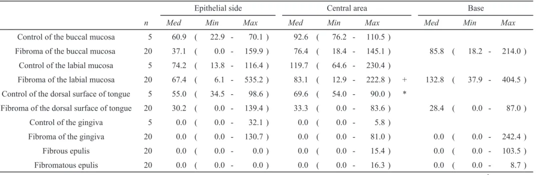
Epithelial side Central area Base
Epithelial side Fibroma of the buccal mucosa Fibroma of the labial mucosa Fibroma of the dorsal surface of tongue Fibroma of the gingiva Fibrous epulis Fibromatous epulis Fibroma of the buccal mucosa
Fibroma of the labial mucosa ns
Fibroma of the dorsal surface of tongue ns ns
Fibroma of the gingiva *** *** *
Fibrous epulis *** *** *** ns
Fibromatous epulis *** *** *** ns ns
Central area Fibroma of the buccal mucosa Fibroma of the labial mucosa Fibroma of the dorsal surface of tongue Fibroma of the gingiva Fibrous epulis Fibromatous epulis Fibroma of the buccal mucosa
Fibroma of the labial mucosa ns
Fibroma of the dorsal surface of tongue ns ns
Fibroma of the gingiva *** *** ns
Fibrous epulis *** *** *** ns
Fibromatous epulis *** *** *** ns ns
Base Fibroma of the buccal mucosa Fibroma of the labial mucosa Fibroma of the dorsal surface of tongue Fibroma of the gingiva Fibrous epulis Fibromatous epulis Fibroma of the buccal mucosa
Fibroma of the labial mucosa ns
Fibroma of the dorsal surface of tongue ns **
Fibroma of the gingiva * *** ns
Fibrous epulis *** *** ** ns
Fibromatous epulis *** *** ** ns ns
㻌 㻌 㻌 㻌
Table 4. Comparison of quantity of elastic fibers among OFH in the same area
ns: Not statistically significant
*p < 0.05, **p < 0.01, ***p < 0.001 (Kruskal-Wallis H test)
Figure legends
Fig. 1 Case of oral fibrous hyperplasia (OFH)
Before excision (a), Excised specimen (b), Hematoxylin-eosin staining (c: ×40)
Fig. 2 Image analysis method (binarization), and Elastica van Gieson (EvG) finding of OFH
The lamina propria is classified into 3 areas based on collagen fibers orientation (1a, 2a; ×40)
(1b-g, 2b-g; ×400). The epithelial side is defined the area of 100μm below the basal lamina (1b,
2b). The central area is below the epithelial side (1c, 2c). The base is defined as the clinical base of
the mass (1d, 2d). The hot spot (yellow squares; 1a, 2a) is defined as the 100μm × 100μm area with
the greatest quantity of elastic fibers (1b-d, 2b-d). After images are binarized using Image J, elastic
fibers are extracted (1e-g, 2e-g).
Elastic fibers show a fine granular appearance on the epithelial side (1b), and elongated
filamentous morphology in the central area and base (1c, 1d). Two cases of fibroma of the labial
mucosa show different patterns of elastic fibers, which appear thick irregular fascicular bundles or
globules on the epithelial side (2b).
Fig. 3 EvG finding in deep layer of lesion
Elastic fibers are observed into the elastic lamina, which constitutes the vascular wall of
myogenic blood vessels, in the submucosal tissues. These elastic fibers are comparatively isolated,
intermingled with hyperplastic collagen fibers bundles, and extend toward the epithelial side in the
lesion (arrow) (×200).
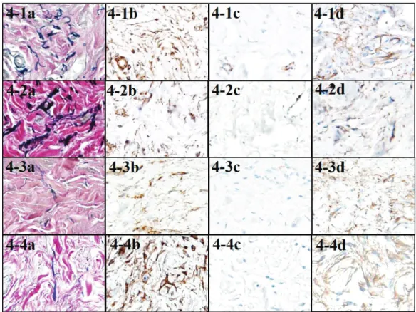
Fig. 4 EvG and immunohistochemical staining of cases in EF+ group
Cases with elastic fibers (EF+ group) include fibroma of the buccal mucosa (1a-d), fibroma of
the labial mucosa (2a-d), fibroma of the dorsal surface of tongue (3a-d), and fibroma of the gingiva
(4a-d). Cytoplasm of spindle cells in all of cases show diffusely positive reactions for Vimentin
(1-4b), and negative for Actin (1-4c). Positive reactions for CD34 are observed in spindle cells, as
well as in undifferentiated mesenchymal cells around the myogenic blood vessels, which intersperse
into connective tissues (1-4d) (×200).
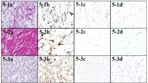
Fig. 5 EvG and immunohistochemical staining of cases in EF- group
Cases without elastic fibers (EFí group) include fibroma of the dorsal surface of tongue (1a-d),
fibroma of the gingiva (2a-d), and fibrous and fibromatous epulis (3a-d). Cytoplasm of spindle cells
in all of cases show positive reactions for Vimentin (1-3b), and negative for Actin (1-3c) and CD34
(1-3d) (×200).
Fig. 6 Comparison of quantity of elastic fibers between control and OFH
There are significantly greater quantities of elastic fibers in the central area of the dorsal surface
of tongue in control than in fibroma (*p < 0.05) (Mann-Whitney U test).
Fig. 7 Comparison of quantity of elastic fibers among OFH in the same area
Significant differences are observed in each area, and the quantity of elastic fibers is
particularly marked in fibroma of the labial mucosa. The lesion with the second greatest quantity of
elastic fibers is fibroma of the buccal mucosa, while fewer elastic fibers are seen in fibrous epulis
and fibromatous epulis (Kruskal-Wallis H test).
Fig. 8 Comparison of quantity of elastic fibers among 3 areas in the same case
Significant differences are observed in fibroma of the buccal mucosa (***p < 0.001) and fibroma
of the labial mucosa (**p < 0.01). Elastic fibers are mainly distributed in the base (**p < 0.01)
(Friedman test).