Fukushima Medical University
福島県立医科大学 学術機関リポジトリ
This document is downloaded at: 2021-11-07T23:07:04Z
Title A case of gastric lipoma resected by endoscopic submucosa dissection with difficulty in preoperative diagnosis
Author(s)
Ichinose, Mizue; Hikichi, Takuto; Kanno, Yukiko; Gunji, Naohiko; Fujita, Masashi; Kuroda, Masahito; Terashima, Kumiko; Sato, Yoshinori; Kawana, Satoshi; Hashimoto, Yuko;
Ohira, Hiromasa; Miyata, Masayuki
Citation Fukushima Journal of Medical Science. 63(3): 160-164
Issue Date 2017
URL http://ir.fmu.ac.jp/dspace/handle/123456789/669
Rights © 2017 The Fukushima Society of Medical Science
DOI 10.5387/fms.2016-19
Text Version publisher
Vol. 63, No. 3, 2017
[Case Report]
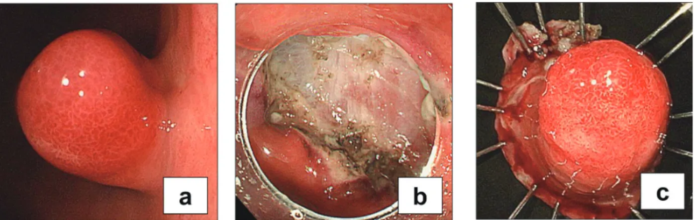
A case of gastric lipoma resected by endoscopic submucosa dissection with difficulty in preoperative diagnosis
Mizue Ichinose
1), Takuto Hikichi
2), Yukiko Kanno
1,3), Naohiko Gunji
1,3), Masashi Fujita
1,3), Masahito Kuroda
1), Kumiko Terashima
1), Yoshinori Sato
1), Satoshi Kawana
4), Yuko Hashimoto
4), Hiromasa Ohira
3)and Masayuki Miyata
1)1)