Fukushima Medical University
福島県立医科大学 学術機関リポジトリ
This document is downloaded at: 2021-11-08T00:40:44Z
Title Utility of peppermint oil for endoscopic diagnosis of gastric tumors
Author(s) Hikichi, Takuto; Irisawa, Atsushi; Sato, Masaki; Watanabe, Ko; Nakamura, Jun; Takagi, Tadayuki; Ikeda, Tsunehiko;
Suzuki, Rei; Ohira, Hiromasa; Obara, Katsutoshi Citation Fukushima Journal of Medical Science. 57(2): 60-65
Issue Date 2011
URL http://ir.fmu.ac.jp/dspace/handle/123456789/302
Rights © 2011 The Fukushima Society of Medical Science
DOI 10.5387/fms.57.60
Text Version publisher
Fukushima J. Med. Sci., Vol. 57, No. 2, 2011
60
引地拓人,入澤篤志,佐藤匡記,渡辺 晃,中村 純,高木忠之,池田恒彦,鈴木 玲,大平弘正,小原勝敏 Corresponding author : Takuto Hikichi E-mail : [email protected]
http://www.jstage.jst.go.jp/browse/fms http://fmu.ac.jp/home/lib/F-igaku/
[Original Article]
UTILITY OF PEPPERMINT OIL FOR ENDOSCOPIC DIAGNOSIS OF GASTRIC TUMORS
TAKUTO HIKICHI
1), ATSUSHI IRISAWA
2), MASAKI SATO
3), KO WATANABE
3), JUN NAKAMURA
3), TADAYUKI TAKAGI
3), TSUNEHIKO IKEDA
3),
REI SUZUKI
3), HIROMASA OHIRA
3)and KATSUTOSHI OBARA
1)1)Department of Endoscopy, Fukushima Medical University Hospital, 2)Department of Gastroenterology, Preparatory Office for Aizu Medical Center, Fukushima Medical University, and 3)Department of Gastro- enterology and Rheumatology, Fukushima Medical University, Fukushima, Japan
(Received June 22, 2011, accepted October 6, 2011)
Abstract : We investigated whether peppermint oil (PO) is useful for endoscopic diagnosis of gas- tric tumors without magnifying endoscopy. Twenty-six patients diagnosed with gastric tumors were examined. Endoscopic images were recorded by white light (WL) and narrow band imaging (NBI) without magnifying endoscopy. After PO administration, images were recorded again by WL and NBI (PO+WL and PO+NBI). The clarity of tumor margins and the effect of PO on the clarity of tumor margins were scored. The mean scores for clarity of tumor margins were 3.6 points in WL, 4 in NBI, 4.3 in PO+WL, and 4.3 in PO+NBI. The scores of WL and NBI showed a tendency to rise by PO. The mean scores for the effect of PO on the clarity of tumor margins were 2.7 in WL and 2.5 in NBI. Therefore, we conclude that PO is useful for endoscopic diagnosis of the margin of gastric tumors.
Key words: peppermint oil, tumor margin, gastric tumor, magnifying endoscopy, chromoendos- copy
INTRODUCTION
To perform endoscopic diagnosis for gastric cancer, observation using white light imaging (WL), so-called conventional endoscopy, is often con- ducted. Whether the lesion is gastric cancer or not is usually discerned according to evidence of irregu- larities and ruggedness obtained by WL. Several stains have been applied in the stomach to improve the WL diagnostic performance. Chromoen- doscopy with indigo carmine (IC), the most popular method among these stains, is a useful technique to detect small gastric cancers and to clarify the tumor margins1−4). However, it is not possible to diagnose the margins of all early gastric cancers correctly using IC method. More recently, diagnoses based on mucosal surface patterns and microvascular pat- terns have been conducted using magnifying endos-
copy with narrow band imaging (NBI) or acetic acid5−9). Reportedly magnifying endoscopy with NBI or acetic acid is useful not only for determining the tumor margins but also for differential diagnosis between benign and malignant lesions. However, NBI magnifying endoscopy cannot be performed in all hospitals because NBI system is very expensive.
On one occasion, we administered a peppermint oil (PO) solution as an antispasmodic agent10) to early gastric cancer in the antrum and the tumor margin became well demarcated. This fact allowed us infer that the PO solution might be useful for the endoscopic diagnosis of early gastric cancers.
Therefore, we investigated whether the PO solution is useful for endoscopic diagnosis of gastric tumors without magnifying endoscopy.
PEPPERMINT OIL FOR ENDOSCOPIC DIAGNOSIS 61
METHODS
Between December 2009 and April 2010, 26 patients (20 men, six women ; mean age 73.5 years
±8.3 SD) who were diagnosed with early gastric cancer and gastric adenoma after endoscopic submu- cosal dissection (ESD) or surgical operation were enrolled. Exclusion criteria were as follows : 1) the lesion was an advanced cancer, 2) the lesion was not a tumor, and 3) endoscopic images could not be evaluated because the lesion was so wide or its images were not clear. All enrolled patients gave their written informed consent to participation in this study.
A 1.6% PO solution was made in the Pharma- cology Division of Fukushima Medical University Hospital by mixing 1.6 ml of PO (Toho Pharmaceu- tical Co. Ltd., Tokyo, Japan) and 0.2 g of sorbitan monostearate (Wako Pure Chemical Industries Ltd., Osaka, Japan), a widely used surfactant, in hot water
until completely dissolved. Subsequently, 100 ml of distilled water was added to the solved PO solu- tion. This solution was put into a plastic bottle and stored at 4 °C.
To dissolve the mucus layer of the stomach, each patient ingested a solution containing 20,000 U pronase (Pronase MS ; Kaken Pharmaceutical Co.
Ltd., Tokyo, Japan) and 80 mg of dimethicone (2% of Balgin antifoaming oral solution ; Kaigen Co. Ltd., Osaka, Japan) and 1 g NaHCO2 in 80 ml water 10 min before the endoscopy. The procedure was con- ducted using a high vision endoscope (GIF- H260 ; Olympus Optical Co. Ltd., Tokyo, Japan) without magnifying observation. Before inserting the scope, pharyngeal anesthesia was achieved using 40 mg of lidocaine spray (8% of Xylocaine pump spray ; AstraZeneca International PLC., Osaka, Japan). One of the five endoscopists (T.H.,
Fig. 1. Clarity of tumor margins
A) B) C)
D) E)
Fig. 1. Clarity of tumor margins. A) 5 points : Margin is clear all around the lesion. B) 4 points : Margin is clear for 3/4 or more of the lesion circumference, but not clear all around the lesion. C) 3 points : Margin is clear for 1/2 or more of the lesion circumference, but not for 3/4 of the lesion circumference. D) 2 points : Margin is clear for less than 1/2 of the lesion circumference. E) 1 point : Margin is clear for only a part of the lesion or unclear all around the lesion.
Table 1. Effects of PO solution on clarity of tumor margins 3 points : It became more clear.
2 points : It was unchanged.
1 point : It became more unclear.
Table 2. Clarity of tumor margins Mean score Median P value
WL 3.6 4
PO+WL 4.3 5 1.00
NBI 4.0 4
PO+NBI 4.3 5 0.97
62 T. HIKICHI et al.
M.S., R.S., K.W., and J.N.) who had performed more than 5,000 upper gastrointestinal (GI) endoscopies and 50 ESD procedures performed endoscopies in this study.
All endoscopic images were recorded using a digital filing system (SolemioENDO ; Olympus Optical Co. Ltd., Tokyo, Japan). First, the gastric lesion was observed using WL alone and more than two images were recorded (WL Group). Second, the lesion was observed using NBI and more than two images were recorded (NBI Group). Then, 50 ml of the PO solution was administered to the lesion through the working channel of the scope. At 30 seconds after administration of the PO solution, the lesion was observed using WL (PO+WL Group) and NBI again (PO+NBI Group).
The tumor margin was finally judged by the resected specimen of ESD or surgical operation.
The recorded endoscopic images were evaluated by one endoscopist (T.H.) for clarity of the tumor mar- gins in each of four groups and the effect of the PO solution on the clarity of the tumor margins. The
clarities of the tumor margins were scored based on a 5- point scale for each image of WL group, NBI Group, PO+WL Group, and PO+NBI Group (Fig. 1).
The scores of WL Group, PO+WL Group, NBI Group, and PO+NBI Group were respectively com- pared. The effects of the PO solution on the clarity of the tumor margins were scored based on a 3-point scale for PO+WL Group against WL Group and PO+NBI Group against NBI Group (Table 1).
Data were analyzed using software (Statcel 2 ; OMS Publication, Tokorozawa, Japan). Differe- nces were evaluated using Wilcoxon rank sum test.
A P value of less than 0.05 was considered signifi- cant.
RESULTS
Of 26 patients, 23 underwent ESD ; and three surgical operations. Final pathological diagnoses were 23 early gastric cancers (20 well or moderately differentiated adenocarcinoma, two differentiated adenocarcinoma combined with undifferentiated
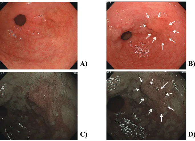
A) B)
Fig. 3. Case 1
C) D)
Fig. 2. Case 1. The lesion was a well-differentiated adenocarcinoma located in the posterior wall of the gastric antrum. It was resected by ESD. Arrows show the tumor margin. A) WL (3 points), B) PO+WL (5 points), C) NBI (4 points), D) PO+NBI (4 points). The effects of the PO solution on the clarity of the tumor margin were 3 points in PO+WL against WL and 2 points in PO+NBI against NBI.
PEPPERMINT OIL FOR ENDOSCOPIC DIAGNOSIS 63
adenocarcinoma, one undifferentiated adenocarci- noma) and three gastric adenomas. The mean tumor size was 18.1 mm (5-35 mm). Of 23 early gastric cancers, the gross type was elevated lesions in 9, flat lesion in 2, and depressed lesion in 12, and the depth of tumor invasion was mucosal layer in 21 and submucosal layer in 2.
The mean scores of the clarities of the tumor margins were 3.6 points in WL Group (median, 4 points), 4 points in NBI Group (median, 4 points), 4.3 points in PO+WL Group (median, 5 points), and 4.3 points in PO+NBI Group (median, 5 points) (Table 2, Fig. 2, 3). The difference between WL Group and PO+WL Group was not statistically significant (P=1.00). The difference between NBI Group and PO+NBI Group was not statistically significant
either (P=0.97). However, scores of WL Group and NBI Group showed a tendency to higher scores by administration of the PO solution.
The mean scores of the effects of the PO solu- tion on the clarity of the tumor margins were 2.7 points in PO+WL group against WL group (median, 3 points) and 2.5 points in PO+NBI Group against NBI Group (median, 3 points) (Table 3, Fig. 2, 3).
The ratios of 3 points were 65.4% (17/26) in PO+WL Group against WL Group and 53.8% (14/26) in PO+NBI Group against NBI Group.
DISCUSSION
Recently, ESD has been developed as a thera- peutic endoscopy method for early stage gastric can-
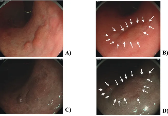
A) B)
C) D)
Fig. 3. Case 2
Fig. 3. Case 2. The lesion was a well-differentiated adenocarcinoma located in the lesser curvature of the lower gastric body. It was resected by ESD. Arrows show the tumor margin. A) WL (3 points), B) PO+WL (5 points), C) NBI (3 points), D) PO+NBI (5 points). The effects of the PO solution on the clarity of the tumor margin were 3 points not only in PO+WL against WL but also in PO+NBI against NBI.
Table 3. Effects of PO solution on clarity of tumor margins Mean score Median Ratio of 3 points
PO+WL against WL 2.5 3 65.4% (17/26)
PO+NBI against NBI 2.7 3 53.8% (14/26)
64 T. HIKICHI et al.
cer in Japan, and is becoming more and more popu- lar. Its use has spread gradually. With ESD, resections of very large gastric tumors of more than 30 mm have also been reported11−13). Therefore, it has become important to precisely identify the extent of lesions. Nowadays, for diagnosing the extent of lesions correctly, the utility of magnifying endoscopy with NBI and acetic acid and chromoen- doscopy with an acetic acid − IC mixture has been reported5−9,14). However, these technologies are not popular because specialized and expensive equipment is necessary.
We reported the utility of PO solution for the control of peristalsis during GI endoscopy10). In fact, PO is a well-known, harmless substance that inhibits GI smooth muscle contractility15). As its major constituent is menthol, PO relaxes GI smooth muscle tissues by reducing the influx of calcium into the muscle cells16). Previous reports were exam- ined to assess the effectiveness of intraluminally administered PO solution as a GI antispasmodic agent for colonoscopy17) and upper GI endos-
copy10,18). Then we applied it for diagnostic and
therapeutic endoscopy. On one occasion, we expe- rienced that the tumor margin located in the antrum was clarified when PO solution was administered to stop the peristaltic movement. Therefore, we inferred that PO solution also has a possibility to be utilized for the diagnosis of gastric tumor. If avail- able, it would be a unique method for the diagnosis of gastric tumor because it is an inexpensive, easy, and safe method. Moreover, it has an antispas- modic effect and can be used for aromatization. We then examined the efficacy of PO solution for endo- scopic diagnosis of gastric tumors without magnify- ing observation.
The present study showed that the margin of gastric tumor became clearer when the PO solution was administered both under WL alone and NBI alone. Although we were unable to determine the mechanism of its action, we infer that the contrast between the tumor and the part around the tumor was emphasized by the PO solution administration because the mucosa around the tumor absorbed the PO solution and then became edematous despite a lack of change in the tumor itself. The mechanism resembles the acetic acid method reported by Yagi et al.6) They noted that the duration of whitening dif- fered among grades of neoplasm and that changes in whitening were observable over time.
In conclusion, the PO solution has a possibility to be useful for endoscopic diagnosis of the margin of gastric tumor. In the future, we plan to study the
clinical significance of the PO solution combined with IC staining or magnifying endoscopy and the mechanism of change in gastric mucosa by PO administration.
REFERENCES
1. Ida K, Tada M. Chromoscopy. In : Sivak MV Jr (ed.). Gastroenterol Endosc, Philadelphia : Saun- ders, 203-220, 1987.
2. Peitz U, Malfertheiner P. Chromoendo scopy : From a research tool to clinical progress. Dig Dis, 20: 111-119, 2002.
3. Canto MI. Staining in gastrointestinal endos- copy : The basics. Endoscopy, 31: 479-486, 1999.
4. Mitooka H, Fujimori T, Maeda S, Nagasako K. Minute flat depressed neoplastic lesions of colon detected by contrast chromoscopy using indigo carmine capsule. Gastrointest Endosc, 41: 453-459, 1995.
5. Yagi K, Nakamura A, Sekine A, Umezu H. Magni- fying endoscopy with narrow band imaging for early differentiated gastric adenocarcinoma. Dig Endosc, 20: 115-122, 2008.
6. Yagi K, Aruga Y, Nakamura A, Sekine A, Umezu H. The study of dynamic chemical magnifying endoscopy in gastric neoplasia. Gastrointest Endosc, 62: 963-969, 2005.
7. Tanaka K, Toyoda H, Kadowaki S, Kosaka R, Shiraishi T, Imoto I, Shiku H, Adachi Y. Features of early gastric cancer and gastric adenoma by enhanced-magnification endoscopy. J Gastroen- terol, 41: 332-338, 2006.
8. Kuznetsov K, Lambert R, Rey JF. Narrow-band imaging : Potential and limitations. Endoscopy, 38: 76-81, 2006.
9. Nakayoshi T, Tajiri H, Matsuda K, Kaise M, Ikegami M, Sasaki H. Magnifying endoscopy combined with narrow band imaging system for early gastric cancer : Correlation of vascular pat- tern with histopathology. Endoscopy, 36: 1080- 1084, 2004.
10. Mizuno Y, Hikichi T, Suzuki T, Ito E, Rai T, Kokubun M, Sato K, Saito M. Peppermint oil is useful as an antispasmodic agent in esophago-gas- tro-duodenoscopy. Fukushima J Med Sci, 57: 9- 16, 2007 (in Japanese with English abstract).
11. Takeuchi Y, Uedo N, Iishi H, Yamamoto S, Tama- moto S, Yamada T, Higashino K, Ishihara R, Tatsuta M, Ishiguro S. Endoscopic submucosal dissection with insulated-tip knife for large mucosal early gastric cancer : A feasibility study (with vid- eos). Gastrointest Endosc, 66: 186-193, 2007.
12. Imagawa A, Okada H, Kawahara Y, Takenaka R, Kato J, Kawamoto H, Fujiki S, Takata R, Yoshino T,
PEPPERMINT OIL FOR ENDOSCOPIC DIAGNOSIS 65
Shiratori Y. Endoscopic submucosal dissection for early gastric cancer : Results and degrees of technical difficulty as well as success. Endoscopy, 38: 987-990, 2006.
13. Oka S, Tanaka S, Kaneko I, Mouri R, Hirata M, Kawamura T, Yoshihara M, Chayama K. Advan- tage of endoscopic submucosal dissection com- pared with EMR for early gastric cancer. Gastro- intest Endosc, 64: 877-883, 2006.
14. Kawahara Y, Takenaka R, Okada H, Kawano S, Inoue M, Tsuzuki T, Tanioka D, Hori K, Yamamoto K. Novel chromoendoscopic method using an acetic acid − indigocarmine mixture for diagnostic accuracy in delineating the margin of early gastric cancers. Dig Endosc, 21: 14-19, 2009.
15. Micklefield GH, Greving I, May B. Effects of peppermint oil and caraway oil on gastroduodenal motility. Phytotherapy Research, 14: 20-23, 2000.
16. Hills JM, Aaronson PI. The mechanism of action
of peppermint oil on gastrointestinal smooth mus- cle. An analysis using patch clamp electrophysi- ology and isolated tissue pharmacology in rabbit and guinea pig. Gastroenterology, 101: 55-65, 1991.
17. Asao T, Mochiki E, Suzuki H, Nakamura J, Hirayama I, Morinaga N, Shoji H, Shitara Y, Kusano H. An easy method for the intraluminal administration of peppermint oil before colonos- copy and its effectiveness in reducing colonic spasm. Gastroint Endosc, 53: 172-177, 2001.
18. Hiki N, Kurosaka H, Tatsutomi Y, Shimoyama S, Tsuji E, Kojima J, Shimizu N, Ono H, Hirooka T, Noguchi C, Mafune K, Kaminishi M. Peppermint oil reduces gastric spasm during upper endos- copy : A randomized, double-blind, double-dummy controlled trial. Gastrointest Endosc, 57: 475- 482, 2003.