Fukushima Medical University
福島県立医科大学 学術機関リポジトリ
This document is downloaded at: 2021-11-08T01:19:07Z
Title 原発部位のCK19発現に基づいた頭頸部扁平上皮癌患者に
おけるOSNA法の診断的可能( 本文 )
Author(s) 鈴木, 政博
Citation
Issue Date 2015-09-28
URL http://ir.fmu.ac.jp/dspace/handle/123456789/480
Rights
© 2015 Wiley Periodicals, Inc. This is the peer reviewed version of the following article: [Head Neck. 2016 Apr;38 Suppl 1:E239-45], which has been published in final form at [https://doi.org/10.1002/hed.23979]. This article may be used for non-commercial purposes in accordance with Wiley Terms and Conditions for Use of Self-Archived Versions.
DOI
Text Version ETD
1
Diagnostic potential of one-step nucleic acid amplification assay in patients with head and neck squamous cell carcinoma based on CK19 expression in a primary lesion
Masahiro Suzuki1, MD, Takashi Matsuzuka1, MD, PhD, Yuko Hashimoto2, MD, PhD, Masakazu Ikeda1, MD, Satoshi Saijo1, MD, Koichi Omori1, MD, PhD
1Department of Otolaryngology, Fukushima Medical University School of Medicine, Fukushima, Japan
2Department of Diagnostic Pathology, Fukushima Medical University School of Medicine, Fukushima, Japan
This is the pre-peer reviewed version of the following article: Head Neck. 2016 Apr;38 Suppl 1:E239-45, which has been published in final form at DOI: 10.1002/hed.23979. This article may be used for non-commercial purposes in accordance with Wiley Terms and Conditions for Use of Self-Archived Versions.
Koichi Omori, MD, PhD
Department of Otolaryngology, School of Medicine, Fukushima Medical University 1 Hikarigaoka, Fukushima City, 960-1295, Japan
Telephone: +81-24-547-1325 Fax: +81-24-547-1325 E-mail: [email protected]
Running title: Cervical metastases using OSNA assay
Key Words: Molecular detection; lymph node metastases; OSNA assay; CK19; head and neck cancer
2 Abstract
Background. This study aimed to investigate the effects of CK19 expression in the primary lesions of head and neck squamous cell carcinoma on the diagnosis of the cervical lymph node (CLN) metastasis using the one-step nucleic acid amplification (OSNA) assay.
Methods. Primary lesions and 54 CLNs were resected from 21 patients with head and neck squamous cell carcinoma between 2009 and 2011. Each CLN was tested by the OSNA assay, and the CK19 mRNA copy number obtained was compared to the corresponding histopathological results.
Results. In the primary lesion CK19-positive group, the sensitivity and specificity of the OSNA assay against hematoxylin-eosin staining were 86% and 100%, respectively. The p value by Fisher’s exact test was less than 0.0001, indicating statistical significance.
Conclusion. These results suggest that OSNA offers similar diagnostic potential to that of histopathological diagnosis of CLN biopsy in patients with a CK19-positive primary lesion.
3 Introduction
Head and neck cancers generally begin in squamous cells, and over 500,000 new cases are diagnosed worldwide each year1. Approximately 60% of the cases are advanced stage III and IV cancer, which have a poor prognosis even with a multimodal therapeutic approach2. Metastasis to lymph nodes also often occurs in the early stages of cancer, and whether neck dissection should be performed as preventive treatment is controversial. Lymph node metastasis is an important prognostic factor in head and neck squamous cell carcinoma; thus, accurate diagnostic techniques are necessary.
Sentinel lymph node biopsy, which enables highly accurate detection of regional lymph node metastasis3, has become a standard diagnostic and therapeutic modality for breast cancer. When a sentinel lymph node biopsy is negative, lymph node dissection can be avoided4. The principles of sentinel lymph node navigation surgery have been expanded to the treatment of other types of cancer including head and neck squamous cell carcinoma5-9.
Lymph node metastasis, including metastasis to the sentinel lymph node, is diagnosed by examining the slices of sectioned specimens pathologically. Micrometastasis may not be detected unless the metastases appear in the sectioned slices.The development of a method to examine a whole lymph node specimen is therefore required. Such a method can be applied to intraoperative diagnosis if the presence or absence of metastasis is determined in a short time.
CK19 is a typical epithelial cell marker expressed in human cancers but not in lymph nodes without metastasis. It is expressed in most, but not all, breast cancer cases10, suggesting there may be a CK19-negative subpopulation among patients with head and neck cancer. The origin of the cancer cell may be elucidated by examining the cytokeratin expression of cancer cells. Moreover, the examination of cytokeratin expression in lymph nodes has a diagnostic significance in metastasis.
The one-step nucleic acid amplification (OSNA) assay for quantitatively detecting CK19 mRNA is a molecular technique that rapidly detects lymph node metastasis and is already in use for intraoperative diagnosis of sentinel lymph node metastasis in breast cancer patients3,11-14. The validity of the OSNA assay for detecting lymph node metastasis has also been reported in patients with head and neck squamous cell carcinoma15,16.
In head and neck squamous cell carcinoma, no report on the OSNA assay yet exists examining the detailed association of CK19 expression between a primary lesion and lymph nodes.
This study intended to investigate the effects of CK19 expression in the primary lesions of head and neck squamous cell carcinoma on the diagnosis of the cervical lymph node (CLN)metastasis by molecular detection with the OSNA assay.
In our current study, clinically non-metastatic cervical lymph nodes were examined using the OSNA assay in order to investigate feasibility of detecting lymph node metastasis.
4 Materials and Methods
Patients
Primary lesions and 54 CLNs resected from 21 patients with head and neck squamous cell carcinoma between 2009 and 2011 at Fukushima Medical University Hospital were examined.
None of the 21 patients had a history of cancer, including squamous cell carcinoma, and had no evidence of distant metastasis. None received treatment prior to surgery.
Characteristics of the 21 patients are summarized in Table 1. Median age was 67 years (range, 38–80 years of age), and most patients were male. T stage was T2 or higher in almost all patients, and clinical N stage was N0 in 52% of patients.
Examination prior to surgery revealed that enlargement and clinically evident metastasis were absent in all 54 CLNs. Two or three lymph nodes were randomly selected from those surgically resected in each patient during neck dissection.
In the primary lesion of oral cavity and maxillary sinus, lymph nodes of cervical level I or II were selected. In the primary lesion of oropharynx, hypopharynx, and larynx, lymph nodes of cervical level II or III were selected.
Intraoperative hematoxylin-eosin (HE) staining of frozen sections revealed metastasis in 9 of the 54 CLNs. Results of HE staining of these frozen sections were in good agreement with those of HE staining of the corresponding permanent sections.
This study was approved by the Institutional Ethics Review Board of Fukushima Medical University.
Immunostaining in primary lesions
Primary lesions excised during surgery were fixed in formalin and embedded in paraffin to prepare 5-m slices. One slice was stained with HE, and the other slices were tested for CK19 expression by immunostaining with the RCK108 monoclonal antibody (DAKO, Hamburg Germany).According to the immunohistochemical assessment using J-score17, when more than 1% of tumor cells were immunostained in primary lesions, it was considered as CK19-positive by the pathologist in our hospital.
Processing for HE staining, CK19 immunostaining, and OSNA assayof CLNs
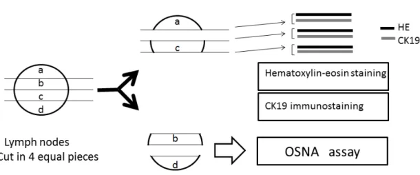
CLNs excised during surgery were divided into four equal portions (a-d, Fig. 1), two of which (b and d) were stored at −80°C until use in the OSNA assay. The other two portions (a and c) were processed in a similar manner to that used for primary lesions. Three pairs of 5-m slices were prepared at each side adjacent to the corresponding portion.
One slice of each pair was stained with HE, and the other was immunostained for CK19 detection
5
in a similar manner to that used for the primary lesions. According to the immunohistochemical assessment using J-Score17, when more than 1% of tumor cells were immunostained in CLNs, it was considered as CK19-positive. The pathologists who performed the HE staining and immunostaining were blinded to the OSNA results.
OSNA assay of CLNs
The OSNA assay of the CLNs was performed according to a previously reported method3. Briefly, two frozen portions (b and d) were homogenized with 4 ml of a buffer (Lynorhag; Sysmex, Kobe, Japan) and centrifuged at 10,000 g at room temperature. Two microliters of the resulting supernatant was analyzed on an automated molecular detection system (RD-100i; Sysmex) using a ready-to-use Lynoamp Kit (Sysmex). Reverse transcription loop-mediated isothermal amplification (RT-LAMP), which completes gene amplification within 30 min, was used for detection of CK19 mRNA18.
Amplicons were detected by real-time monitoring of turbidity caused by increasing levels of magnesium pyrophosphate (a byproduct of amplification). A total of six primers were used to perform highly specific reactions. Amplification of one specimen required less than 20 min. Full processing of three specimens was completed in approximately 30 min.
Each assay tested a positive control containing 5,000 copies/l of CK19 mRNA, which is considered to be equivalent to the amount of CK19 mRNA in 2 mm3 of breast cancer cells, and a negative control containing 0 copies/l of CK19 mRNA for calibration. Results are expressed as concentrations of CK19 mRNA. Specimens of head and neck squamous cell cancer containing
<1.31×10² copies/l of CK19 mRNA were considered CK19 negative, and those containing
≥1.31×10² copies/l of CK19 mRNA were considered CK19 positive15. Each CLN was tested by the OSNA assay, and the CK19 mRNA copy number obtained was compared to the corresponding histopathological results.
Statistical analyses
Sensitivity and specificity were calculated. Fisher’s exact test was used to test for statistical significance (p < 0.05).
Results
Results of all 21 primary lesions and 54 CLNs are summarized in Table 2. Immunostaining confirmed that 22 CLNs were from 9 patients with CK19-positive primary lesions (primary lesion CK19-positive group), whereas 32 were from 12 patients with CK19-negative primary lesions (primary lesion CK19-negative group).
Of the 54 CLNs, CK19 was positive by immunostaining in 6 CLNs, whereas CK19 was negative
6
in 48 CLNs. In the primary lesion CK19-positive group, CK19 was positive by immunostaining in 6 CLNs (27%), whereas CK19 was negative in 16 CLNs (73%). In the primary lesion CK19- negative group, CK19 was determined to be negative by immunostaining in all 32 CLNs.
The OSNA assay detected 8 positive CLNs (range, 1.31×10²–9.5×10⁵ copies/l; mean 1.6×10⁴ copies/l) and 46 negative CLNs (<1.31×10² copies/l; some were under the detection limit).
Six CLNs were CK19 positive by immunostaining and positive by the OSNA assay, 46 CLNs were CK19 negative by immunostaining and negative by the OSNA assay, and 2 CLNs were CK19 negative by immunostaining and positive by the OSNA assay.
The sensitivity and specificity of the OSNA assay against CK19 immunostaining were 100%
(95% CI: 68–100%) and 96% (95% CI: 92–96%), respectively (Fig.2). The p value by Fisher’s exact test was less than 0.0001, indicating statistical significance.
In the primary lesion CK19-positive group, 7 CLNs (32%) contained metastatic cells diagnosed by HE staining, whereas 15 (68%) did not, and 6 CLNs (27%) were positive by the OSNA assay, whereas 16 (73%) were negative. One CLN containing metastatic cells diagnosed by HE staining was negative by CK19 immunostaining and the OSNA assay (mRNA level under the detection limit). The sensitivity and specificity of the OSNA assay against HE staining in this group were 86% (95% CI: 61–86%) and 100% (95% CI: 88–100%), respectively (Fig.3). The p value by Fisher’s exact test was less than 0.0001, indicating statistical significance. A representative case containing a primary lesion with HE staining and CK19 positive by immunostaining, and metastatic LN with HE staining and CK19 positive by immunostaining is demonstrated (Fig.4).
In the primary lesion CK19-negative group, 2 CLNs (6%) contained metastatic cells diagnosed by HE staining, whereas 30 (94%) did not, and 2 CLNs (6%) were positive by the OSNA assay, whereas 30 (94%) were negative. In the 2 CLNs with metastatic cells diagnosed by HE staining, one was positive by the OSNA assay (1.31×10² copies/l), whereas the other was negative (6 copies/l). Additionally, one of CLNs that did not contain metastatic cells diagnosed by HE staining was positive by the OSNA assay (1.4×10³ copies/l). In this group, the sensitivity and specificity of the OSNA assay against HE staining was 50% (95% CI: 10–86%) and 97% (95%
CI: 94–99%), respectively (Fig.5). A representative case containing a primary lesion with HE staining and CK19 negative by immunostaining, and non-metastatic LN with HE staining and CK19 negative by immunostaining is demonstrated (Fig.6).
Discussion
Recently, sentinel lymph node biopsy, which enables highly accurate detection of regional lymph node metastasis3, has become a standard diagnostic and therapeutic modality for breast cancer.
When the sentinel lymph node biopsy is negative, lymph node dissection can be avoided4, thereby also reducing the risk for postoperative complications. The principles of sentinel lymph node
7
navigation surgery, originally used in breast cancer treatment, have been expanded to the treatment of other types of cancer including head and neck squamous cell carcinoma5-9.
Although detailed pathological examination of the sentinel lymph node offers highly accurate information on metastasis19, a considerable number of slices are required for high accuracy of pathological results20. Preparation of specimens is time consuming, and the examination of a high number of slices, especially during intraoperative examination, is a heavy burden on pathologists12.
It is impossible to observe an entire lymph node by histopathological examination, and diagnostic results may differ depending on slices examined. However, molecular biological techniques can investigate global events in the lymph node and are superior in this respect to histopathological examination. Several procedures, such as the RT-LAMP method18, have been considered for rapid gene detection. Another such technique is RT-PCR21, although extraction and purification of RNA involve time-consuming and difficult steps as well as require trained technicians and specifically equipped laboratories. Thus, it is difficult to use RT-PCR as a rapid intraoperative test at ordinary testing laboratories.
For these reasons, there is a need for a new accurate intraoperative test that rapidly detects metastasis in a simple manner in sentinel lymph node biopsy. One such candidate is the rapid OSNA assay, which is a rapid semi-quantitative assay for intraoperative detection of CK19 mRNA3. The OSNA assay does not require extraction and preparation of mRNA, and thus the entire gene amplification procedure is completed in 30 min, more quickly than for conventional PCR. The accuracy of the OSNA assay in breast, gastric, and colorectal cancer has already been verified in multicenter clinical trials12-14,22,23. In head and neck squamous cell carcinomas as well, the OSNA assay has been reported as a valid method for detecting lymph node metastasis15,16. CK19 is a typical epithelial cell marker widely expressed in human cancer, and expression of marker was observed in more than 90% of breast cancers24. Increased expression of CK19 associated with head and neck carcinogenesisand CK19 was expressed in 82% of the specimens from head and neck cancer patients25.The CK19 positive rate in cancerous tissue of oral squamous cell carcinoma patients was 91% detected using immunohistochemistry26. It is considered to be a highly sensitive and useful marker for detecting lymph node metastasis in various types of cancer, including head and neck squamous cell carcinoma27,28.
This protein is expressed in most, but not all, breast cancer cases. Although the OSNA assay has proven useful in breast cancer, this prevalent expression of CK19 suggests there is a risk of false negatives by the OSNA assay when testing CK19-negative patients that have breast cancer10 and, similarly, those with head and neck squamous cell carcinoma.
Sentinel lymph node biopsy detects metastasis in patients even when not clinically evident.
So that our experimental conditions were as similar to current clinical practice as possible, we
8
examined patients with no clinical CLN metastasis by the OSNA assay and histopathology of primary lesions and lymph nodes. This approach tested the accuracy of the OSNA assay in head and neck squamous cell carcinoma. Although the two methods detect different events (expression of CK19 mRNA by the OSNA assay versus expression of CK19 protein by immunohistochemistry), in most cases in this study, the results of the two methods were in good agreement, suggesting a strong correlation between CK19 protein expression and the presence of CK19 mRNA.
Immunostaining revealed that less than half (43%) of subjects had a CK19-positive primary lesion.
A previous study of patients with lingual cancer reported a rate of primary lesions with positive immunostaining for CK19 of 47%29, supporting the notion that CK19 is not an epithelial cell marker highly expressed in all patients with head and neck squamous cell carcinoma. Additionally, we found that lymph nodes positive for CK19 by immunostaining were found only when primary lesions were also stained positive for CK19. It is highly likely that positive immunostaining for CK19 in lymph nodes is associated with the characteristics of primary lesions.
In patients with a primary lesion CK19-positive by immunostaining, the sensitivity and specificity of the OSNA assay against HE staining of the lymph nodes were 86% and 100%, respectively.
On the other hand, in patients with a primary lesion CK19-negative by immunostaining, the sensitivity and specificity of the OSNA assay were 50% and 97%, respectively. Thus, the accuracy of the OSNA assay was higher in patients with a CK19- positive primary lesion by immunostaining than in those with a CK19-negative primary lesion by immunostaining. This finding suggests that positive staining for CK19 in primary lesions is a prerequisite for detecting lymph node metastasis by the OSNA assay.
Among patients with a primary lesion CK19-positive by immunostaining, one lymph node had discrepant results for metastasis between HE staining (positive) and the OSNA assay (negative).
This lymph node immunostaining was also negative for CK19, suggesting possible discrepancies in CK19 expression between the primary lesion and lymph nodes. None of the patients with a primary lesion CK19 negative by immunostaining had lymph nodes positively immunostained for CK19. Expression levels of CK19 mRNA in two lymph node metastases (diagnosed by HE staining) were 1.31×10² copies/l (cut-off value) and 6 copies/l. This indicates that detecting lymph node metastasis by the OSNA assay is highly unlikely when primary lesions are negatively stained for CK19. On the other hand, in one lymph node, the OSNA result was positive even though metastasis was absent. This may have been due to contamination of epithelial tissue specimens before proceeding with the OSNA assay. Contamination can occur during experimental procedures, but CLN specimens themselves have been reported to contain thyroid tissue (1.5%)30 and salivary gland tissue (0.9%)31. This contamination is a problem not limited to tests for metastasis in the head and neck region and needs to be accounted for when testing for metastasis
9
using any epithelial gene.The other possible reason being is that approximately half of each CLN was provided for the OSNA assay, and the other half was used for histological analysis. There is a possibility that metastatic cells were present in only the specimen for OSNA assay.
This study revealed that CK19 expression is not high in head and neck squamous cell carcinoma, suggesting that not all patients can benefit from the OSNA assay for detecting CLN metastasis.
The study further suggests that the OSNA assay may be valid only in patients with a primary lesion positive for CK19 by immunostaining. Although this was an experimental study, our findings indicate that to select patients who might benefit from the OSNA assay in the clinical setting, it is preferable to examine CK19 expression at the time of preoperative pathological diagnosis of the primary lesion.
In the head and neck squamous cell carcinomas, using the OSNA assay, a detailed relationship of the CK19 expression between a primary lesion and lymph nodes was examined.
These results suggest that the OSNA assay offers similar diagnostic potential to that of the histopathological diagnosis of CLN biopsy in patients with a CK19-positive primary lesion.
It is desirable to actually examine the utility of the OSNA assay for the metastatic diagnosis of a sentinel lymph node in the patients with head and neck squamous cell carcinoma in the next step study.
10
11
12
Figure 1 Processing for HE staining, CK19 immunostaining, and the OSNA assay in CLN.
Preparation of lymph nodes for the OSNA assay and histopathological examination. Lymph nodes were divided into four pieces (a, b, c, and d), and two pieces (b, d) were homogenized and subjected to the OSNA assay. A pair of sections obtained from the cut surface of another two pieces (a, c) was examined with HE staining and CK19 immunostaining.
13 Figure 2
14 Figure 3
15
Figure 4 Histopathological and Immunohistological findings in a hypopharynx case. (A) Primary lesion of HE staining .Well-differentiated squamous carcinoma (×100). (B) Primary lesion of CK19- positive by immunostaining (×100). (C) Metastatic lymph node of HE staining (×40). (D) Metastatic lymph node of CK19-positive by immunostaining(×40).
16 Figure 5
17
Figure 6 Histopathological and Immunohistological findings in an oral cavity case. (A) Primary lesion of HE staining .Well-differentiated squamous carcinoma (×100). (B) Primary lesion of CK19-negative by immunostaining (×100). (C) Non-metastatic lymph node of HE staining (×40). (D) Non-metastatic lymph node of CK19-negative by immunostaining (×40).
18 REFERENCES
1) Ferlay J, Shin HR, Bray F, Forman D, Mathers C, Parkin DM. Estimates of worldwide burden of cancer in 2008: GLOBOCAN 2008. Int J Cancer 2010;127(12):2893–2917.
2) Seiwert TY, Cohen EEW. State-of-the-art management of locally advanced head and neck cancer. Br J Cancer 2005;92(8):1341–1348.
3) Tsujimoto M, Nakabayashi K, Yoshidome K, et al. One-step nucleic acid amplification for intraoperative detection of lymph node metastasis in breast cancer patients. Clin Cancer Res 2007;13: 4807–16.
4) Murer K, Huber GF, Haile SR, Stoeckli SJ. Comparison of morbidity between sentinel node biopsy and elective neck dissection for treatment of the n0 neck in patients with oral squamous cell carcinoma. Head Neck 2011;33:1260–4.
5) Alkureishi LW, Ross GL, Shoaib T, et al. Sentinel node biopsy in head and neck squamous cell cancer: 5-year follow-up of a European multicenter trial. Ann Surg Oncol 2010;17:2459–64.
6) Kohno N, Ohno Y, Kihara K, et al. Feasibility of sentinel lymph node radiolocalization in neck node-negative oral squamous cell carcinoma patients. ORL J Otorhinolaryngol Relat Spec 2003;65:66–70.
7) Kosuda S, Kusano S, Kohno N, et al. Feasibility and cost-effectiveness of sentinel lymph node radiolocalization in stage N0 head and neck cancer. Arch Otolaryngol Head Neck Surg 2003;129:1105–9.
8) Matsuzuka T, Kano M, Ohtani I, Miura T, Shishido F, Omori K. Impact of sentinel node navigation technique for carcinoma of tongue with cervical node metastases. Auris Nasus Larynx 2005;32:59–63.
9) Terada A, Hasegawa Y, Goto M, et al. Sentinel lymph node radiolocalization in clinically negative neck oral cancer. Head Neck 2006;28:114–20.
10) Vilardell F, Novell A, Martin J, et al. Importance of assessing CK19 immunostaining in core biopsies in patients subjected to sentinel node study by OSNA. Virchows Arch 2012;460:569–
575.
19
11) Tamaki Y, Sato N, Homma K, et al. Routine clinical use of the one-step nucleic acid amplification assay for detection of sentinel lymph node metastases in breast cancer patients:
results of a multicenter study in Japan. Cancer. 2012.
12) Tamaki Y, Akiyama F, Iwase T, et al. Molecular detection of lymph node metastases in breast cancer patients: results of a multicenter trial using the one-step nucleic acid amplification assay.
Clin Cancer Res 2009;15(8):2879–84.
13) Schem C, Maass N, Bauerschlag DO, et al. One-step nucleic acid amplification-a molecular method for the detection of lymph node metastases in breast cancer patients; results of the German study group. Virchows Arch 2009;454(2):203–10.
14) Visser M, Jiwa M, Horstman A, et al. Intra-operative rapid diagnostic method based on CK19 mRNA expression for the detection of lymph node metastases in breast cancer. Int J Cancer 2008;122(11):2562–7.
15) Matsuzuka T, Takahashi K, Kawakita D, et al. Intraoperative molecular assessment for lymph node metastasis in head and neck squamous cell carcinoma using one-step nucleic acid amplification (OSNA) assay. Ann Surg Oncol 2012;19(12):3865–70.
16) Goda H, Nakashiro K, Oka R, et al. One-step nucleic acid amplification for detecting lymph node metastasis of head and neck squamous cell carcinoma. Oral Oncol 2012;48(10):958–63.
17) Kurosumi M. Immunohistochemical assessment of hormone receptor status using a new scoring system (J-Score) in breast cancer. Breast Cancer 2007;14(2):189–93.
18) Notomi T, Okayama H, Masubuchi H, et al. Loop-mediated isothermal amplification of DNA. Nucleic Acids Res 2000;28:E63.
19) Dowlatshahi K, Fan M, Anderson JM, Bloom KJ. Occult metastases in sentinel lymph nodes of 200 patients with operable breast cancer. Ann Surg Oncol 2001;8:675–81.
20) Turner RR, Giuliano AE, Hoon DS, Glass EC, Krasne DL. Pathologic examination of sentinel lymph node for breast carcinoma. World J Surg 2001;25:798–805.
20
21) Germani RM, Civantos FJ, Elgart G, Roberts B, Franzmann EJ. Molecular markers of micrometastasis in oral cavity carcinomas. Otolaryngol Head Neck Surg 2009;141(1):52–8.
22) Yaguchi Y, Sugasawa H, Tsujimoto H, et al. One-step nucleic acid amplification (OSNA) for the application of sentinel node concept in gastric cancer. Ann Surg Oncol 2011;18:2289–96.
23) Yamamoto H, Sekimoto M, Oya M, et al. OSNA-based novel molecular testing for lymph node metastases in colorectal cancer patients: results from a multicenter clinical performance study in Japan. Ann Surg Oncol 2011;18:1891–8.
24) Shao MM, Chan SK, Yu AM, Lam CC, Tsang JY, Lui PC, et al. Keratin expression in breast cancers. Virchows Arch 2012;461(3):313–22.
25) Xu XC, Lee JS, Lippman SM, Ro JY, Hong WK, Lotan R. Increased expression of cytokeratins CK8 and CK19 is associated with head and neck carcinogenesis. Cancer Epidemiol Biomarkers Prev 1995;4(8):871–6.
26) Zhong LP, Chen WT, Zhang CP, Zhang ZY. Increased CK19 expression correlated with pathologic differentiation grade and prognosis in oral squamous cell carcinoma patients. Oral Surg Oral Med Oral Pathol Oral Radiol Endod 2007;104:377–84.
27) Tao L, Lefèvre M, Ricci S, et al. Detection of occult carcinomatous diffusion in lymph nodes from head and neck squamous cell carcinoma using real-time RT-PCR detection of cytokeratin 19 mRNA. Br J Cancer 2006;94:1164–9.
28) Xu Y, Zhao X, Guan M, Li B, Zhou Y, Zhou F. Determination of lymph node micrometastases in patients with supraglottic carcinoma. Acta Otolaryngol 2007;127:1188–95.
29) Yamauchi K, Fujioka Y, Kogashiwa Y, Kohno N. Quantitative expression study of four cytokeratins and p63 in squamous cell carcinoma of the tongue: suitability for sentinel node navigation surgery using one-step nucleic acid amplification. J Clin Pathol 2011;64(10):875–9.
30) Shinohara M, Harada T, Nakamura S, Oka M, Tashiro H. Heterotopic salivary gland tissue in lymph nodes of the cervical region. Int J Oral Maxillofac Surg 1992;21(3):166–71.
31) León X, Sancho FJ, García J, Sañudo JR, Orús C, Quer M. Incidence and significance of
21
clinically unsuspected thyroid tissue in lymph nodes found during neck dissection in head and neck carcinoma patients. Laryngoscope 2005;115(3):470-4.