博士学位論文
論文題目
The mechanism of lipid droplet biogenesis
by syntaxin 17
氏名
木村 葉那
The mechanism of lipid droplet biogenesis by syntaxin 17
Hana Kimura
Abbreviation list
ACSL; acyl-CoA synthetase light chain
ADRP; adipose differentiation-related protein (Perilipin-2)
CDP- ethanolamine pathway; Cytidine diphosphate- ethanolamine pathway CHD; C-terminal hairpin-like hydrophobic domain
CHD+C; CHD with the C-terminal cytoplasmic region CM; crude mitochondria
CNX; calnexin
DGAT; diacylglycerol acyltransferase ER; endoplasmic reticulum
FCS; fetal calf serum
FRAP experiment; fluorescence recovery after photobleaching experiment GFP; green fluorescent protein
LD; lipid droplet
LPCAT; lisophosphatidylcholine acyltransferases MAM; mitochondria-associated membrane MS; microsomes
OA; oleic acid
OAG; 1-oleoyl-2-acetyl-sn-glycerol PE; phosphatidylethanolamine PLA; proximity ligation assay PNS; postnuclear supernatant PS; phosphatidylserine
PSD pathway; PS decarboxylation pathway SNAP; Synaptosomal-associated protein SNARE; SNAP receptor
Stx17 (NC); siRNA that targets the 3’ non-coding region of Stx17 Stx17; syntaxin 17
TAG; triacylglycerol
Tip47/M6PRBP1; mannose-6-phosphate receptor-binding protein 1 (Perilipin-3:) TMD; transmembrane domain
CONTENTS
1. General introduction ・・・・・・ 6
1.1. Phospholipids synthesis on the MAM ・・・・・・ 6 1.2. Syntaxin 17 ・・・・・・ 7
1.3. Lipid droplet ・・・・・・ 7
1.4. Proteins located to the surface of LDs ・・・・・・ 8 2. Introduction ・・・・・・ 9
3. Results ・・・・・・ 11
3.1. Stx17 is required for LD biogenesis ・・・・・・ 11
3.2. Depletion of Stx17 causes aberrant distribution of ACSL3 on the LD surface ・・・・・・ 12
3.3. Stx17 regulates the redistribution of ACSL3 to LDs ・・・・・・ 13 3.4. Stx17 interacts with ACSL3 through its SNARE domain ・・・・・・ 15 3.5. The GATE domain of ACSL3 is important for the interaction with the SNARE
domain of Stx17 ・・・・・・ 16
3.6. Overexpression of ACSL3 can compensate for Stx17 depletion ・・・・・・ 16 3.7. SNAP23 localizes to the MAM ・・・・・・ 17
3.8. SNAP23 and ACSL3 bind exclusively to Stx17 ・・・・・・ 18
3.9. MAM, but not tethering between MAM and mitochondria, is important for LD formation ・・・・・・ 19
3.10. Transient binding of Stx17 to ACSL3 during LD formation ・・・・・・ 19 3.11. Figures and legends・・・・・・ 21
4. Discussion ・・・・・・ 48
5. Materials and methods ・・・・・・ 51
5.1. Chemicals and antibodies ・・・・・・ 51 5.2. Cell culture ・・・・・・ 51
5.3. Plasmids and transfection ・・・・・・ 51 5.4. RNA interference ・・・・・・ 52
5.5. Immunoprecipitation ・・・・・・ 52 5.6. TAG measurement ・・・・・・ 53
5.9. FRAP ・・・・・・ 54
1. General introduction
In eukaryotic cells, there are “small compartments” called organelles, which are separated by membranes of the lipid bilayer and accomplish each role independently. Vesicle-mediated transport, one of the intracellular communication systems, plays essential roles for constant transportation of a plethora of proteins between different organelles. As such, organelles correctly exert unique functions. In addition to the vesicular transport system, recent studies have revealed that contacts between organelles are also important as the alternate communication system.
The endoplasmic reticulum (ER) is a continuous membrane structure expanding from the cell center to the cell periphery, but has multiple sub-compartments, which are different in the function and morphology. These sub-compartments make the ER possible to have multiple functions. Recently, it has revealed that the ER has many contact sites with other organelles, such as mitochondria, plasma membrane, Golgi apparatus, and endosomes, and these contact sites are used for communication between the organelles through the exchange of Ca2+ and lipids. The mitochondria-associated membrane (MAM), which is a part of the ER in close vicinity to mitochondria, has a lot of physiological functions in cells, such as mitochondrial division, synthesis of phospholipids, and the regulation of apoptosis and autophagy.
1.1. Phospholipids synthesis on the MAM
synthesized on the MAM, on the PSD pathway, it must be exported from the ER and imported to mitochondria. PE, which is synthesized by decarboxylation in mitochondria, is returned to the ER again to be converted to phosphatidylcholine (PC) due to methylation in liver cells (Vance, 2008). Therefore the MAM plays a role in efficient phospholipid synthesis in cooperation with mitochondria. The transport of PS and PE occurs directly through membrane contacts and is therefore independent of vesicular transport.
1.2. Syntaxin 17
SNARE (SNAP receptor) proteins are required for the pathway of vesicular transport. Syntaxin 17 (Stx17) is known as a t-SNARE in the smooth ER, but also located in the MAM and mitochondria. MAM-located Stx17 regulates mitochondrial division, Ca2+ signaling together with mitochondria, and autophagy. Recent studies strongly suggest that Stx17 is a key player on the MAM for multi-physiological events through changing interaction partners depending on cellular contexts (Hamasaki et al., 2013; Arasaki et al., 2014).
Interestingly, Stx17 expression is ubiquitous, but particularly abundant in steroigenic cells (Steegmaier et al., 2000). Therefore, we focused on the function of Stx17 in LD formation and discovered that LDs are decreased in the Stx17 -depleted cells.
1.3. Lipid droplet
In animal cells, there are lipid droplets (LDs) that store neutral lipids, such as triacylglycerol (TAG) and sterol esters. It has been thought that LDs are inactive structures that serve as a storehouse of excessive esters made of lipids, because these storages of TAG and sterol esters in LDs are used as sources for the construction of the lipid bilayer of the other organelles. The mechanism of LD formation and the process of redistribution of lipids and proteins between LDs and the other organelles are still unclear (Guo et al., 2009).
Pol et al., 2014). Mature LDs are detached from the ER and transported to a juxtanuclear position depending on microtubule localization (Pfisterer et al., 2014). In addition to this flux, in liver cells, LDs are secreted into the ER lumen to form VLDLs (very low-density lipoprotein) that are transported out of cells through the secretory pathway (Sturly and Hussain, 2012).
1.4. Proteins located to the surface of LDs
2. Introduction
LDs are ubiquitous organelles that store neutral lipids such as TAG and sterol esters, and play central roles in energy and lipid metabolism (Walther and Farese, 2012). LDs are dynamic and diverse organelles, their size and number depending on cellular energy and the metabolic state, and their protein and lipid compositions varying with the cell type and the degree of LD maturation in individual cell types (Ohsaki et al., 2014; Pol et al., 2014; Thiam and Beller, 2017). Moreover, LDs are in contact with many organelles, including the ER, mitochondria, and peroxisomes (Gao and Goodman, 2015; Ohsaki et al., 2017). Recent studies revealed that LDs have additional functions, such as in ER stress responses, protein storage, protein degradation, and viral replication (Stordeur et al., 2014; Welte, 2015).
LDs are unique among cellular organelles in that they are surrounded by a phospholipid monolayer. LD formation starts in the ER at pre-defined or random sites (Kassan et al., 2013; Thiam and Forêt, 2016), with the formation of lipid lenses of around 50 nm in the intermembrane space of the ER lipid bilayer (Choudhary et al., 2015). The formation of lenses and their enlargement as a consequence of lateral fusion and/or accumulation of more lipids generate curvature of the ER membrane. At this stage ACSL3, an enzyme that provides acyl-CoA for LD formation, moves within the ER and becomes concentrated at emerging LD sites through its amphipathic α helices (Kassan et al., 2013; Poppelreuther et al., 2012). This enzyme is critical for LD expansion and maturation. Loss or overexpression of other ACSL family members does not affect LD biogenesis (Kassan et al., 2013), highlighting the importance of ACSL3-mediated local synthesis of acyl-CoA for LD expansion. ACSL3 belongs to the class I LD proteins (Kory et al., 2016), which are characterized by their presence in the ER without LDs and translocation to the LD surface during LD formation or after reconnection of LDs to the ER via membrane bridges (Wilfling et al., 2013). Most class I proteins have hydrophobic regions with hairpin-like structures (Kory et al., 2016). Emerging LDs expand and then are recognized by proteins such as perlipins. Perlipins are class II proteins containing amphipathic α helices or other hydrophobic domains that move from the cytosol to the LD surface (Kory et al., 2016).
autophagy in starved cells (Diao et al., 2015; Hamasaki et al., 2013; Itakura et al., 2012; Takáts et al., 2013). In mitochondrial division and autophagosome formation, Stx17 functions not as a SNARE, but as a scaffold at the MAM (Arasaki et al., 2015; Hamasaki et al., 2013). For these functions and the MAM localization, the C-terminal hairpin-like hydrophobic domain (CHD) and the subsequent cytoplasmic tail of Stx17, but not its SNARE domain, are important (Arasaki et al., 2015). The MAM has versatile functions (Vance, 2014), including the synthesis of neutral lipids as well as phospholipids (Rusiñol, et al., 1994; Stone et al., 2009).
3. Results
3.1. Stx17 is required for LD biogenesis
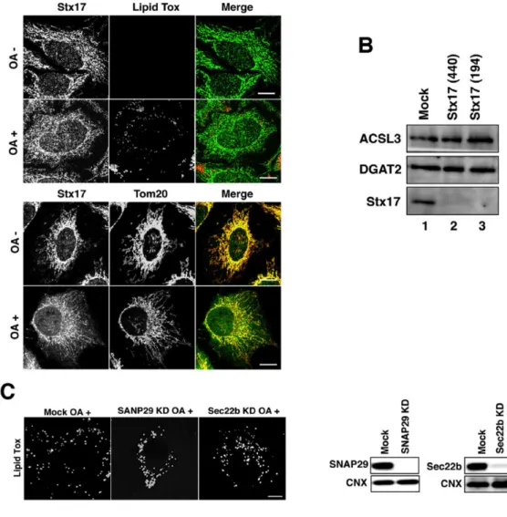
Although Stx17 is ubiquitously expressed, it is abundantly expressed in steroidogenic and hepatic cells (Steegmaier et al., 2000), both of which have large numbers of LDs. This and the MAM localization of Stx17 prompted us to examine the role of Stx17 in LD biogenesis. We first examined whether Stx17 is required for LD biogenesis by silencing the protein. We used two siRNAs (siRNA 440 and 194) that were able to effectively knockdown Stx17 (Arasaki et al., 2015, and Fig. 1A), and found that the size and number of LDs were significantly reduced in hepatic cells (HepG2 and Huh7 cells) (Fig. 1B, left two columns). OA is known to induce LD formation and thereby increase the number of LDs in cells, such as HeLa cells, that have only a few LDs under normal culture conditions. Incubation of HeLa cells with 150 µM OA for 16 hr caused LD formation concomitantly with a slight change in Stx17 distribution (Fig. 2A). Stx17 in OA-treated cells exhibited somewhat diffuse staining compared to that in OA-untreated cells, and colocalization of Stx17 with mitochondria was reduced. Stx17 silencing also blocked OA-induced LD formation in HeLa cells (Fig. 1B, right column), without affecting the expression levels of two important neutral lipid synthesizing enzymes, ACSL3 and DGAT2 (Fig. 2B). Of note is that Stx17 was not localized on the surface of LDs (Fig. 1B, top row, and Fig. 2A), suggesting that Stx17 does not directly participate in LD biogenesis, but rather has a regulatory role. In accordance with the inhibition of LD formation, TAG synthesis was blocked in Stx17-silenced HeLa cells (Fig. 1C). The specific involvement of Stx17 in LD formation was demonstrated by the finding that depletion of SNAP29, a Stx17 partner in autophagy (Itakura et al., 2012), or Sec22b, a partner in membrane trafficking (Steegmaier et al., 2000), did not affect LD formation (Fig. 2C).
To verify the physiological importance of Stx17 in LD biogenesis, we examined the effect of Stx17 silencing on the differentiation of 3T3-L1 preadipocytes into adipocytes. As differentiation progressed, the expression level of Stx17 markedly increased (Fig. 3A). Silencing of Stx17 inhibited adipocyte differentiation, as demonstrated by a reduced increase in Oil-Red O staining (Fig. 3B).
siRNAs used (Fig. 1E,F). We examined the ability of several Stx17 mutants to compensate for Stx17 depletion. No rescue was observed for Stx17 K254C in which Lys254 in the middle of the CHD was replaced by Cys, the CHD+C mutant, or the ΔSNARE mutant (Fig. 1E,F), suggesting that both the SNARE domain and the CHD with the C-terminal cytoplasmic region, the latter of which is required for the MAM localization (Arasaki et al., 2015), are involved in LD biogenesis.
3.2. Depletion of Stx17 causes aberrant distribution of ACSL3 on the LD surface
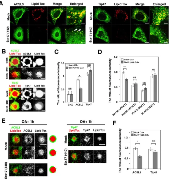
ACSL3, an abundant acyl-CoA synthetase family member on LDs in certain cells (Brasaemle et al., 2004; Fujimoto et al., 2004), redistributes from a microdomain of the ER to the LD surface during LD formation, and the enzymatic activity of ACSL3 plays a crucial role in LD expansion (Fujimoto et al., 2007; Kassan et al., 2013). Because TAG synthesis was markedly compromised in Stx17-silenced cells, we reasoned that Stx17 depletion might have caused ACSL3 dysfunction. Therefore, we examined the localization of ACSL3 in Stx17-silenced cells. As reported previously (Kassan et al., 2013), ACSL3 was detected on the surface of LDs exhibiting a circular distribution in mock-treated cells (Fig. 4A, left, upper row, B, top row). In Stx17-silenced cells, on the other hand, ACSL3 exhibited a crescent-like distribution on the surface of LDs (Fig. 4A, left, lower row, B, second row). Importantly, there was no difference in the circular distribution of Tip47/PLIN3, a LD-localized protein that redistributes from the cytosol, between mock- and Stx17-silenced cells (Fig. 4A, right, B, third and bottom rows). Quantification revealed that the recruitment of ACSL3 to the surface of LDs was specifically suppressed by Stx17 silencing (Fig. 4C).
As ACSL1 and ACSL4 also localize to LDs in certain cells (Pol et al., 2014), we examined the effect of silencing of these ACSLs. ACSL1 was not colocalized with LDs, whereas ACSL4 showed some colocalization in HeLa cells (Fig. 5A). Silencing of either protein did not significantly affect LD formation, whereas ACSL3 depletion blocked LD formation (Fig. 5B).
significantly affected upon Stx17 depletion (Fig. 4D). It should be noted that no significant translocation defect was observed for LPCAT2, the closest homologue of LPCAT1 among the LPCAT family proteins (Shindou and Shimizu, 2009).
3.3. Stx17 regulates the redistribution of ACSL3 to LDs
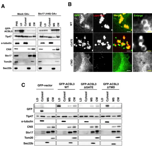
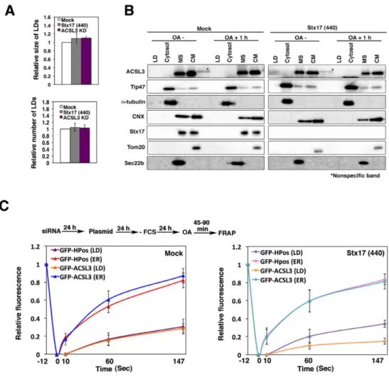
We then performed subcellular fractionation experiments using HeLa cells transiently expressing GFP-ACSL3 and found that GFP-ACSL3 was predominantly recovered in the LD fraction (Fig. 8B, left). Upon Stx17 depletion, the amount of GFP-ACSL3 recovered in the LD fraction was reduced, along with its appearance in the microsomal and crude mitochondrial fractions (Fig. 8B, right), the latter of which contained the MAM (Vance, 2014).
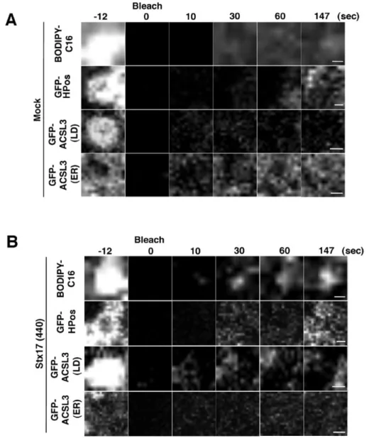
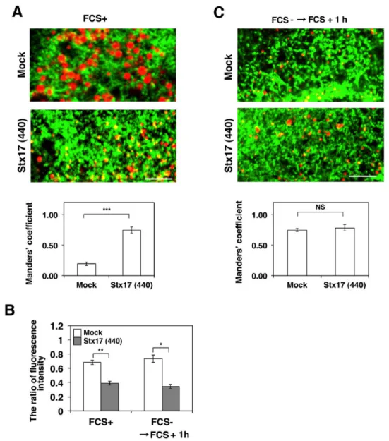
Because it is not clear whether LDs recovered in the LD fraction represent LDs detached from the ER or loosely associated with the ER, we performed FRAP experiments at an early sage of LD formation. HeLa cells were incubated in the absence of fetal calf serum (FCS) for 24 hr to allow the consumption of preformed LDs, and then incubated with OA for 1 hr to induce the formation of new LDs. The position relative to the ER, size, and number of LDs in Stx17-depleted cells was indistinguishable from those in mock-treated cells (Fig. 6A, lower row and Fig. 9A), suggesting that Stx17 is not required for the early stage of LD formation. It should be noted that similar results were obtained in cells depleted of ACSL3 (Fig. 9A), consistent with the notion that ACSL3 is required for LD maturation and expansion (Fujimoto et al., 2007; Kassan et al., 2013). In both mock-treated and Stx17-silenced cells, neither Tip47 nor ACSL3 was detected in the LD fraction (Fig. 9B), suggesting that nascent LDs are fully connected with the ER. At this time point, Tip47 fully decorated LDs, whereas many ACSL3 exhibited a disrupted distribution around the surface of LDs in Stx17-silenced cells (Fig. 4E,F), as observed in cells incubated with OA for 16 hr (Fig. 4B,C). FRAP analysis showed that the signals for GFP-ACSL3, as well as those of GFP-HPos, were recovered after photobleaching in mock-treated cells (Fig. 9C, left). Similarly, the signals of GFP-HPos were recovered in Stx17-silenced cells (Fig. 9C, right). However, little recovery of the signal was observed for GFP-ACSL3, verifying that GFP-ACSL3 redistribution to LDs is impaired in Stx17-depleted cells.
ACSL3 from the microsomal and crude mitochondrial fractions to LDs (Fig. 10C). As in the case of HeLa cells (Fig. 9C), the signals of GFP-HPos and GFP-ACSL3 were similarly recovered in mock-treated cells (Fig. 10D, upper panel and Fig. 12B), whereas the recovery of GFP-ACSL3 fluorescence was much lower than that of GFP-HPos in Stx17-silenced cells (Fig. 10D, lower panel and Fig. 12B), consistent with the subcellular fractionation data (Fig. 10C). The results of FRAP experiments were essentially the same as those obtained using HeLa cells. In mock-treated cells, little fluorescence recovery after photobleaching was observed for both GFP-HPos and GFP-ACSL3 (Fig. 10B,D, upper panel and Fig. 12A), whereas GFP-HPos fluorescence, but not GFP-ACSL3 fluorescence, was considerably recovered in Stx17-depleted cells (Fig. 10B,D, lower panel and Fig. 12B). We then examined the early stage of LD formation. To this end, Huh7 cells were fasted for 24 hr followed by incubation with FCS for 1 hr. The position of LDs relative to the ER was similar between mock-treated and Stx17-silenced cells (Fig. 11C). It should be noted that the amounts of Tip47 in the LD fraction were comparable in mock-treated and Stx17-silenced cells, suggesting the specific absence of ACSL3 in LDs in Stx17-silenced cells.
3.4. Stx17 interacts with ACSL3 through its SNARE domain
Because the redistribution of ACSL3 to LDs was suppressed in Stx17-silenced cells, Stx17 might regulate the localization of ACSL3 through protein-protein interaction. To test this possibility, we performed immunoprecipitation and proximity ligation assay (PLA). Significant amounts of endogenous ACSL3 (Fig. 13A) and GFP-ACSL3 (Fig. 14A) were co-precipitated with FLAG-Stx17 wild-type and the K254C mutant, but not with the ∆SNARE mutant. Similar results were obtained for PLA (Fig. 13B and Fig. 14B), suggesting that Stx17 interacts with ACSL3 via its SNARE domain. PLA combined with the ER-LD and mitochondrial markers (GFP-HPos and Su9-GFP) revealed that the interaction between Stx17 and ASCL3 mainly occurs close to mitochondria, rather than to HPos-positive ER membranes regardless of whether OA is present or not (Fig. 13C).
defect and the binding to Stx17.
Taking advantage of in situ nature of PLA, we examined whether substrate/inhibitor binding to ACSL3 affects the interaction between ACSL3 and Stx17. The proximity between Stx17 and ACSL3 was suppressed by triacsin C, a competitive inhibitor for ACSL (Omura et al., 1982) (Fig. 13D). Because substrate acyl-CoA seemed not to be efficiently incorporated into cells, we used an indirect way, i.e., blockage of TAG synthesis by DGAT inhibitors. We reasoned that inhibition of DGAT 1 and 2 in the presence of OA would allow the synthesis of acyl-CoA catalyzed by ACSL but inhibit its consumption, leading to the accumulation of acyl-CoA in cells, although we could not exclude the possibility that some acyl-CoA synthesized is consumed by oxidation in mitochondria or secreted into medium (Rambold et al., 2015). DGAT1/2 inhibitors enhanced the proximity between Stx17 and ACSL3 (Fig. 13D). These results suggest that the interaction of ACSL3 with Stx17 is dependent on the substrate/inhibitor occupancy in the active site of ACSL3.
3.5. The GATE domain of ACSL3 is important for the interaction with the SNARE domain of Stx17
We next examined which domains of ACSL3 are required for the interaction with Stx17. ACSL3 has a transmembrane domain (TMD) and a GATE domain (Fig. 13E), the latter of which is implicated in the control of access of the fatty acid substrate to the catalytic site of each of the two subunits (Hisanaga et al., 2004; Soupene et al., 2010). GFP-ACSL3 wild-type was found to decorate LDs (Fig. 8C, top row). On the other hand, a dot-like distribution at the edge of LDs was observed for the ΔGATE mutant (middle row), and expression of the ΔTMD mutant reduced the number of LDs (bottom row). Subcellular fractionation confirmed the absence of the ΔGATE and ΔTMD mutants in the LD fraction (Fig. 8D). Binding experiments revealed that both the TMD and GATE domains of ACSL3 are required for the interaction with Stx17 (Fig. 13E), although the ΔGATE mutant as well as the wild-type protein may be close to Stx17 in the absence of OA (Fig. 13F). These findings combined with the fact that the SNARE domain of Stx17 is required for LD formation (Fig. 1E,F) suggest that the interaction of ACSL3 with Stx17 is important for the redistribution of ACSL3 to LDs.
3.6. Overexpression of ACSL3 can compensate for Stx17 depletion
to nascent LDs, e.g., increasing the amount of ACSL3 escaping from the MAM, transient overexpression of ACSL3 may compensate for Stx17 depletion. When transiently overexpressed (approximately 4-fold excess over endogenous ACSL3), FLAG-tagged ACSL3 was found to translocate LDs and restore LD formation in Stx17-silenced cells (Fig. 15A), implying that Stx17 is dispensable for LD biogenesis if ACSL3 is abundantly expressed. This finding may be relevant to the fact that transiently expressed GFP-ACSL3 was efficiently transported to LDs (Fig. 8A) compared to endogenous ACSL3 (Fig. 6D, 10A,C).
The ACSL3 overexpression experiments also suggest that Stx17 depletion-induced defect in LD formation is not due to secondary effects such as the shortage of diacylglycerol, substrate for DGAT, or ER structure disruption. To exclude the possibility that diacylglycerol is not adequately supplied in Stx17-silenced cells due to metabolic disturbance, we used membrane-permeable diacylglycerol, 1-oleoyl-2-acetyl-sn-glycerol (OAG). In mock-treated cells, addition of OAG stimulated OA-induced LD formation, and this LD formation was blocked by DGAT1/2 inhibitors, implying that OAG incorporated into cells were converted to TAG to produce LDs (Fig. 15B, left). In contrast, LD formation was not stimulated upon the addition of OAG in Stx17-depleted cells (Fig. 15B, right). These results exclude the possibility that impaired LD formation in Stx17-silenced cells is attributed to the lack of diacylglycerol and are consistent with the view that the supply of acyl-CoA on site of LD biogenesis is blocked in Stx17-silenced cells.
Finally, we examined the ability of Stx17-silenced cells to form and/or retain LDs when TAG is fully present within cells. To this end, we incubated Stx17-sileced cells with TAG complexed with bovine serum albumin. A previous study showed that TAG and other lipids and fatty acids, when compelxed with bovine serum albumin or low-density lipoprotein, are taken up by a variety of cells (Hammeed, 2013). When HeLa cells were incubated with TAG, LDs were formed (Fig. 15C). This LD formation was not inhibited by triacsin C or DGAT1/2 inhibitors, suggesting that TAG incorporated into cells directly, not through degradation and resynthesis induced LD formation. The number and size of LDs formed were indistinguishable between mock-treated and Stx17-silenced cells. These results suggest that Stx17-silenced cells retain the capacity to form LDs.
3.7. SNAP23 localizes to the MAM
redistribution by regulating the Stx17-ACSL3 interaction. We focused on SNAP23 because previous studiers showed the involvement of this protein in LD formation and localization (Boström et al., 2007; Jägerström et al. 2009), and our interactome analysis identified SNAP23 as an Stx17-interacting protein (data not shown). We examined whether SNAP23, like Stx17, localizes to the MAM, in addition to the ER and mitochondria. As shown in Fig. 16A, SNAP23 was found to be highly enriched in the MAM fraction. Of note, a significant amount of ACSL3 was also recovered in the MAM fraction.
Silencing of SNAP23 (Fig. 16B) markedly reduced the size of LDs, as previously reported (Boström et al., 2007; Jägerström et al. 2009), with a disrupted TAG amount and distribution of ACSL3 on LDs (Fig. 16C,D). SNAP23 depletion caused a change in the distribution of Stx17 from a mitochondria-like pattern to a pattern with punctate structures (Fig. 16E, left, second and fourth rows). Our previous study showed the distribution of Stx17 is significantly perturbed upon treatment of cells with a low concentration (30 µg/ml) of digitonin (Arasaki et al., 2015), a reagent that can efficiently extract cholesterol (Oliferenko et al., 1999). This likely reflects the fact that the MAM is enriched in cholesterol and sphingolipids, thus resembling lipid raft-like structures (Chipuk et al., 2012; Hayashi and Su, 2007; Sano et al., 2009). We found that punctate Stx17-positive structures were relatively resistant to 30 µg/ml digitonin treatment compared to the mitochondria-like distribution of Stx17 in mock-treated cells (Fig. 16E, right). This suggests that Stx17 failed to gain access to raft-like structures in the absence of SNAP23.
3.8. SNAP23 and ACSL3 bind exclusively to Stx17
We next determined the region of Stx17 responsible for the interaction with SNAP23 by immunoprecipitation (Fig. 17A) and PLA (Fig. 17B). As expected, the interaction of Stx17 with SNAP23 was abolished by deletion of the SNARE domain of Stx17 (Fig. 17A, lane 8, B). Interestingly, the interaction was drastically reduced by mutation at Lys254 located in the middle of the CHD, even though this mutant retained the SNARE domain (Fig.17A, lane 7,B).
GFP-ACSL3 wild-type, but not mutants lacking the ability to bind Stx17, i.e., the ΔGATE and ΔTMD mutants, the proximity of Stx17 to SNAP23 was suppressed (Fig. 17E). These results suggest that SNAP23 and ACSL3 compete for Stx17 binding.
3.9. MAM, but not tethering between MAM and mitochondria, is important for LD formation
Next, we examined the effect of depletion of PACS2, a multifunctional sorting protein that is required for maintaining MAM integrity (Simmen et al., 2005) and Mfn2, a key tether for ER-mitochondria (Naon et al., 2016). As shown in Fig. 18A, Mfn2 depletion did not affect OA-induced LD formation, whereas PACS-2 was found to be required for LD formation. Consistent with these results, the proximity signal for FLAG-Stx17 and ACSL3was reduced upon depletion of PACS-2, but not Mfn2, and OA increased the signal (Fig. 18B).
We assessed Stx17 milieu in cells depleted of PACS-2 or Mfn2 by means of digitonin sensitivity. Although some punctate Stx17-positive structures were observed in Mfn2-silenced cells, they were diminished upon digitonin treatment (Fig. 18C, middle row). Similar to that observed in SNAP23-depleted cells, prominent punctate Stx17-positive structures were resistant to digitonin treatment (Fig. 18C, bottom row), suggesting that Stx17 fails to localize to raft-like structures in cells depleted of PACS-2 as well as SNAP23. Alternatively, raft-like structures might have been disrupted upon depletion of PACS-2.
3.10. Transient binding of Stx17 to ACSL3 during LD formation
Given that SNAP23 and ACSL3 compete for Stx17 binding, one attractive hypothesis for the regulation of the redistribution of ACSL3 from the ER to nascent LDs by Stx17 and SNAP23 is that at the onset of LD formation ACSL3 first binds to Stx17, and then SNAP23 competitively binds to Stx17, chasing out ACSL3 to allow its redistribution to LD sruface. We monitored the change of binding partner during LD maturation. Immunoprecipitation (Fig. 19A) and PLA (Fig. 19B) revealed that the binding of ACSL3 to Stx17 augmented at a 1-hr incubation with OA and then declined, whereas the binding of ACSL3 to SNAP23 increased up to 6 hr.
3. 11. Figures and legends
Fig. 1. LD formation and TAG synthesis are impaired in Stx17-silenced cells.
A: HepG2, Huh7, and HeLa cells were mock-transfected or transfected with siRNA Stx17 (440), (194), or (NC) targeting the 3’ non-coding region of Stx17. After 72 h the protein amounts of Stx17 and α-tubulin were determined using their antibodies. B: HepG2, Huh7, and HeLa cells were
Fig. 2. Stx17 is specifically related to LD formation.
Fig. 3. Silencing of Stx17 suppresses differentiation of 3T3-L1 preadipocytes.
Fig. 4. Aberrant distribution of ACSL3 on LDs in Stx17-silenced cells.
A, B: HeLa cells were mock-transfected or transfected with siRNA Stx17 (440), treated with OA for 16 h, fixed, and then stained with Lipid Tox and an antibody against ACSL3 or Tip47. Bars in normal images, 5 µm, and in enlarged images, 1 µm. C, F: Quantitation of data in (B) and (E). The fluorescence intensities of CNX, ACSL3, and Tip47 surrounding LDs relative to that of Lipid Tox were plotted. The bar graph shows the means ± SEM (n = 3). D: HeLa cells were mock-transfected or transfected with siRNA Stx17 (440). After 48 h, cells were
Fig. 5. ACSL3 specifically regulates LD biogenesiss and Stx17 interacts with ACSL3. A: HeLa cells were incubated with OA for 16 h, fixed, and then stained with Lipid Tox and an antibody against each ACSL species. Bars in normal images, 5 µm, and in enlarged images, 1 µm. B: HeLa cells were mock-transfected or transfected with siRNA targeting each ACSL species, treated with OA for 16 h, fixed, and then stained with Lipid Tox. Bar, 5 µm.
Fig. 6. Stx17 regulates the redistribution of ACSL3 to LDs in HeLa cells.
Fig. 7. LDs are closed to the ER in Stx17-silenced cells. The images in FRAP experiments.
Fig. 8. Localization of GFP-ACSL3 and its mutants.
Fig. 9. Analysis of nascent LDs in HeLa cells.
Fig. 10. Stx17 regulates the redistribution of ACSL3 to LDs in Huh7 cells.
Fig. 11. Aberrant distribution of ACSL3 on LDs in Stx17-silenced Huh7 cells.
Fig. 12. LDs are also closed to the ER in Stx17-silenced cells. The images in FRAP experiments in Huh7 cells.
Fig. 13. The SNARE domain of Stx17 and GATE domain of ACSL3 are indispensable for the interaction between Stx17 and ACSL3.
Fig. 14. Stx17 binding and localization of ACSL3 mutants.
Fig. 15. Overexpression of ACSL3 can compensate for Stx17 depletion.
Fig. 16. SNAP23 localizes to the MAM.
A: Subcellular fractionation of HeLa cell lysates was performed as described under Materials and methods. Equal amounts of proteins in individual fractions were subjected to SDS-PAGE and analyzed with the indicated antibodies. PNS, postnuclear supernatant; MS, microsomes;
Mt, mitochondria. B: HeLa cells were mock-transfected or transfected with SNAP23 (837) or (534), and protein levels were determined with antibodies against SNAP23 and CNX. C: Alternatively, the amount of TAG was determined. D: Mock-treated and SNAP23-depleted cells were treated with OA for 16 hr, and then stained with an anti- ACSL3 antibody and Lipid Tox. Bars, 5 µm. The bar graph on the right shows the relative ACSL3 staining intensity surrounding LDs. Values are the means ± SEM (n = 3). E: HeLa cells stably
Fig. 17. Exclusive binding of SNAP23 and ACSL3 to Stx17.
Fig. 18. MAM is important for LD formation and Stx17-ACSL3 interaction.
Fig. 19. Stx17 changes the binding partner from ACSL3 to SNAP23 during LD maturation.
4. Discussion
The protein and lipid compositions of LDs vary not only with the cell type but also in the degrees of LD maturation and consumption in individual cell types (Ohsaki et al., 2014; Pol et al., 2014; Thiam and Beller, 2017). The importance of the recruitment of proteins to LDs in LD biogenesis is highlighted by the finding that depletion of LD-localized ACSL3 impaired LD formation, whereas depletion of non-LD-localized members of the ACSL family, ACSL1 and ACSL4, had no impact on LD biogenesis (Kassan et al., 2013 and this study), suggesting that on site supply of acetyl-CoA is necessary for the growth of LDs. Therefore, how proteins selectively bind to the LD surface, which is surrounded by a phospholipid monolayer, in a maturation stage-dependent manner is one of the most key questions to be addressed in LD biogenesis. In this study, we demonstrated that Stx17, a SNARE protein localized in the MAM, regulates the translocation of ACSL3 and perhaps LPCAT1 from the ER to nascent LDs. On the other hand, the distribution of some other class I LD proteins such as DGAT2 and class II LD proteins such as Tip47/PLIN3, which translocate from the ER and cytosol to LDs, respectively, was found to be independent of Stx17. These findings suggest that Stx17 selectively interacts with and regulates the localization of a subset of proteins for LD biogenesis.
structures. SNAP23 is known to be palmitoylated at several residues located in the middle of the protein (Vogel and Roche, 1999), and this modification is required for the membrane attachment and/or raft association of SNAP23 (Salaün et al., 2005). Palmitoylation is a signal for targeting of proteins to raft structures (Salaun et al., 2010) and the MAM (Lynes et al., 2012). Finally, ACSL3 enters the phospholipid monolayer surrounding LDs.
Although the SNARE domain of Stx17 is important for LD biogenesis, we do not favor the idea that Stx17 participates in the formation of bridges that connect the ER and nascent LDs or, as proposed previously for Stx5 (Boström et al., 2007), LD fusion. After initial formation, LDs retain functional connection with the ER through membrane bridges (Jacquier et al., 2011; Wilfling et al., 2013), and a subset of proteins (class I proteins: Kory et al., 2016) translocate to LDs likely through these bridges (Ingelmo-Torres et al., 2009; Wilfling et al., 2013; Zehmer et al., 2008). FRAP experiments after maturation of LDs revealed that LDs are not connected with the ER in mock-treated cells, whereas in Stx17-silenced cells LDs are still in contact with the ER, as shown by the recovery of fluorescence of BODIPY FL-C16 and GFP-HPos, although the link between the LDs and the ER may be susceptible to mechanical force such as cell homogenization. Our idea that Stx17 is not involved in the formation of ER-LD bridges is supported by the finding that knockdown of Sec22b, a cognate v-SNARE for Stx17 in membrane fusion (Steegmaier et al., 2000), does not affect LD formation (Fig. 2).
5. Materials and methods 5.1. Chemicals and antibodies
Triacsin C and digitoninwere obtained from Enzo Life Sciences, Wako Chemicals, respectively. OA, TAG, OAG, DGAT1 inhibitor (PF-04620110), and DGAT2 inhibitor (PF-06424439) were obtained from Sigma-Aldrich. OA, TAG, and OAG were dissolved in DMSO, mixed with bovine serum albumin, and used. Lipid Tox and BODIPY FL-C16 were obtained from Thermo Fisher Scientific. The following antibodies were purchased from Sigma-Aldrich: monoclonal FLAG (No. F3165) and polyclonal FLAG (No. F7425). The following antibodies were obtained from BD Bioscience Pharmingen: CNX (No. 610523), Tom20 (No. 612278), and Tim23 (No. 611223). The following antibodies were purchased from Proteintech: Tip47 (No. 10694-1-AP), and SNAP29 (No. 12704-1-AP). The following antibodies were purchased from Abcam: polyclonal ACSL1 (No. ab76702) and α-tubulin (No. ab15246). Polyclonal and monoclonal antibodies against ACSL3 were obtained from GeneTeX (No. GTX112431) and Abnova (No. H00002181-B01P), respectively. The following antibodies were from the indicated sources: ACSL4 (Santa Cruz Biotechnology, H-53: sc-134507), HA (Sigma-Aldrich, H6908), and Alexa Fluor® 488 and 594 goat anti-mouse and -rabbit antibodies (Thermo Fisher Scientific, No. A-11001, A-11005, A-11008, and A-11012). Rabbit antibodies against Sec22b (Hirose et al., 2004), Stx5 (Mizoguchi et al., 2000), and Stx3 were produced in this laboratory and affinity purified. An antibody against Stx17 was prepared as described previously (Arasaki et al., 2015) and used for immunofluorescence analysis. For immunoblotting, an anti-Stx17 antibody (Sigma-Aldrich; No. HPA001204) was used.
5.2. Cell culture
293T, HepG2, Huh7, and 3T3-L1 cells were grown in DMEM supplemented with 50 IU/ml penicillin, 50 µg/ml streptomycin, and 10% FCS. HeLa cells (RIKEN, RCB0007) were cultured in α-MEM supplemented with the same materials plus 2 mM L-glutamine. Stable transfectants were prepared as described previously (Arasaki et al., 2015). For the induction of LDs in HeLa cells, OA was added at a final concentration of 150 µM.
5.3. Plasmids and transfection
Albert Pol (IDIBAPS). The plasmid for FLAG-ACSL3 was constructed by inserting the cDNA for GFP-ACSL3 into the EcoRI/SmaI site of pFLAG-CMV-6a vector. The plasmids for FLAG-DGAT1/2, 3x-HA-LPCAT1/2, and GFP-SU9 were gifts from Dr. Robert V. Farese (Harvard University), Dr. Christoph Thiele (University of Bonn), Dr. Naotada Ishihara (Kurume University), respectively. Deletion mutants of ACSL3 were constructed by inverse PCR. Transfection was carried out using LipofectAMINE Plus, LipofectAMINE2000 (Thermo Fisher Scientific), or PEI (Longo et al., 2013) according to the manufacturer’s protocol.
5.4. RNA interference
The following siRNAs were used:
Stx17 (440) : 5’-GGUAGUUCUCAGAGUUUGAUU-3’ Stx17 (194) : 5’-CGAUCCAAUAUCCGAGAAAUU-3’ Stx17 (NC) : 5’-GGAAAUUAAUGAUGUAAGA-3’ Stx17 (421) : 5’-CACACUGGGGAGGCUGAAGCU-3’ Sec22b: 5’-CAGCATTGGATTCAAAGGCTA-3’ SNAP29: 5’-UAUCAUCCAGCUUUCUAAGGUUUGG-3’ ACSL1: 5’-AAGGAUGCUUUGCUUAUUCGA-3’ ACSL3: 5’-CCGAAGUGUGGGACUACAAUA-3’ ACSL4: 5’-AUGCAUCAUAGCAAUUUGAUA-3’
siRNAs were purchased from Japan Bio Services. HeLa, HepG2, Huh7, and 3T3-L1 cells were grown on 35-mm dishes, and siRNAs were transfected at a final concentration of 200 nM using Oligofectamine, LipofectAMINE2000, or LipofectAMINE2000 RNAi Max (Thermo Fisher Scientific) according to the manufacturer’s protocol.
5.5. Immunoprecipitation
5.6. TAG measurement
TAG measurement was performed using a triglyceride quantification colorimetric kit (BioVison: No-k662) according to the manufacturer’s protocol. The OD 570 nm was measured and TAG amount was calculated using a standard curve.
5.7. Immunofluorescence microscopy and image analysis
For immunofluorescence microscopy, cells were fixed with 4% paraformaldehyde for 20 min at room temperature followed by permeabilization in 0.2% Triton X-100 in PBS for 10 min at room temperature or with ice-cold methanol for 1 min at room temperature, and blocked with 2% bovine serum albumin in PBS for 10 min. The cells were incubated with a primary antibody (ACSL3 (1:50), Tip47 (1:50), Stx17 (1:50), FLAG (1:300), Tom20 (1:300), ACSL1 (1:50), or ACSL4 (1:20)) for 1 hr at 37℃, followed by three washes in PBS and incubation with Alexa Fluor 488- or 594 conjugated anti-mouse or anti-rabbit IgG antibody (1:200) for 1 hr at 37℃. When stained with Lipid Tox, the cells were incubated with Lipid Tox (dissolved in DMSO) for 30 min in PBS at room temperature. After washing in PBS, they were mounted with mounting medium (Dako) and observed using a 100 × oil immersion objective lens (UPlan FI: NA = 1.3) under a laser scanning confocal microscope (OLYMPUS Fluoview FV300) with a pinhole of 3 AU. All images were single confocal sections.
ImageJ software (NIH) was used to determine the size of LDs and the fluorescence intensity ratio between Lipid Tox and LD proteins. In each LD, the intensities of circular Lipid Tox fluorescence and surrounding FITC fluorescence were measured. This analysis was performed for randomly chosen 30 LDs in each cell, and 30 cells were analyzed in each experiment. Experiments were repeated three times.
5.8. Electron microscopy
LD surface covered by contacted ER to the total perimeter of the LD. ER-LD contact length and the whole circumference of each LD were measured with ImageJ software.
5.9. FRAP
FRAP experiments were performed with an Olympus Fluoview 1000 laser scanning microscope equipped with a stage-top incubator (37°C, 5% CO2). To monitor LDs during
FRAP experiments, cells expressing GFP constructs or labeled with BODIPY FL-C16 were incubated with Lipid Tox in Opti-MEM supplemented with 10% FCS before photobleaching. The region containing several LDs was photobleached using a 488-nm laser at 100% laser power for 2 s. After photobleaching, images were obtained at 0.5 s intervals. In each experiment 30 cells were used, and more than 3 LDs in each cell were bleached. Experiments were repeated three times.
5.10. Differentiation of 3T3-L1 cells and measurement of intensity of Oil Red O staining Differentiation was induced using an AdipoInducer Reagent (for animal cell) kit (Takara Bio, MK429) according to the manufacturer’s protocol. Briefly, 3T3-L1 cells were mock-transfected or transfected with siRNA Stx17 (421). At 72 hr after transfection, cells were incubated with DMEM supplemented with 50 IU/ml penicillin, 50 µg/ml streptomycin, and 10% FCS, plus 10 µg/ml insulin solution, 2.5 µM dexamethasone, and 0.5 mM 3-isobutyl-1-methylxanthine, for 48 hr. The medium was replaced with DMEM containing 10 µ g/m insulin, and then incubated for ~9 days. For Oil Red O staining, cells were fixed, washed with 60% isopropanol for 1 min, and then incubated with a 60% Oil Red O solution (99% isopropanol/0.3% Oil Red O) for 15 min at 37℃. After washing with 60% isopropanol and drying, Oil Red O was dissolved in 100% isopropanol and the OD at 500 nm was measured.
5.11. PLA
5.12. Subcellular fractionation
6. Statistical analyses
7. References
Arasaki K, Shimizu H, Mogari H, Nishida N, Hirota N, Furuno A, Kudo Y, Baba M, Baba N, Cheng J, Fujimoto T, Ishihara N, Ortiz-Sandoval C, Barlow LD, Raturi A, Dohmae N, Wakana Y, Inoue H, Tani K, Dacks JB, Simmen T, Tagaya M. (2015) A role for the ancient SNARE syntaxin 17 in regulating mitochondrial division. Dev Cell 32:304-17 Boström P, Andersson L, Rutberg M, Perman J, Lidberg U, Johansson BR, Fernandez-Rodriguez J, Ericson J, Nilsson T, Borén J, Olofsson SO. (2007) SNARE proteins mediate fusion between cytosolic lipid droplets and are implicated in insulin sensitivity. Nat Cell Biol 9:1286-1293.
Brasaemle DL, Dolios G, Shapiro L, Wang R. (2004) Proteomic analysis of proteins associated with lipid droplets of basal and lipolytically stimulated 3T3-L1 adipocytes. J Biol Chem 279:46835-46842.
Cartwright BR, Binns DD, Hilton CL, Han S, Gao Q, Goodman JM. (2015) Seipin performs dissectible functions in promoting lipid droplet biogenesis and regulating droplet morphology. Mol Biol Cell 26:726-739.
Chipuk JE, McStay GP, Bharti A, Kuwana T, Clarke CJ, Siskind LJ, Obeid LM, Green DR. (2012) Sphingolipid metabolism cooperates with BAK and BAX to promote the mitochondrial pathway of apoptosis. Cell 148:988-1000.
Choudhary V, Ojha N, Golden A, Prinz WA. (2015) A conserved family of proteins facilitates nascent lipid droplet budding from the ER. J Cell Biol 211:261-271.
Diao J, Liu R, Rong Y, Zhao M, Zhang J, Lai Y, Zhou Q, Wilz LM, Li J, Vivona S, Pfuetzner RA, Brunger AT, Zhong Q. (2015) ATG14 promotes membrane tethering and fusion of autophagosomes to endolysosomes. Nature 520:563-566.
Fei W, Zhong L, Ta MT, Shui G, Wenk MR, Yang H. (2011) The size and phospholipid composition of lipid droplets can influence their proteome. Biochem Biophys Res Commun. 415:455-462.
Fujimoto Y, Itabe H, Kinoshita T, Homma KJ, Onoduka J, Mori M, Yamaguchi S, Makita M, Higashi Y, Yamashita A, Takano T. (2007) Involvement of ACSL in local synthesis of neutral lipids in cytoplasmic lipid droplets in human hepatocyte HuH7. J Lipid Res 48:1280-1292.
Gao Q, Goodman JM. (2015) The lipid droplet-a well-connected organelle. Front Cell Dev Biol. 3:49.
Grippa A, Buxó L, Mora G, Funaya C, Idrissi FZ, Mancuso F, Gomez R, Muntanyà J, Sabidó E, Carvalho P. (2015) The seipin complex Fld1/Ldb16 stabilizes ER-lipid droplet contact sites. J Cell Biol 211:829-844.
Guo Y, Cordes K R, Farese R V, Walther T C. (2009) Lipid droplets at a glance. J Cell Sci 122:749–752.
Hamasaki M, Furuta N, Matsuda A, Nezu A, Yamamoto A, Fujita N, Oomori H, Noda T, Haraguchi T, Hiraoka Y, Amano A, Yoshimori T. (2013) Autophagosomes form at ER-mitochondria contact sites. Nature 495:389-393.
Hameed R. (2013) Analysis of lipid uptake and processing in cultured cells. PhD Thesis: Universität Bonn. http://hss.ulb.uni-bonn.de/2013/3112/3112.pdf
Hayashi T, Su TP. (2007) Sigma-1 receptor chaperones at the ER-mitochondrion interface regulate Ca2+ signaling and cell survival. Cell 131:596-610.
Hirose H, Arasaki K, Dohmae N, Takio K, Hatsuzawa K, Nagahama M, Tani K, Yamamoto A, Tohyama M, Tagaya M. (2004) Implication of ZW10 in membrane trafficking between the endoplasmic reticulum and Golgi. EMBO Journal 23: 1267-1278.
Hisanaga Y, Ago H, Nakagawa N, Hamada K, Ida K, Yamamoto M, Hori T, Arii Y, Sugahara M, Kuramitsu S, Yokoyama S, Miyano M. (2004) Structural basis of the substrate specific two-step catalysis of long chain fatty acyl-CoA synthetase dimer. J Biol Chem 279:31717-31726.
Ingelmo-Torres M, González-Moreno E, Kassan A, Hanzal-Bayer M, Tebar F, Herms A, Grewal T, Hancock JF, Enrich C, Bosch M, Gross SP, Parton RG, Pol A. (2009) Hydrophobic and basic domains target proteins to lipid droplets. Traffic 10:1785-1801.
Itakura E, Kishi-Itakura C, Mizushima N. (2012) The hairpin-type tail-anchored SNARE syntaxin 17 targets to autophagosomes for fusion with endosomes/lysosomes. Cell 151:1256-1269.
Jacquier N, Choudhary V, Mari M, Toulmay A, Reggiori F, Schneiter R. (2011) Lipid droplets are functionally connected to the endoplasmic reticulum in Saccharomyces cerevisiae. J Cell Sci 124:2424-2437.
33:934-940.
Kassan A, Herms A, Fernández-Vidal A, Bosch M, Schieber NL, Reddy BJ, Fajardo A, Gelabert-Baldrich M, Tebar F, Enrich C, Gross SP, Parton RG, Pol A. (2013) Acyl-CoA synthetase 3 promotes lipid droplet biogenesis in ER microdomains. J Cell Biol 203:985-1001.
Kennedy E P, Weiss S B. (1956) The function of cytidine coenzymes in the biosynthesis of phospholipides. J Biol Chem 222:193-214.
Krajick K. (2006) Great balls of fat. Science 1232–1234.
Kory N, Farese RV Jr, Walther TC. (2016) Targeting fat: mechanisms of Protein localization to lipid droplets. Trends Cell Biol 26:535-546.
Kuerschner L, Moessinger C, Thiele C. (2008) Imaging of lipid biosynthesis: how a neutral lipid enters lipid droplets. Traffic 9:338-352.
Lands W E M. (1958) Metabolism of glycerophospholipides: a comparison of lecithin and triglyceride sythesis. J Biol Chem 231:883–888.
Longo PA, Kavran JM, Kim MS, Leahy DJ. (2013). Transient mammalian cell transfection with polyethylenimine (PEI). Methods Enzymol 529:227-240.
Magré J, Delépine M, Khallouf E, Gedde-Dahl T Jr, Van Maldergem L, Sobel E, Papp J, Meier M, Mégarbané A, Bachy A, Verloes A, d'Abronzo FH, Seemanova E, Assan R, Baudic N, Bourut C, Czernichow P, Huet F, Grigorescu F, de Kerdanet M, et al. (2001) Identification of the gene altered in Berardinelli-Seip congenital lipodystrophy on chromosome 11q13. Nat Genet 28:365-370.
McLelland GL, Lee SA, McBride HM, Fon EA. (2016) Syntaxin-17 delivers PINK1/parkin-dependent mitochondrial vesicles to the endolysosomal system. J Cell Biol 214:275-291.
Mizoguchi T, Nakajima K, Hatsuzawa K, Nagahama M, Hauri HP, Tagaya M, Tani K. (2000) Determination of functional regions of p125, a novel mammalian Sec23p-interacting protein. Biochem Biophys Res Commun. 279:144-149.
Moessinger C, Klizaite K, Steinhagen A, Philippou-Massier J, Shevchenko A, Hoch M, Ejsing CS, Thiele C. (2014) Two different pathways of phosphatidylcholine synthesis, the Kennedy Pathway and the Lands Cycle, differentially regulate cellular triacylglycerol storage. BMC Cell Biol 15:43.
they catalyze the formation of phosphatidylcholine. J Biol Chem 286:21330-21339. Myhill N, Lynes E M, Nanji J A, Blagoveshchenskaya A D, Fei H, Carmine Simmen K,
Cooper T J, Thomas G, Simmen T (2008). The subcellular distribution of calnexin is mediated by PACS-2. Mol Biol Cell 19:2777-2788.
Naon D, Zaninello M, Giacomello M, Varanita T, Grespi F, Lakshminaranayan S, Serafini A, Semenzato M, Herkenne S, Hernández-Alvarez MI, Zorzano A, De Stefani D, Dorn GW 2nd, Scorrano L. (2016) Critical reappraisal confirms that Mitofusin 2 is an endoplasmic reticulum-mitochondria tether. Proc Natl Acad Sci U S A. 113:11249-11254.
Ohsaki Y, Kawai T, Yoshikawa Y, Cheng J, Jokitalo E, Fujimoto T. (2016) PML isoform II plays a critical role in nuclear lipid droplet formation. J Cell Biol 212:29-38.
Ohsaki Y, Suzuki M, Fujimoto T. (2017) The lipid droplet and the endoplasmic reticulum. Adv Exp Med Biol 997:111-120.
Ohsaki Y, Suzuki M, Fujimoto T. (2014) Open questions in lipid droplet biology. Chem Biol 21:86-96.
Oliferenko S, Paiha K, Harder T, Gerke V, Schwärzler C, Schwarz H, Beug H, Günthert U, Huber LA. (1999) Analysis of CD44-containing lipid rafts: Recruitment of annexin II and stabilization by the actin cytoskeleton. J Cell Biol 146:843–854.
Omura S, Tomoda H, Xu QM, Takahashi Y, Iwai Y. (1986) Triacsins, new inhibitors of acyl-CoA synthetase produced by Streptomyces sp. J Antibiot (Tokyo) 39:1211–1218. Pagac M, Cooper DE, Qi Y, Lukmantara IE, Mak HY, Wu Z, Tian Y, Liu Z, Lei M, Du X,
Ferguson C, Kotevski D, Sadowski P, Chen W, Boroda S, Harris TE, Liu G, Parton RG, Huang X, Coleman RA, Yang H. (2016) SEIPIN regulates lipid droplet expansion and adipocyte development by modulating the activity of glycerol-3-phosphate acyltransferase. Cell Rep 217:1546-1559.
Pfisterer S G, Bakula D, Frickey T, Cezanne A, Brigger D, Tschan M P, Proikas-Cezanne T. (2014) Lipid droplet and early autophagosomal membrane targeting of Atg2A and Atg14L in human tumor cells. J Lipid Res 55:1267-78
Pol A, Gross SP, Parton RG. (2014) Biogenesis of the multifunctional lipid droplet: lipids, proteins, and sites. J Cell Biol 204:635-646.
53:888-900.
Rambold AS, Cohen S, Lippincott-Schwartz J. (2015) Fatty acid trafficking in starved cells: regulation by lipid droplet lipolysis, autophagy, and mitochondrial fusion dynamics. Dev Cell 32:678-692.
Rusiñol AE, Cui Z, Chen MH, Vance JE. (1994) A unique mitochondria-associated membrane fraction from rat liver has a high capacity for lipid synthesis and contains pre-Golgi secretory proteins including nascent lipoproteins. J Biol Chem 269:27494-27502.
Salaün C, Gould GW, Chamberlain LH. (2005) The SNARE proteins SNAP-25 and SNAP-23 display different affinities for lipid rafts in PC12 cells. Regulation by distinct cysteine-rich domains. J Biol Chem 280:1236-1240.
Salaün C, Greaves J, Chamberlain LH. (2010) The intracellular dynamic of protein palmitoylation. J Cell Biol 91:1229-1238.
Salo VT, Belevich I, Li S, Karhinen L, Vihinen H, Vigouroux C, Magré J, Thiele C, Hölttä-Vuori M, Jokitalo E, Ikonen E. (2016) Seipin regulates ER-lipid droplet contacts and cargo delivery. EMBO J 35:2699-2716.
Shindou H, Shimizu T. (2009) Acyl-CoA:lysophospholipid acyltransferases. J Biol Chem 284:1-5.
Signorell A, Gluenz E, Rettig J, Schneider A, Shaw M K, Gull K, Bütikofer P. (2009) Perturbation of phosphatidylethanolamine synthesis affects mitochondrial morphology and cell-cycle progression in procyclic-form Trypanosoma brucei. Mol Microbiol 72:1068–1079.
Simmen T, Aslan JE, Blagoveshchenskaya AD, Thomas L, Wan L, Xiang Y, Feliciangeli SF, Hung CH, Crump CM, Thomas G. (2005) PACS-2 controls endoplasmic reticulum-mitochondria communication and Bid-mediated apoptosis. EMBO J. 24:717-729.
Somwar R, Roberts CT Jr, Varlamov O. (2011) Live-cell imaging demonstrates rapid cargo exchange between lipid droplets in adipocytes. FEBS Lett 585:1946-1950.
Soupene E, Dinh NP, Siliakus M, Kuypers FA. (2010) Activity of the acyl-CoA synthetase ACSL6 isoforms: role of the fatty acid Gate-domains. BMC Biochem 11:18.
Steenbergen R, Nanowski T S, Beigneux A, Kulinski A, Young S G, Vance J E. (2005) Disruption of the phosphatidylserine decarboxylase gene in mice causes embryonic lethality and mitochondrial defects. J Biol Chem, 280:40032–40040.
Stone SJ, Levin MC, Zhou P, Han J, Walther TC, Farese RV Jr. (2009) The endoplasmic reticulum enzyme DGAT2 is found in mitochondria-associated membranes and has a mitochondrial targeting signal that promotes its association with mitochondria. J Biol Chem 284:5352-5361.
Stordeur C, Puth K, Sáenz JP, Ernst R. (2014) Crosstalk of lipid and protein homeostasis to maintain membrane function. Biol Chem 395:313-326.
Sturley S L, Hussain M M. (2012) Lipid droplet formation on opposing sides of the endoplasmic reticulum. J Lipid Res 53:1800–1810.
Suzuki M, Murakami T, Cheng J, Kano H, Fukata M, Fujimoto T. (2015) ELMOD2 is anchored to lipid droplets by palmitoylation and regulates adipocyte triglyceride lipase recruitment. Mol Biol Cell 26:2333-2342.
Szymanski KM, Binns D, Bartz R, Grishin NV, Li WP, Agarwal AK, Garg A, Anderson RG, Goodman JM. (2007) The lipodystrophy protein seipin is found at endoplasmic reticulum lipid droplet junctions and is important for droplet morphology. Proc Natl Acad Sci U S A. 104:20890-20895.
Tagaya M, Simmen T (Editors) (2017) Organelle contact sites: from molecular mechanism to disease. Springer Switzerland, in press.
Takáts S, Nagy P, Varga Á, Pircs K, Kárpáti M, Varga K, Kovács AL, Hegedűs K, Juhász G. (2013) Autophagosomal Syntaxin17-dependent lysosomal degradation maintains neuronal function in Drosophila. J Cell Biol 201:531-539.
Tauchi-Sato K, Ozeki S, Houjou T, Taguchi R, Fujimoto T. (2002) The surface of lipid droplets is a phospholipid monolayer with a unique fatty acid composition. J Biol Chem 277:44507–44512.
Thiam AR, Beller M. (2017) The why, when and how of lipid droplet diversity. J Cell Sci 130:315-324.
Thiam AR, Forêt L. (2016) The physics of lipid droplet nucleation, growth and budding. Biochim Biophys Acta 1861:715-722.
Vance, J. E. (2008) Phosphatidylserine and phosphatidylethanolamine in mammalian cells: two metabolically related aminophospholipids. J Lipid Res 49:1377–1387.
phosphatidylethanolamine in mammalian cells. Mol Cell Biol Lipids 1831:543–554. Vance J E. (2014) MAM (mitochondria-associated membranes) in mammalian cells: lipids
and beyond. Biochim Biophys Acta 1841:595-609.
Vogel K, Roche PA. (1999) SNAP-23 and SNAP-25 are palmitoylated in vivo. Biochem Biophys Res Commun 258:407-410.
Walther TC, Farese RV Jr. (2012) Lipid droplets and cellular lipid metabolism. Annu Rev Biochem 81:687-714.
Wang C W, Miao Y H, Chang Y S. (2014) Control of lipid droplet size in budding yeast requires the collaboration between Fld1 and Ldb16. J Cell Sci 127:1214-28.
Wang H, Becuwe M, Housden BE, Chitraju C, Porras AJ, Graham MM, Liu XN, Thiam AR, Savage DB, Agarwal AK, Garg A, Olarte MJ, Lin Q, Fröhlich F, Hannibal-Bach HK, Upadhyayula S, Perrimon N, Kirchhausen T, Ejsing CS, Walther TC, Farese RV. (2016) Seipin is required for converting nascent to mature lipid droplets. Elife pii: e16582.
Welte MA. (2015) Expanding roles for lipid droplets. Curr Biol 25:R470-481.
Wilfling F, Wang H, Haas JT, Krahmer N, Gould TJ, Uchida A, Cheng JX, Graham M, Christiano R, Fröhlich F, Liu X, Buhman KK, Coleman RA, Bewersdorf J, Farese RV Jr, Walther TC. (2013) Triacylglycerol synthesis enzymes mediate lipid droplet growth by relocalizing from the ER to lipid droplets. Dev Cell 24:384-399.
8. Acknowledgements