Instructions for use
Title Oncolytic Adenovirus Utilizing RNA Stabilization Mechanism Can Synergize with Chemotherapy
Author(s) HOSSAIN, Elora
Citation 北海道大学. 博士(医理工学) 甲第14280号
Issue Date 2020-09-25
DOI 10.14943/doctoral.k14280
Doc URL http://hdl.handle.net/2115/79487
Type theses (doctoral)
File Information Hossain̲Elora.pdf
Hokkaido University Collection of Scholarly and Academic Papers : HUSCAP
Thesis
Oncolytic Adenovirus Utilizing RNA Stabilization Mechanism Can Synergize with Chemotherapy
RNA
安定化メカニズムを応用した腫瘍溶解性アデノウイル スとそのがん化学治療法との相乗効果September 2020 Hokkaido University
Hossain Elora
Thesis
Oncolytic Adenovirus Utilizing RNA Stabilization Mechanism Can Synergize with Chemotherapy
RNA
安定化メカニズムを応用した腫瘍溶解性アデノウイル スとそのがん化学治療法との相乗効果September 2020 Hokkaido University
Hossain Elora
Table of content
Topic Page no
Basic/reference paper list 1
List of presentations 4
Chapter I 5
1.Intorduction 6
1.2. The conventional method of cancer treatment 6 1.3. Gene Therapy
7 1.4. Oncolytic virotherapy
7 1.5. Oncolytic Adenovirus
10 1.6. Engineering oncolytic adenovirus by inserting ARE in the 3’
untranslated region 12
1.6.1. Construction of Ad-fosARE
12 1.6.2. What makes Ad-fosARE is cancer-selective?
13 1.7. Hypothesis
16 1.8. Objectives
17 Chapter II
20 2.1. Introduction
21 2.2. Material and Method:
22 2.3 Results
27 2.3.1.Cancer-Selective replication of Ad-fosARE in cancer cells
27 2.3.2. HuR-mediated mRNA stabilization is essential for adenovirus
replication 29
2.3.3. In vitro cytolytic potentiality of Ad-fosARE
32 2.3.4. Comparison of the oncolytic effects of Ad-fosARE and dl1520
36 2.4. Discussion
38 Chapter III
39
3.1. Introduction
40 3.2. Materials and method
41 3.3. Results
45 3.3.1. The potentiality of paclitaxel for synergism
45 3.3.2. Synergistic effect of oncolytic adenovirus and paclitaxel.
48 3.3.3. Effects of paclitaxel on Ad-fosARE viral protein synthesis and
mRNA stabilization 50
3.3.4. Combination treatment Increases the Level of
PosttranslationallyModified Tubulin 52
3.3.5 Virus exit assay
52 3.3.6. Effects of paclitaxel on the cell lysis activity of the virus
54 3.3.7. In vivo synergistic effect of Ad-fosARE and Paclitaxel in
murine flank tumor 55
3.3.8. PTX induces in vivo virus propagation.
56 3.4. Discussion
58 4. Future prospects and Conclusion
60 Acknowledgement
61 References
62 Supplementary data
70
List of Table
List of Figures
Topic Page
Chapter I
Figure 1.1 Cancer-selective replication of oncolytic virus 8
Figure 1.2. Structure of Ad-fosARE virus 12
Figure 1.3. Adenosine- and uridine-rich elements (ARE) 13 Figure 1.4. Degradetion and Stabilization of ARE mRNA. 14
Figure 1.5. HuR and stabilization of mRNA 15
Figure 1.6. Cancer selective replication of Ad-fosARE 16 Chapter II
Figure 2.1. Expression of early and late virus protein in Ad-fosARE 27 Figure 2.2. The production efficiency and selectivity of Ad-fosARE. 28 Figure 2.3. HuR relocalization in Ad-fosARE infected cells and mRNA
stabilization
30 Figure 2.4. Effect of HuR knockdown on virus replication. 32
Figure 2.5. Cell lysis activity of Ad-fosARE 33
Figure 2.6. Cytopathic activity of Ad-fosARE 34
Figure 2.7. Assessment of cell death by apoptosis 35 Figure 2.8. Comparison of the virus production and cell lysis activity of
Ad-fosARE with dl1520 and WT300.
37
Chapter III
Figure 3.1. Effects of paclitaxel on cytoplasmic HuR exportation. 46
Topic Page
Table 1.1: Oncolytic viruses used as an anti-cancer drug 8 Table 1.2: Current status of oncolytic virotherapy in Japan. 9
Table 1.3: List of acronyms 18
Table 2.1: Cell lines 22
Table 2.2: Antibodies used in this study. 23
Figure 3.2. Western blot analysis of expression of coxsackie-adenovirus receptor (CAR) at different time-points.
47
Figure 3.3. To assess the viral replication and cell lysis activity of combination treatment Ad-fosARE and PTX.
49
Figure 3.4. Effects of paclitaxel on Ad-fosARE replication. 50 Figure 3.5. Expression of E1AmRNA and stabilization of E1a mRNA
after combination treatment.
51
Figure 3.6. Effects of MTs stabilization on viral replication and viral exit. 53 Figure 3.7. Assessment of cell lysis activity of combination treatment. 54 Figure 3.8. In vivo tumor lysis potential of the combination therapy of
Ad-fosARE and PTX in human cervical cancer xenograft nude mice.
55
Figure 3.9. Effect of PTX on Ad-fosARE propagation in vivo. 56
Figure 3.10. Diagrammatic summary of the study 60
Supplementary data 70
Figure S.1. Time course of expression of virus protein in Ad-fosARE infected cells.
71
Figure S.2. Calculation of combination index for Ad-fosARE and PTX. 72 Figure S.3. E1A mRNA expression in Ad-fosARE treated cells. 73 Figure S.4. Level of posttranslationally modified MTs after combination treatment.
74
Figure S.5. Intracellular virus propagation after combination treatment. 75
1
Basic/Reference Paper List
1. Basic papers: 1 Number of titles: 1
(1) Title:
Advantages of Using Paclitaxel in Combination with Oncolytic Adenovirus Utilizing RNA Destabilization Mechanism
(2) Author(s): Elora Hossain, Umma Habiba, Aya Yanagawa-Matsuda, Arefin Alam, Ishraque Ahmed, Mohammad Towfik Alam, Motoaki Yasuda and Fumihiro Higashino
(3) Academic journal in which the basic paper has been published : Cancers 2020, 12(5), 1210; https://doi.org/10.3390/cancers12051210 (4) Month/year of publication May 2020
(5)Impact factor: 6.162
2. Reference papers
Equally contributed first author Number of titles: 1
(1) Title:
Conditionally Replicative Adenovirus Controlled by the Stabilization System of AU-rich Elements-Containing mRNA
(2) Author(s):
† Equally contributed first author
Yohei Mikawa†, Mohammad Towfik Alam†, Elora Hossain†, Aya Yanagawa- Matsuda, Tetsuya Kitamura, Motoaki Yasuda, Umma Habiba, Ishraque Ahmed, Yoshimasa Kitagawa, Masanobu Shindoh and Fumihiro Higashino (3) Academic journal in which the basic paper has been published :
Cancers 2020, 12(5), 1205; https://doi.org/10.3390/cancers12051205 (4) Month/year of publication May 2020
(5)Impact factor: 6.162
2 Number of titles: 2
(1) Title
Enhanced oncolytic activity of E4orf6-deficient adenovirus by facilitating nuclear export of HuR
(2) Author(s)
Ishraque Ahmed , Mohammad Towfik Alam b, Aya Yanagawa-Matsuda , Elora Hossain, Tetsuya Kitamura , Kazuyuki Minowa and Fumihiro Higashino
(3) Academic journal in which the basic paper has been published:
BBRC, 2020, 529(2);494-499 https://doi.org/10.1016/j.bbrc.2020.04.147 (4) Month/year of publication: July, 2020
5) Impact factor: 2.704
Number of titles: 3
(1) Title
Cisplatin enhances the oncolytic activity of E4orf6 deleted adenovirus through HuR relocalization
(2) Author(s)
Umma Habiba, Elora Hossain, Aya Yanagawa-Matsuda, Abu Faem Mohammad Almas Chowdhur5, Masumi Tsuda, Asad-uz- Zaman, Shinya Tanaka, Fumihiro Higashino
(3) Academic journal in which the basic paper has been published Cancers 2020, 12(4), 809; https://doi.org/10.3390/cancers12040809
(4) Month/year of publication: May 2020
5) Impact factor: 6.162
3 Number of titles: 4
(1) Title
HuR translocation to the cytoplasm of cancer cells in actin-independent manner
(2) Author(s)
Umma Habiba, Takeshi Kuroshima, Aya Yanagawa-Matsuda, Tetsuya Kitamura,
AFMA Chowdhury, Jumond P. Jehung, Elora Hossain, Hidehiko Sano, Yoshimasa Kitagawa, Masanobu Shindoh, Fumihiro Higashino
(3) Academic journal in which the basic paper has been published Experimental Cell Research 369 (2018) 218–225
(4) Month/year of publication: May 2018 5) Impact factor: 3.246
06/12/2020
Ph.D. candidate: Hossain Elora
4 List of presentations
Part of this study was presented in the following academic conferences:
Oral Presentation:
1. Hossain E, Habiba U, Matsuda AY, Kitamura T, and Higashino F. Microtubule stabilizer can enhance the activity of Oncolytic virus.
The 5th Hokkaido University cross-department symposium. O-12. Sapporo, Japan, November 6th 2019.
2.
Hossain E, Habiba U, Kitamura T, Matsuda AY, Higashino F, Efficacy of Ad+AU virus. Umea University and Hokkaido University exchange program.Sapporo, November 20th, 2019.
Poster Presentation:
3. Hossain E, Habiba U, Matsuda AY, Kitamura T, Higashino F. Evaluation of the efficacy of newly developed oncolytic adenovirus.
The 108th JSP annual meeting. Tokyo International Forum department. P-236.
TOKYO, Japan, May 2019.
4. Hossain E, Habiba U, Matsuda AY, Kitamura T, Higashino F, Application study of newly developed adenovirus.
The 4th Hokkaido University cross-department symposium. P-59.Sapporo, Japan, January 2019.
5
Chapter I
General Introduction
6 1. Introduction
Almost 70% of death are caused by noncommunicable diseases (NCDs) in the world.
This includes heart disease, stroke, cancer, diabetes, and chronic lung diseases (WHO 2020). Cancer is the uncontrolled cell growth with the potential to invade or spread to other parts of the body. Agents that may cause a normal body cell to develop abnormality can cause cancer; there are many causative agents such as chemical or toxic compound exposures, radiation, pathogens, and genetic mutations ( Anand et al., 2008; Davis et al., 2020). There are some common environmental elements that contribute to cancer including tobacco (25–30%), food habit and obesity (30–35%), infectious disease (15–20%), radiation (both ionizing and non-ionizing, up to 10%), sedentary lifestyle, and pollution (Anand et al., 2008; Islami et al.,2018; Cohen et al., 2019). The most common type of cancers is; breast cancer, skin cancer, lung cancer, colon cancer, prostate cancer, lymphoma, carcinoma, sarcoma, melanoma, and leukemia.
To date, cancer is the second-leading cause of globally (WHO 2018), excluding any kind of endemic diseases like COVID-19. Despite the advancement in treatment options for cancer, it remains the leading cause of mortality and morbidity worldwide.
Hopefully, the survival rates are improving for many types of cancer, thanks to improvements in cancer screening and cancer treatment. Besides the conventional treatment approaches like chemotherapy or radiotherapy, new modalities have emerged, such as immunotherapy and oncolytic virotherapy.
1.2 The conventional method of cancer treatment
There are many methods for treating cancer, depending on the type of cancer, grading, staging, types of treatment are available, and the goals of treatment. The cancer treatment categories include a range of procedures, machines, drugs, and/or approaches.
The most commonly available treatment options are; surgery, chemotherapy, radiotherapy, targeted therapy and gene therapy.
The treatment option may be the combination of two existing modalities to target as many cancer cells as possible. The goal is to achieve the highest level of potential efficacy with the lowest level side effect.
7 1.3 Gene Therapy
Gene therapy holds unique promise byoffering a means of specific targeting of tumor genes that promotemalignant behavior and treatment resistance (Wang et al., 2016). Usually, cancer develops by abnormal growth factor signaling, which initiates a cell to grow and divide develops constitutively active. These are occurred by oncogenes via mutations in proto-oncogenes such as a growth factor receptor and resulted in the abnormal cell signaling pathways. Hence, to successfully treat cancer, the oncogene must be destroyed; otherwise, cancer will continue to grow and spread (Gene Therapy Review 2020).
To date, successful trials of gene therapies have been reported in patients with chronic lymphocytic leukemia, acute lymphocytic leukemia, brain tumors, andothers.
However, only a few gene therapy products are available for clinical use such as ONYX-015 andGendicine for refractory head andneck cancer. (Wang et al., 2016).
There are various types of gene therapy, recognized by ongoing clinical trials worldwide. The aim of these strategies is to destroy cancer cells without harmful effects on normal cells. This can be achieved by the following approaches.
Gene therapy approaches:
1. Oncolytic virotherapy
2. Expression of the tumor suppressor gene 3. Immunotherapy
4. Gene-directed enzyme prodrug therapy 5. Expression of genes that initiate cell death
1.4 Oncolytic virotherapy
Oncolytic virotherapy is a promising anti-cancer therapy. From the past few decades, it has been known that some viruses can kill cancer cells if they are able to replicate inside them. Currently, oncolytic viruses (OVs) are using an anti-cancer drug (Table-1). OVs therapy was first demonstrated in the 1990s when a recombinant thymidine kinase (TK)-negative herpes simplex virus (HSV-1) showed tumor cell killing capacity (Fu et al., 2019). OVs infect and kill cancer cells with little to no effect on healthy tissue (Hao et al., 2019; Jenner et al., 2018). To date, research is being
8
conducted around the world using both non-engineered and engineered OVs to explore their efficacy against multiple types of cancer. Natural OVs or with genomic editing can target and replicate certain types of tumor cells. However, these unedited viruses are not tumor-specific, or they can harm normal cells as well (Figure 1.1), such as Reovirus, Senecavirus, Vaccinia virus, and adenovirus (Fu et al., 2019).
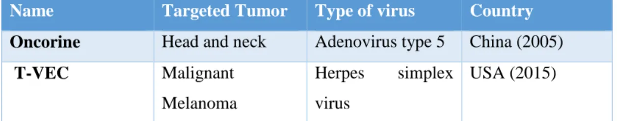
Table 1: Oncolytic viruses used as an anti-cancer drug
Source: www.sunwaybio.com, www.imlygic.com
Figure 1.1. Cancer-selective replication of the oncolytic virus
On the other hand, the engineered virus can selectively target and/or replicate in specific cancer cells. Such as adenovirus, reovirus, measles, herpes simplex, newcastle disease virus, and vaccinia have been clinically tested as oncolytic agents (Donnelly et al., 2012). Most current oncolytic viruses are engineered for tumor selectivity. There are many ways to develop OVs, either modifying viral genes by mutation or by the
Name Targeted Tumor Type of virus Country Oncorine Head and neck Adenovirus type 5 China (2005)
T-VEC Malignant
Melanoma
Herpes simplex virus
USA (2015)
9
insertion of cancer-specific agents (Abul El Hassan et al., 2004). The current status of oncolytic virotherapy in Japan is discussed in Table 2.
Table 2: Current status of oncolytic virotherapy in Japan.
Precursor virus
Virus Phas
e
Tumor Institute Status
HSV-1 T-VEC (talimogene laherparepvec;
IMLYGIC®;
OncoVEXGM -CSF
I Unresectable Stage
IIIB–IV malignant melanoma
Multicenter Underway
HSV-1 G47Δ I–IIa Glioblastoma The University of Tokyo
Completed
HSV-1 II Glioblastoma The University
of Tokyo
Underway I Castration-
resistant prostate cancer
The University of Tokyo
Completed
I/IIa Olfactory neuroblastoma
The University of Tokyo
Underway I/IIa Malignant
mesothelioma
The University of Tokyo
Planned
HSV-1 HF10
(Canerpaturev
;
C-REV)
I Metastatic breast cancer
Nagoya University
Completed
I Recurrent head and neck cancer
Nagoya University
Completed I Pancreatic cancer Nagoya
University
Completed I Malignant solid
tumors
Mie University Terminate d
I Advanced unresectable pancreatic cancer
Nagoya University
Completed
I Solid tumors with superficial
lesions
National Cancer Center Hospital
Completed
II Unresectable or metastatic melanoma
Multicenter Underway
I unresectable pancreatic cancer
Multicenter Planned
10 Adenoviru
s
Telomelysin (OBP-301)
I/II Esophageal cancer Okayama University
Underway I Esophageal cancer Unpublicized Planned I Advanced solid
tumors
National Cancer Center Hospital East
Planned
Surv.m-CRA I Advanced solid tumors
Kagoshima University
Underway Sendai
virus (HVJ)
Sendai virus particle (HVJ-E;
GEN0101;
TSD-0014)
I/II Castration- resistant prostate cancer
Osaka University
Completed
I Castration- resistant prostate cancer
Osaka University
Underway
I/II Malignant melanoma
Osaka University
Underway I Malignant
melanoma
Osaka University
Underway
(Taguchi 2018)
1.5 Oncolytic Adenovirus
Among all the oncolytic viruses, using adenoviruses (Ads) is much more attractive because of their high proliferation ability in vitro. Ads were first isolated in 1953 from human adenoid cells (Rowe et al.,1953). The Ads are non‐enveloped viruses with double‐stranded linear DNA about 35kbp length. Human Ads are classified into seven subgroups (A–G). Among them, serotype 5 (Ad5), which belongs to subgroup C, is more extensively used in gene therapy and virotherapy (Rux & Burnett, 2004). The viral genome consists of early (E) and late (L) genes (Berk et al., 2006). The early genes include E1, E2, E3, and E4 and they regulate viral replication through modification of host cells (Russel et al., 2000). The early gene 1A, a subunit of the E1 gene, is the first expressed gene after infection of modulating the cell cycle of host cells and regulate the expression of cellular and viral genes (Abudoureyimu et al., 2019). E1B gene usually codes two proteins called E1B-55k and -19kDa. The primary function of E1B is to prevent apoptosis in adenovirus infected cells. E1B-55k blocks p53 and halt its action from inducing apoptosis (Debbas et al., 1993). The late genes encode structural proteins L1,
11
L2, L3, L4, and L5. These proteins package the viral DNA into the virion in the final stages of replication.
The adenovirus life cycle is separated by the DNA replication process into two phases: an early and a late phase. The early phase occurs inside the nucleus and produces early genes. These early genes products produce early viral mRNA via transcription and transfer to the cytoplasm of the host cell. After further translation of mRNA produces early viral proteins. E2F protein force the host cell into S-phase of the cell cycle. The late phase produces the late genes which undergo transcription to produce late viral mRNA and after the final translation of mRNA form the late viral structural protein.
After completion of mRNA translation, all the virion proteins get assembled and viral DNA packaged into the capsid, and this process is called maturation. Finally, the progeny virus released by cell lysis or budding to infect the adjacent cells.
There are three main tactics to develop tumor-selective oncolytic adenoviruses as follows.
1.Targeting Capsid Modification
The capsid is the external surface of the virus and is composed of hexon, penton, and projectile fibers (Knipe and Howly, 2001). These capsid proteins can be modified so that the virus will only target cancer cells. Caspid modified virus upregulates the attachment with the primary receptor and also, they showed potent anti-cancer effects by IV injection.
2. Selective Replication by deletion of an Antitumor Gene
Oncolytic adenovirus can be engineered by the deletion of certain genes, which are vital to viral replication in normal cells but dispensable in tumor cells. Such as “E1B- 55k deleted” H101 virus (Bischoff et al., 1996). H101 virus can replicate only in the absence of the p53 gene; thus, it fails to replicate in normal cells.
12
3. Selective Replication by Insertion or Arming of Certain Gene, cytokines, Drugs or Antigen
Adenovirus can be modified by inserting genes, such as a transcriptional promoter only activated in cancer cells or arming with certain drugs or receptor genes.
These viruses are modified so that they will replicate only in the presence of certain characteristics. They normally triggered replication in cancer cells and leading to cancer cell death (Gene Therapy Review, 2020).
1.6 Engineering oncolytic adenovirus by inserting ARE in the 3’-untranslated region
We developed an adenovirus designated Ad-fosARE by inserting AU-rich element (ARE) of c-fos gene in the 3’-untranslated regions (3’-UTR) of the E1A gene (Figure 1.2).
Figure 1.2. Structure of Ad-fosARE virus 1.6.1 Construction of Ad-fosARE
Ad-fosARE was constructed using a pXhoIC plasmid and pAdenoX-PRLS (Clontech). pXhoIC has the E1 region of the type 5 human adenovirus genome. A 69- base pair of synthesized ARE fragment (5’- ttttattgtg tttttaattt atttattaag atggattctc agatatttat atttttattt tattttttt -3’) of the c-fos gene was inserted into the HpaI site in the 3’-
13
UTR of the E1A gene in pXhoIC. A fragment of the E1 region was then amplified by PCR, and inserted into the deleted E1 region of the adenovirus genome in the pAdenoX- PRLS by in-fusion technique (Fw: 5’ gtaactataacggtcatttgtctagggccgcggggactt-3’, Rv:
5’-attacctctttctccgccacgcccacacatttcagtacc-3’). The entire genome of Ad-fosARE was isolated by cutting using PacI and the fragment was then transfected into 293 cells.
Virus particles were concentrated by several rounds of viral infection and cells were collected in order to prepare a virus lysate by subjecting them to three cycles of freezing and thawing. The titers of the infectious unit (ifu) of Ad-fosARE were determined using the Adeno-XTM Rapid Titer Kit (Clontech) and 293 cells according to the manufacturer’s instructions.
1.6.2 What makes Ad-fosARE is cancer-selective?
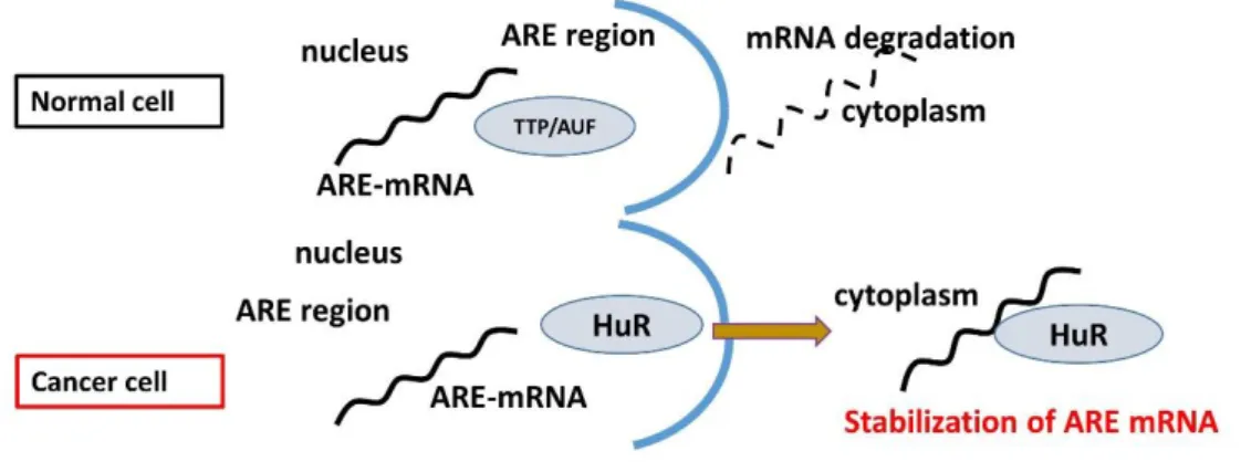
AREs
AREs are the targets for rapid degradation of mRNA and the fate of ARE- mRNA is controlled by several RNA-binding proteins, such as HuR. In cancer cells, there is an abundance of cytoplasmic HuR exportation, so mRNA can stabilize.
Figure 1.3. Adenosine- and uridine-rich elements (AREs)
AREs are usually located in the 3’-UTR of mRNAs and are the best-known determinants of mRNA instability (Figure 1.3). Studies on the expression of cytokines and proto-oncogenes have shown that many 3’-UTRs contained AREs and subsequently, some of their translation are controlled by these AREs (Espel et al., 2005, Kruys et al., 1987,1989). AREs are primarily defined by their ability to promote rapid de-adenylation-dependent mRNA decay (Kruys et al., 1989). There is relatively lesser sequence similarity among AREs, the c-fos mRNA with the most typical ARE contains
14
AUUUA pentamers and/or U-rich sequences (Espel et al., 2005; Chen et al., 1995).
AREs control mRNA degradation and translation via interactions with RNA binding proteins, which specifically bind to ARE. mRNAs carrying AREs are considered unstable and are translated only for a short period, as in the case for transcription factors, cytokines, oncogenes, and certain RNA transcripts of DNA viruses (Chen et al., 1995; Nadar et al., 2011).
Cellular RNA binding proteins, such as Tristetraprolin (TTP), ARE/poly (U)- binding/degradation factor 1 (AUF 1), Hu antigen R (HuR), and T-cell internal antigen- 1 (TIA-1), bind to mRNAs that contain AREs. ARE binding proteins (AREBPs) either aim for the mRNA to enter the degradation pathway, (TTP and AUF1) (Nadar et al., 2011, DeMaria et al., 1996), or protect the mRNA from degradation (HuR), or suppress their translation (HuR and TIA) (Yeh et al., 2008) (Figure 1.4).
Figure 1.4. Degradation and Stabilization of ARE mRNA.
15 HuR and stabilization of mRNA
HuR is a member of the embryonic lethal abnormal vision (ELAV) family of RNA- binding proteins, which is ubiquitously expressed by the most type of cells.
Intracellularly, HuR is localized mainly in the nucleus but shuttles between the nucleus and the cytoplasm (Habiba et al., 2016, Fan et al.,1998). Cytoplasmic HuR protects ARE-mRNAs against rapid degradation by binding with ARE (Brennan et al., 2001).
In normal cells, HuR transiently relocalizes to the cytoplasm under stress conditions.
In contrast, in cancer cells, HuR is constitutively localized in the cytoplasm and speculated to be involved in the ARE-mRNAs stabilization and transformation of cancer cells (Silanes et al., 2003,2005, Kuroshima et al., 2011).
Figure 1.5. HuR and stabilization of ARE-mRNA
16 1.7 Hypothesis
For Ad-fosARE replication, cytoplasmic HuR relocalization and ARE-mRNA stabilization are necessary. In normal cells, where there is no cytoplasmic HuR, E1A mRNA is degraded soon after its synthesis due to the existence of ARE. On the other hand, it is stabilized in cancer cells, resulting in the replication of Ad-fosARE and lyse cancer cells. This is the theory that makes Ad-fosARE cancer-selective.
Figure 1.6. Cancer-selective replication of Ad-fosARE
17 1.8 Objectives:
General objective
To develop a potential conditionally replicative adenovirus (CRADs) by inserting c- fos ARE in the 3’ UTRs of the E1A gene.
Specific Objective:
1. To investigate the cancer-selective replication and cytolytic activity of Ad- fosARE.
2. To evaluate the dependency on stabilized ARE-mRNA for Ad-fosARE replication by manipulating HuR.
3. To compare the efficacy of Ad-fosARE with dl1520 and WT300 in terms of less normal cell damage.
4. To investigate the synergistic effect of the combination treatment with chemotherapy.
5. To assess the increased mRNA stabilization by chemotherapy.
6. To find out the effect of the upregulated coxsackie-adenovirus receptor (CAR) expression and stabilized microtubules on viral uptake and release.
7. To investigate in vivo cytolytic activity of Ad-fosARE and synergistic effect of combination treatment.
18 Table-3: List of Acronyms
List of Acronym
Ad Adenovirus
Ad5 Adenovirus serotype S
ARE Adenylate-uridylate-rich element
AUF-1 ARE/poly (U)-binding/degradation factor CAR Coxsackie-adenovirus receptor
CPE Cytopathic effect
CRADs Conditionally replicative adenovirus
DNA Deoxyribonucleic acid
ELAV Embryonic lethal abnormal vision
GAPDH Glyceraldehyde 3 phosphate dehydrogenase
HuR Human Antigen R
HS Heat shock
HSV Herpes simplex virus
ifu Infectious unit
i.t. Intra-tumoral
i.p. Intraperitoneal
i.v Intra-venus
IMRT Intensity-modulated radiotherapy
KD Knockdown
MOI Multiplicity of infection
OV Oncolytic Virus
PARP Poly(ADP-ribose )polymerase PBS Phosphate buffered saline PCR Polymerase chain reaction
PTX Paclitaxel
mRNA Messenger RNA
MTs Microtubules
NCDs Noncommunicable diseases
19
RBP RNA binding protein
RNA Ribonucleic acid
RT-PCR Real-time polymerase chain reaction
SDS Sodium Dodecyl Sulphate
SDS-P AGE SDS polyacrylamide gel electrophoresis siRNA small interfering RNA
TIA-1 T-cell internal antigen-1
TK Thymidine kinase
TTP Tristetraprolin
vp viral particle
UTRs Untranslated region
WT Wild type
20
Chapter II
Oncolytic Potential of Conditionally Replicative Adenovirus Controlled by the
Stabilization System of AU-rich Elements-
Containing mRNA
21 2.1. Introduction
Viral therapy based on conditionally replicating adenoviruses (CRAds) is a promising cancer therapeutic strategy, as these viruses selectively replicate and lyse cancer cells (Alemany et al., 2000, Kim et al., 2000). CRAds are obtained by, either modifying viral gene by mutation, and by insertion of cancer-specific agents (Abou El Hassan et al., 2004). We developed an adenovirus designated Ad-fosARE by inserting c-fos AU-rich elements (ARE) in the 3'-untranslated region (3'-UTR) of the E1A gene.
In this study, we investigated the potential of adenovirus Ad-fosARE as oncolytic virus. The replication of this virus was markedly high in cancer cells compared with that in normal cells. The propagation and cytolytic activity of this virus in cancer cells were similar to dl1520 (equivalent to H101), which is clinically applied (Bischoff et al., 1996, Kasuya et al., 2005). But in normal cells viral replication and cytotoxicity of Ad- fosARE were less than E1B55k-deleted adenovirus. These findings indicate that Ad- fosARE is a potential oncolytic virus.
22 2.2. Material and Method:
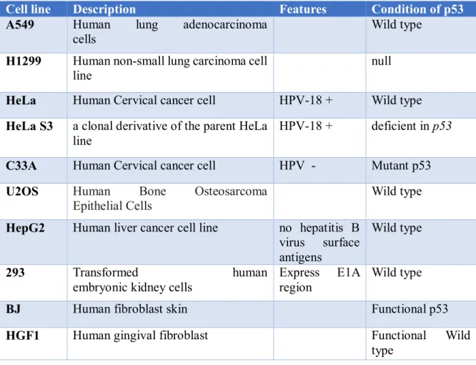
2.2.1 Cell lines:
Cell lines used in this study are described in Table-2.1. Cells were acquired from the American Type Culture Collection (ATCC; Manassas, VA, USA) and cultured in Dulbecco's modified Eagle's medium (DMEM; Sigma-Aldrich; Merck KGaA, Darmstadt, Germany) including 10% fetal bovine serum (FBS; Biowest, Nuaille, France) with antibiotics at 37 °C in a 5% CO2 atmosphere in humidified conditions.
Table 2.1: Cell lines
Cell line Description Features Condition of p53
A549 Human lung adenocarcinoma cells
Wild type H1299 Human non-small lung carcinoma cell
line
null HeLa Human Cervical cancer cell HPV-18 + Wild type HeLa S3 a clonal derivative of the parent HeLa
line
HPV-18 + deficient in p53 C33A Human Cervical cancer cell HPV - Mutant p53 U2OS Human Bone Osteosarcoma
Epithelial Cells Wild type
HepG2 Human liver cancer cell line no hepatitis B virus surface antigens
Wild type
293 Transformed human
embryonic kidney cells
Express E1A region
Wild type
BJ Human fibroblast skin Functional p53
HGF1 Human gingival fibroblast Functional Wild
type 2.2.2.Adenoviruses:
Newly constructed Ad-fosARE, Wild-type adenovirus type 5 (WT300) (generous gift from Dr T. Shenk; Princeton University), E1B55k-deleted mutant adenovirus (dl1520) (generous gift from Dr A.J. Berk; University of California) were used in this study.
23 2.2.3.Drug, reagents, and antibodies
Actinomycin D (fromStreptomyces species) were purchased from Sigma-Aldrich (USA). Antibodies used in this study are described in Table-2.2
Table 2.2: Antibodies used in this study.
1st antibodies 2nd
antibodies Molecular
weight Dilutions Source
E1A Mouse 30~50 kDa 1:1000 Santa Cruz
Biotechnology
E1B Mouse 55 kDa 1:10 Gift from T Shenk
L133 Rabbit 110,70,60
kDa
1:5000 Gift from T Dobner
CAR Mouse 46 kDa 1:200 Santa Cruz
Biotechnology
HuR Mouse 36 kDa 1:5000 Santa Cruz
Biotechnology
ACTIN HRP - 43 kDa 1:5000 Santa Cruz
Biotechnology β-TUBULIN Mouse 52 kDa 1:5000 Millipore Corp
LAMIN B Goat 1:5000 Santa Cruz
Biotechnology
PARP Rabbit 46kDa 1:1000 Cell Signaling
Technology Acetylated
tubulin
Mouse 52kDa 1:1000 Millipore Corp
Detyrosinated tubulin
Rabbit 52kDa 1:1000 Millipore Corp
24
2.2.5. Preparation of Ad-fosARE, WT300 and dl1520 virus stock
To prepare virus stock, Ad-fosARE, WT300 and dl1520-infected 293 cells were harvested by centrifugation to avoid aspirate non-adherent cells, and then they were subjected to three cycles of freezing and thawing. Viral titers infectious units (ifu/ml) were counted using the Adeno-X™ Rapid Titer kit (Clontech Laboratories, Inc., Mountain View, CA, USA) according to the manufacturer's directions. Virus particles (vp/ml) were determined by a QuickTiter Adenovirus Quantitation kit (Cell Biolabs, San Diego, CA, USA).
2.2.6. Western blot analysis
Cells were lysed with RIPA buffer (150 mM NaCl; 25 mM Tris-HCl, pH 7.6;
1%Nonidet P-40; 1% sodium deoxycholate; 0.1% SDS) containing protease inhibitors.
An equal amount (20 µg) of total protein were separated by 10% sodium dodecyl sulfate-polyacrylamide gel electrophoresis (SDS-PAGE) and transferred onto polyvinylidene difluoride membranes (Millipore, Billerica, MA, USA). Fractionation buffer (10 mM Tris-HCl, pH 7.6, 150 mM NaCl, 1.5 mM MgCl2, 0.5% Nonidet P-40, protease inhibitor cocktail) was used to separate the cells into the cytoplasmic and nuclear fractions, followed by vigorous shaking for 5 min and centrifugation at 12,000 rpm for 30 s. The supernatant was used as the cytoplasmic protein fraction. To assess the accuracy of cell fractionation, cytoplasmic (β-tubulin) and nuclear (lamin B) proteins were detected. Bands were visualized using SuperSignal West Femto Maximum Sensitivity Substrate (Thermo Fisher Scientific, Inc., Waltham, MA, USA).
2.2.7.In vitro virus proliferation assay
Cancer cells and normal cells were seeded into 12-well plates at a density of 5×10⁴ cells/well 24 h before the infection. Cells were infected with Ad-fosARE, dl1520, or WT300 at an MOI of 10 ifu/cell and the medium was not changed after virus infection.
After incubation at 37 °C for 72 h, cells were collected, and a virus lysate was prepared as described above. Viral titers (ifu/ml) were determined using the Adeno-X Rapid Titer kit (Clontech Laboratories).
25 2.2.8. HuR depletion
For HuR knockdown, Lipofectamine RNAiMAX (Invitrogen; Thermo Fisher Scientific) was used to transfect HeLa cells with 20 nM each siRNA targeting HuR 1 (5'-AAG UGC AAA GGG UUU GGC UUU UU-3') or HuR 2 (5'-AAU CUU AAG UUU CGU AAG UUA UU-3') or HuR 3 (5'-UUC GUA AGU UAU UUC CUU UAA UU-3') with a negative control siRNA (5'-TCT TAA TCG CGT ATA AGG CTT-3';
Qiagen, Hilden, Germany). After 48 h of transfection, HeLa cells were infected with Ad-fosARE. After 48 h of infection, all cells were scrapped off, and the virus lysate was prepared by three freeze-thaw cycles. Viral titers were detected by using the Adeno-X Rapid Titer kit (Clontech Laboratories) and 293 cells.
For heat shock treatment, HeLa cells were kept at 43 °C for 2 h and immediately infected with Ad-fosARE. And after 24 h the infected cells were heat-shocked (2 h) again. Cells were incubated at 48 h after infection, and the viral titers were determined as described above.
2.2.9. RNA extraction and quantitative RT-PCR
Total RNA was extracted using the RNeasy Mini Kit (250) from QIAGEN and the RNA was subjected to reverse transcription by ReverTra Ace qPCR RT master mix with genomic DNA remover (Toyobo, Osaka, Japan). Quantitative RT-PCR was performed in the DNA Engine Opticon2 (MJ Research, Watertown, MA, USA), and cDNA was amplified using the following primers: Ad5 E1A; 5'-gaa cca ccta ccc ttc acg-3', 5'-ccg cca aca tta cag agt cg-3', GAPDH; 5'-atc ctg ggc tac act gag ca-3', 5'-tgc tgt agc caa att cgt tg-3'. GAPDH was used for normalization. To evaluate the E1A mRNA, HeLa cells were heat-shocked and cellular RNA of each cell was used for quantitative RT-PCR. To estimate the half-life of total E1AmRNA, cells were treated with 5µl/mL Actinomycin D 60, 120, or 180 min. The total cellular RNA of each cell was used for quantitative RT-PCR.
2.2.10. Cytopathic effect (CPE) assay and cell viability assay
Cancer and normal cells were seeded on 24-well plates (5×10⁴ cells/well). 24 h later, the cells were infected with Ad-fosARE at a multiplicity of infection (MOI) of 1, 10,50, and 100 ifu/cell and maintained for an additional 7 days. Then cells were fixed and stained with Coomassie brilliant blue.
A 2-3-bis [2-methoxy-4-nitro-5-sulfophenyl]-2H-tetrazolium-5-carboxanilide inner
26
salt assay was used to observe the cytolytic activity. Cells were seeded on 96-well plates at 3×10³ cells/well. 24 h later, the cells were infected with Ad-fosARE at an MOI of 100 ifu/cell. Cytolytic activity was assessed using an XTT assay on days 1, 3, 5, and 7 with the Cell Proliferation kit II (Roche Diagnostics, Basel, Switzerland) according to the manufacturer's protocol.
2.2.11. Immunocytochemistry
Cells were grown on coverslips in 35-mm dishes at 60–80% confluence and treated with virus and drug for Hoechst 33342 staining for detection of cell death. After treatment, cells were rinsed once with phosphate buffer saline (PBS), fixed for 20 min at RT with 4% paraformaldehyde in PBS, blocked and permeabilized in 2% BSA plus 0.1% Triton X-100 in PBS at room temperature. Cell nuclei were stained with Hoechst 33342 before mounted on slides by using Mountant permafluor (Thermo scientific, FM 111212A). Cells were observed using an IX71 inverted microscope (Olympus). Image acquisition was performed with the Olympus FluoView Software (FV10-ASW Viewer, ver.4.2).
27 2.3 Results
2.3.1. Cancer-Selective replication of Ad-fosARE
The Adenoviral genome consists of four early genes (E1–E4) and five late genes (L1–L5). Early genes are responsible for virus replication, and late genes code capsid proteins. After viral infection, the E1 gene is expressed first and then controls the expression of other viral genes (Abudoureyimu et al., 2019). To examine the expression of E1A (30~50 kDa) protein, two kinds of cancer cells (HeLa, A549) and normal cells (BJ) were infected with Ad-fosARE at an MOI of 100 (ifu/cell) for 96 h (Figure 2.1).
E1A expression was found 72 h after infection in cancer cell HeLa and A549 (Supplementary Figure 1). But in BJ cells, we could not get clear E1A expression. A similar result was found for E1B-55k. In this study, we used WT300 as a positive control because it shows viral protein expression both in cancer and normal cells and non-treated cells as a negative control. We also assessed Actin as a loading control.
28
Figure 2.1. Expression of early and late virus protein in Ad-fosARE and WT300- infected cells. (A) HeLa, A549 and BJ cells were infected by both viruses for 96 h and the expression of early proteins E1A and E1B were examined. (B) Late protein hexon, penton, and fiber were estimated by western blot analysis. Actin expressions were also assessed as the internal control.
We also examined the expression of late viral protein hexon, penton and fiber (Figure 2. 1). Cells were treated with Ad-fosARE 96 h and the whole lysate was collected and subjected to western blot analysis. The results were similar to E1A expression. In cancer cells, we found both E1B-55k and late gene products but not in normal BJ cells.
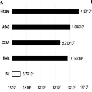
To examine the production efficiency of Ad-fosARE, cancer cells (HeLa, C33A, A549 and H1299) and normal cells (BJ) were infected with at an MOI of 10 ifu/cell and virus titers generated after 48 h and detected by staining the hexon protein of virus particles. In cancer cells, the propagation of Ad-fosARE was very high. On the other hand, the titer in normal cells was 3 to 4 logs lower than those in cancer cells (Figure 2.2). These results indicate that Ad-fosARE can selectively replicate in cancer cells.
A B
29
Figure 2.2. The production efficiency of Ad-fosARE. Cancer (HeLa, C33A, A549 and H1299) cells and normal (BJ) cells were infected with Ad-fosARE at an MOI of 10 ifu/cell and virus production was determined 48 h after infection. (A) Each titer (ifu/ml) is shown on the graph. Data are shown above as the mean ± standard deviation of three independent experiments. (B) The hexon staining in 293 cells used to count virus titers.
2.3.2. HuR-mediated mRNA stabilization is essential for adenovirus replication In order to investigate whether Ad-fosARE replicates in an ARE-mRNA stabilization-dependent manner, Ad-fosARE replication was examined using conditions that decrease ARE-mRNA stabilization and conditions that enhance stabilization conversely. To evaluate virus production in HuR-depleted cells, where ARE-mRNA cannot be stabilized, we treated cells with heat shock (HS), as a previous survey revealed that heat shock transiently decreases HuR abundance (Abdelmohsen et al., 2009). HS treatment of HeLa cells for 2 h resulted in reduced expression of HuR protein in the cytoplasm of cells and also shortened the half-life of E1A mRNA containing ARE (Figure 2.3.C bottom). Likewise, HS-treated cells showed a significant reduction in virus production compared to the non-treated cells (Figure 2.3.B).
30 A
B
C
31
Figure 2.3. The effects of HuR-depletion on E1A mRNA stabilization and Ad- fosARE replication. (A) HeLa cells were heat-shocked at 43˚C for 2 h and the amount of HuR in the total, the nuclear and cytoplasmic fraction was determined using western blot analysis. β-tubulin and lamin expression were also identified as the markers of the cytoplasm and nuclear fractions. (B) Virus production was then assessed, and the values were compared to the non-treated cells (middle). (C) HeLa cells were infected with Ad-fosARE (MOI 10 ifu/cell) and heat-shocked immediately after infection at 43˚ C for 2 h.
Transcriptions were inhibited by actinomycin D 5µg/mL for 60, 120 and 180 minutes. After the indicated periods, cells were harvested and extracted for total cellular RNA. mRNA levels are quantified by quantitative RT-PCR experiments using GAPDH as a normalization control. Graphs depict the percentage of remaining E1A mRNA levels with GAPDH mRNA and compared with the standards of normalized mRNA species measured immediately after the addition of actinomycin D and which were set as 100%.
The half-lives of the mRNAs (min) are indicated as t½ (bottom). Data are shown above as the mean ± standard deviation of three independent experiments.
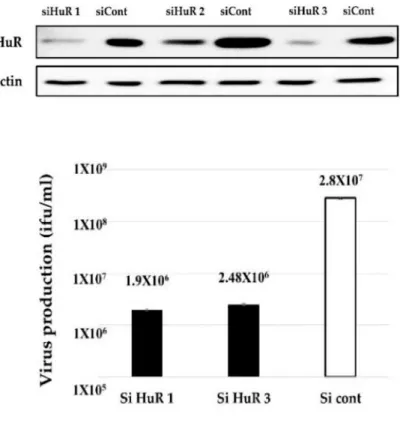
Since HS treatment affects many factors other than HuR degradation in cells, we confirmed the HuR-depletion by HuR knockdown (KD) (Figure 2.4). We found that virus propagation was decreased in siRNA-treated cells compared to control siRNA- treated cells (Figure 2.4). Taken together, these results indicate that HuR is essential for Ad-fosARE replication and it replicates in an ARE-mRNA stabilization-dependent manner. Our previous report showed that HuR depletion (HS and HuR KD) causes the downregulation of the replication of WT300 because HuR is required for full replication of adenovirus (Jehung et al., 2017). However, since the downregulation rate of WT300 was lower than that of Ad-fosARE, and this is because the dependency of HuR of Ad-fosARE is significantly higher than that of WT300.
32
Figure 2.4. Effect of HuR knockdown on virus replication. HeLa cells were transfected with a siRNA targeting HuR and a negative control siRNA, and via western blot analysis, the expression of HuR was estimated. (top) HuR KD-HeLa cells were infected with Ad-fosARE, and viral titers were determined as described in materials and methods after 48 h of virus infection.
(bottom) Data are shown above as the mean ± standard deviation of three independent experiments.
2.3.3. In vitro cytolytic potentiality of Ad-fosARE
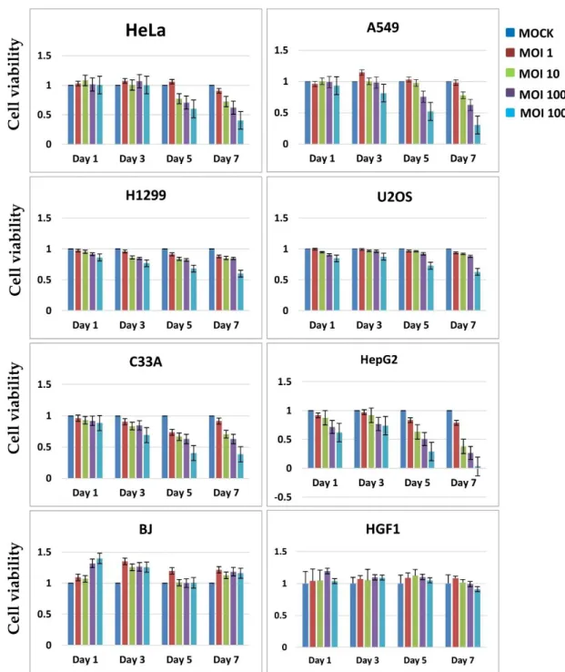
To estimate the cell lysis activity of Ad-fosARE, we examined cell viability using the XTT assay. Cancer cells (HeLa, A549, U2OS, H1299, C33A, H1299, and HepG2 ) and normal cells (BJ and HGF1) were infected with Ad-fosARE at an MOI of 1,10,100 and 1000 (ifu/cell) and the XTT assay was performed 1, 3, 5 and 7 days after infection (Figure 2.5). Most of the cancer cells died after 7 days in a time-dependent manner. In contrast, most normal cells infected with the virus did not die even after 7 days.
33
Figure 2.5. Cell lysis activity of Ad-fosARE. Cell metabolic activities of Ad- fosARE-infected cells were measured to estimate the cell lysis activity using the XTT assay. Cancer (HeLa, A549, C33A, H1299, U2OS and HepG2) cells and normal (BJ and HGF1) cells were infected with the virus at an MOI of 1,10,100 and 1000 ifu/cell and cell viabilities were estimated 1, 3, 5 and 7 days
34
post-infection. Data are shown above as the mean ± standard deviation of three independent experiments.
To assess the cell lysis activity of Ad-fosARE further, we examined the cell death of virus-infected cells using a cytopathic effect (CPE) assay. Six different cancer cell lines (HeLa, C33A, A549, H1299, HepG2 and U2OS) and two normal cell lines (BJ and HGF1) were infected with virus at MOIs of 1, 10, 50 or 100 ifu/cell.
Cytotoxicity was assessed by staining the remaining cells with Coomassie brilliant blue 7 days after infection (Figure 2.6). The virus killed all cancer cells in a dose-dependent manner, although most normal cells survived in any dose. These results determine the in vitro cancer-selective cytolytic activity of Ad-fosARE.
Figure 2.6. Cytopathic activity of Ad-fosARE. Cancer (HeLa, A549, C33A, H1299, U2OS and HepG2) cells and normal (BJ and HGF1) cells were
35
infected with the virus at the MOIs indicated and cells were stained using Coomassie brilliant blue 7 days post-infection. Living cells stained blue.
Next, we explored the pathway behind Ad-fosAREF induced cell death in cancer cells. As various pathways of adenovirus-induced cell death such as apoptosis, autophagy has been reported (Abou El Hassan et al., 2004. Tazawa et al., 2017), we examined the proteins expressed specifically for each cell death. Although LC3, expressed autophagy, was not detected in Ad-fosARE infected cells (data not shown), only the processing of the caspase substrate PARP was detected by western blot analysis on various days post-infection (Figure 2.7.A). PARP expression in cisplatin-treated cells was used as a positive control (Anke et al., 2019).
Figure 2.7. Assessment of cell death by apoptosis. (A) The expression of the cleavage of PARP was assessed by western blot analysis to detect the cell lysis activity mediated by Ad-fosARE. (B) HeLa and A549 cells were infected with A
B
36
Ad-fosARE with MOI 100 ifu/cell. 96 h after infection, the apoptosis of cells was also analyzed by Hoechst 33324 staining. Nuclear fragmentation and chromatin clumping were observed in the virus treated cells (Yellow arrows).
The number of apoptotic cells was quantitated in the histogram (right panel).
Scale bar: 50µm. Three replicates represented each assay.
We also observed the apoptosis by Hoechst 33324 staining. After the cells were infected with the virus with MOI 100 ifu/cell, then cells were stained with Hoechst 33324 to find the morphological changes of cell apoptosis in both control and Ad- fosARE infected cells. Morphological changes of cell apoptosis, such as condensation of chromatin and nuclear fragmentations, were clearly found in Ad-fosARE infected cells (Figure 2.7.B). These results suggest that Ad-fosARE infection-induced apoptosis.
2.3.4. Comparison of the oncolytic effects of Ad-fosARE and dl1520 Since an E1B-55k gene deleted-adenovirus dl1520 is already applied clinically (Bischoff et al., 1996; Kasuya et al., 2005), we next compared the oncolytic activity and viral replication of Ad-fosARE with that of the dl1520. Ad-fosARE and dl1520 were infected at an MOI of 10 ifu/cell, using cancer cells, (HeLa, C33A, A549 and H1299) and normal BJ cells, and virus titers were estimated 48 h after infection. As shown in Figure 2.8.A, Ad-fosARE production was higher than dl1520 in all cancer cells. We also compared the cytolytic activity by XTT assay as presented in Figure 2.8.B. Cytolytic activity of Ad-fosARE and dl1520 in cancer cells were more or less similar. But in normal cells, Ad-fosARE caused less damage compared to dl1520.
37
Figure 2.8. Comparison of the virus production and cell lysis activity of Ad-fosARE with dl1520 and WT300. (A) Cancer (HeLa, C33A, A549 and H1299) cells and normal (BJ) cells were infected with Ad-fosARE and dl1520 at MOI of 10 ifu/cell. Virus production was assessed 48 h after infection. Each titer (ifu/ml) is shown on the graph.
(B) Cell viabilities were estimated 7 days post-infection. Data are shown above as the mean ± standard deviation of three independent experiments.
38 2.4. Discussion
Among the currently investigated oncolytic viruses, the use of oncolytic adenovirus strains appears to be the most promising. In the present study, we demonstrated the oncolytic efficacy of the Ad-fosARE with ARE of the c-fos gene, in the 3'UTR of the E1A gene. Our results showed that the replication efficiency of this virus in cancer cells was higher than in normal cells, and also, was better than dl1520 equivalent to H101, which is clinically applied (Bischoff et al., 1996, Kasuya et al., 2005).
We investigated the requirement of mRNA stabilization system for Ad-fosARE replication. Since Ad-fosARE possesses an ARE downstream of E1A gene, an increased expression level of E1A mRNA was expected in cancer cells, where ARE-mRNA is stabilized, thereby promoting virus growth. To confirm such ARE-mRNA-dependent virus growth and cell lysis, HuR was depleted by heat shock and knockdown to avoid ARE-mRNA exportation and stabilization. As expected, the virus replication decreased in response to inhibited ARE-mRNA stabilization. Also, the half-life of E1A mRNA was low in HuR-depleted cells compared to control cells. These data indicate that Ad- fosARE replicates in HuR dependent manner. Since, in the vast majority of cancer cells, ARE-mRNAs relocate to the cytoplasm with HuR and are constitutively stabilized (López et al. 2003), these facts suggest that Ad-fosARE has the potential to be effective for many types of cancer cells.
In a previous study, an adenovirus including the ARE of COX-2 gene in the 3'-UTR of the E1A gene was shown to be a conditionally replicating virus (Ahmed et al., 2003) and that virus replicated selectively in RAS/P-MAPK-activated cancer cells because ARE-mRNA was stabilized under the activated RAS pathway. In the case of Ad- fosARE, cytolytic activity was not dependent on the RAS/P-MAPK pathway, because the virus exhibited oncolytic activity for HeLa cells, which carry a normal ras gene.
These findings indicate that Ad-fosARE is effective for cancer cells carrying unmutated ras. These data indicate that Ad-fosARE is effective in more cancers than previously shown viruses.
Since the stability of ARE-mRNA has been reported in many biological or pathological events, such as inflammation, viral infection, hypoxia, and UV irradiation, Ad-fosARE has potential in the treatment of diseases with these biological features.
39
Chapter III
Synergism of Oncolytic Adenovirus Utilizing
RNA Stabilization Mechanism and Paclitaxel
40 3.1 Introduction
Oncolytic adenovirus can be used either as a standalone therapeutic modality or in combination with other approaches to overcome or enhance viral activity in the tumor microenvironment. Paclitaxel (PTX) is the most common anticancer agent used against many cancers, but the emergence of drug resistance is a major drawback. PTX is a microtubule (MTs) stabilizing drug (Ingemarsdotter et al., 2010; McGuire et al., 1996) and inhibits cell replication by enhancing the polymerization of tubulin monomers into stabilized microtubule bundles that are unable to reorganize into the proper structures during mitosis (McGuire et al., 1996; Horwitz et al., 1992). MT networks are very important for adenovirus entry, nuclear trafficking, development of replication compartments, and controls broader aspects of infected-cell behavior (Kelkar et al., 2004). After entering through the receptor, the viral capsid interacts with the motor protein dynein and trafficking to the nucleus in a microtubule-dependent manner (Leopold et al., 2007). Virus infection and consequent signaling alter the MT dynamics of the host cell, thus making MTs stable for the intracellular transport of the virus (Warren et al., 2016).
PTX can increase the expression of coxsackie and adenovirus receptor (CAR) (Seidman et al., 2001; Fok et al., 2007). Adenoviral fiber knob of the capsid binds with cell surface receptor CAR and initiates gene transfer (Abdolazimi et al., 2007). CAR is a 46-k transmembrane glycoprotein and belongs to the immunoglobulin superfamily, which is the primary receptor for adenovirus (Abdolazimi et al., 2007; Tomko et al., 1997; Bergelson et al., 1997). Furthermore, PTX increases the HuR export to the cytoplasm in cancer cells and thus facilitates mRNA stabilization (Badawi et al., 2018).
Some studies have shown that the ubiquitously expressed HuR can utilize either the actin- or microtubule-dependent cytoskeleton for the transport of mRNA cargo (Cheng et al., 2013; Doller et al.,2008; Holt et al., 2008). MTs are essential for cell motility and the transport of organelles like; vesicles and are relevant for long-distance transport of protein and mRNA (Holt et al., 2008; Blower et al., 2013; Eberhardt et al., 2016).
In this study, I show that Ad-fosARE can synergies with a microtubule stabilizer PTX. Use of heat shock or a knockdown depleted HuR, which further down-regulated Ad-fosARE replication was indicating that Ad-fosARE replicates in an ARE-mRNA stabilization-dependent manner. PTX promotes the nuclear export of HuR, which can
41
enhance the propagation of Ad-fosARE, in cancer cells but not in normal cells.
Furthermore, it up-regulated the expression of CAR and increases virus internalization.
The oncolytic activity of Ad-fosARE was enhanced synergistically by PTX through the increase of E1A expression and enhancement of viral apoptotic activity both in vitro and in vivo. These results indicate that PTX offers many benefits to Ad-fosARE.
3.2 Materials and method 3.2.1 Cell Lines
A549 (human lung cancer cell line) and HeLa, HeLa S3 (human cervical carcinoma cell lines), 293 (human embryonal kidney cells transformed by the adenovirus E1 gene) and BJ (human foreskin fibroblast cell line) were used in this study. Cells were acquired from the American Type Culture Collection (ATCC; Manassas, VA, USA) and cultured in Dulbecco's modified Eagle's medium (DMEM; Sigma-Aldrich; Merck KGaA, Darmstadt, Germany) including 10% fetal bovine serum (FBS; Biowest, Nuaille, France) with antibiotics at 37 °C in a 5% CO2 atmosphere in humidified conditions.
3.2.2 Cytopathic effect (CPE) assay and cell viability assay
Cancer and normal cells were seeded on 24-well plates (5×10⁴ cells/well). 24 h later, the cells were treted with PTX (4 nM) and, additional 4 h after, they were infected with Ad-fosARE at a multiplicity of infection (MOI) of 1, 10 ,50, and 100 ifu/cell and maintained for an additional 7 days. Then cells were fixed and stained with Coomassie brilliant blue.
A 2-3-bis [2-methoxy-4-nitro-5-sulfophenyl]-2H-tetrazolium-5-carboxanilide inner salt assay was used to observe the cytolytic activity. Cells were seeded on 96-well plates at 3×10³ cells/well. 24 h later, the cells were treted with PTX (4 nM) and, additional 4 h after, they were infected with Ad-fosARE at a MOI of 10 ifu/cell. Cytolytic activity was assessed using an XTT assay on days 1, 3, 5, and 7 with the Cell Proliferation kit II (Roche Diagnostics, Basel, Switzerland) according to the manufacturer's protocol.
42 3.2.3 Western blot analysis
Cells were treated with 4 nM PTX and were lysed with RIPA buffer (150 mM NaCl; 25 mM Tris-HCl, pH 7.6; 1%Nonidet P-40; 1% sodium deoxycholate; 0.1%
SDS) containing protease inhibitors. Equal amount (20 µg) of total protein were separated by 10% sodium dodecyl sulfate-polyacrylamide gel electrophoresis (SDS- PAGE) and transferred onto polyvinylidene difluoride membranes (Millipore, Billerica, MA, USA). Fractionation buffer (10 mM Tris-HCl, pH 7.6, 150 mM NaCl, 1.5 mM MgCl2, 0.5% Nonidet P-40, protease inhibitor cocktail) was used to fractionate the cytoplasmic and nuclear proteins, followed by vigorous shaking for 5 min and centrifugation at 12,000 rpm for 30 s. The supernatant was used as the cytoplasmic protein fraction. To assess the accuracy of cell fractionation, cytoplasmic (β-tubulin) and nuclear (lamin B) proteins were detected. For detection of surface protein CAR, the protocol described in Andolazimi et al., was used (Andolazimi et al., 2007).
3.2.4 Immunocytochemistry
HeLa, A549 and BJ Cells were grown on coverslips in 35-mm dishes at 60–80%
confluence and treated with PTX (4 nM) for 4 h. Immediately following the treatment, cells were rinsed once with phosphate buffer saline (PBS), fixed for 20 min at room temperature with 4% paraformaldehyde in PBS, blocked and permeabilized in 2% BSA plus 0.1% Triton X-100 in PBS at room temperature. Subsequently, the fixed cells were incubated with HuR and Tubulin (abcam 6160) primary antibody for overnight at 4 °C followed by Alexa Fluor 488 and Alexa Fluor 568 secondary antibodies for 1 h at room temperature respectively. Cell nuclei were counterstained with DAPI.
For Hoechst 33342 staining, cells were infected with Ad-fosARE (MOI 10 ifu/cell) and treated with PTX (4 nM). After these treatments, cells were rinsed once with phosphate buffer saline (PBS), fixed for 20 min at room temperature with 4%
paraformaldehyde in PBS, blocked and permeabilized in 2% BSA plus 0.1% Triton X- 100 in PBS at room temperature. Cell nuclei were stained with Hoechst 33342 before mounted on slides by using Mountant permafluor (Thermo scientific, FM 111212A).
Cells were observed using an IX71 inverted microscope (Olympus). Image acquisition was performed with the Olympus FluoView Software (FV10-ASW Viewer, ver.4.2).
43 3.2.5 In vitro virus proliferation assay
Cancer cells and normal cells were seeded into 12-well plates at a density of 5×10⁴ cells/well 24 h before the infection. After 4 h of treatment of PTX (4 nM), cells were infected with Ad-fosARE at an MOI of 10 ifu/cell, and the medium was not changed after virus infection. After incubation at 37 °C for 72 h, cells were collected, and a virus lysate was prepared as described above. Viral titers (ifu/ml) were determined using the Adeno-X Rapid Titer kit (Clontech Laboratories). For virus exit assay, the medium was collected from virus-infected cells and centrifuged for 5 min at 4000 rpm then the supernatant was tittered (Ingemarsdotter et al., 2010).
3.2.6 RNA extraction and quantitative RT-PCR
Total RNA was extracted using the RNeasy Mini Kit (250) from QIAGEN and the RNA was subjected to reverse transcription by ReverTra Ace qPCR RT master mix with genomic DNA remover (Toyobo, Osaka, Japan). Quantitative RT-PCR was performed in the DNA Engine Opticon2 (MJ Research, Watertown, MA, USA), and cDNA was amplified using the following primers: Ad5 E1A; 5’-gaa cca ccta ccc ttc acg-3’, 5’-ccg cca aca tta cag agt cg-3’, GAPDH; 5’-atc ctg ggc tac act gag ca-3’, 5’-tgc tgt agc caa att cgt tg-3’. GAPDH was used for normalization. To evaluate the E1A mRNA, HeLa cells were infected with Ad-fosARE, WT300 and treated with PTX for 24, 48, 72 and 96 h and cellular RNA of each cell was used for quantitative RT-PCR. To estimate the half-life of total E1AmRNA, cells were treated with 5µl/mL Actinomycin D 60, 120, or 180 min. The total cellular RNA of each cell was used for quantitative RT-PCR.
3.2.7 In vivo analysis
Female BALB/c nu/nu mice (5-week-old and 20-22 g) were purchased (Hokudo, Sapporo, Japan) and kept in a specific pathogen-free environment. The temperature was maintained at 26-28 °C, 10 h/14 h light/dark cycle; food and water were given ad libitum. Flank tumor was established by injecting HeLa S3 cells (1×10⁶ cells/mouse) subcutaneously into the flanks of mice and permitted to grow to ~5-6 mm in diameter.
The mice were assigned into four groups randomly (five per group) and PTX (0.4 mg/100 µl) or vehicle (cremophor EL/dehydrated ethanol/saline) was given by intraperitoneal (i.p) injection. 1×10⁶ ifu (100 µl) of Ad-fosARE or the same volume of PBS was injected intratumorally (i.t) thrice (days 1, 4, and 7) after PTX injection. The
44
perpendicular diameters of the tumors were recorded every 2 or 3 days. Tumor volumes were measured using the following equation: Volume (mm3) = A × B2 × 0.5 (A is considered the longest diameter, B is considered shortest diameter). The body weight and motor activity of each animal was monitored as indicators of general health and toxicity. The mice were sacrificed by cervical dislocation after 21 days of injection of the virus as the tumors on the control group began to ulcerate. All techniques executed in this study involving animals were performed according to the ethical standards of Animal Care and Use Committee of the Hokkaido University. Sapporo, Japan (Permission number for the animal experiment: 19-0099).
3.2.8. Immunohistochemistry
The xenografted tumors were removed, fixed with 4% formaldehyde, and embedded for hematoxylin & eosin staining and immunohistochemistry. Next paraffin- embedded specimens were sectioned in 4-µm-thick. The immunohistochemical detection of Adenovirus 5 (Abcam, ab6982) was performed using anti-Adenovirus Type 5 (Rabbit Adenovirus antibody, dilution, 1:1,000; catalog no.,ab6982; Abcam) in PBS in a humidified chamber at 4 °C overnight. The sections were then subjected to anti- rabbit secondary antibody (H1901, Nichirei Bioscience, Tokyo, Japan) at 37 °C for 30 min, followed by antibody detection using a peroxidase conjugated streptavidin- diaminobenzidine (DAB) readout system (DAKO), and counterstaining with DAPI.
Rinses were carefully performed with several changes of PBS between each stage of the procedure (5 min washes repeated three times). Images were randomly captured using a nanozoomer slide scanner and NDPViewer (NanoZoomer 2.0 HT, version 2.3.27, Hamamatsu, Shizuoka, Japan).
45 3.3 Results
3.3.1 The potentiality of paclitaxel for synergism
It is reported that low concentration PTX increases the cytoplasmic exportation of HuR (36kDa) (Badawi et al., 2018), so we examined whether a low dose of PTX (4 nM) induces the cytoplasmic HuR exportation, as the upregulation of the cytoplasmic HuR expression has to benefit for the replication of Ad-fosARE. We treated cancer cells with PTX and incubated for 4, 8 and 24 hours and collected and fractionated the cytoplasmic and nuclear protein. We found an increase in cytoplasmic HuR exportation in cancer cells by western blot analysis (Figure 3.1.A). Normal cells BJ were also treated with PTX and checked by western blot analysis, but after 24 hours of treatment, cytoplasmic HuR were decreased contrary. HuR exportation was also confirmed using confocal microscopy. HuR localization PTX treated HeLa, A549 and BJ cells was observed using immunostaining method. In cancer cells, cytoplasmic HuR increased in the PTX treated cells compared to control cells (Figure 3.1.B). But in the case of normal cells BJ, PTX treatment diminished cytoplasmic HuR compared to control cells. These results indicate that PTX has the potential to increase cytoplasmic HuR in cancer cells but not in normal cells.
46
A .
B
47
Figure 3.1.Effects of paclitaxel on cytoplasmic HuR exportation. (A) Cancer (HeLa and A549) cells and normal (BJ) cells were treated with 4nM PTX.
Cells were separated into the cytoplasmic and nuclear fractions and HuR localization was estimated by western blot analysis. The level of HuR protein was monitored in total cell lysates (TP) and cytoplasmic (CP) and nuclear extracts (NP). Tublin, Lamin and Actin are the inner control of each fraction.
(B) Confocal microscopy was performed to visualize changes in HuR exportation. HeLa, A549 and BJ cells were treated with PTX (4 nM) and were stained with HuR and Tublin after fixation and permeabilization as described in materials and methods. In all cases, cell nuclei were visualized by staining with DAPI. Bars indicate 20 µm. Data shown are from a single experiment representative of three repeats giving similar results.
Coxsackie-adenovirus receptor (CAR), which is the primary affinity receptor for adenovirus also upregulated by PTX . The first interaction of adenovirus with cells occurs through the virus fiber protein with its cell surface receptor CAR. This binding leads to internalization of the virus by endocytosis and trafficking along dynein (motor protein of microtubules) toward the nucleus (Seidman et al., 2001; Fok et al., 2007). To check the CAR (46kDa) expression, HeLa cells were treated with 4nM PTX for a different time course, and after treatment, CAR expression was examined by western blot analysis. As shown in Figure 3.2, CAR expression increased after treatment with PTX.
Figure 3.2. Western blot analysis of expression of coxsackie-adenovirus receptor (CAR) at different time-points. After treatment with PTX, total HeLa