Japan Advanced Institute of Science and Technology
JAIST Repository
https://dspace.jaist.ac.jp/Title
Cryopreservation of a two-dimensional monolayer using a slow vitrification method with
polyampholyte to inhibit ice crystal formation
Author(s)
Matsumura, Kazuaki; Kawamoto, Keiko; Takeuchi, Masahiro; Yoshimura, Shigehiro; Tanaka, Daisuke; Hyon, Suong-Hyu
Citation ACS Biomaterials Science & Engineering, 2(6): 1023-1029
Issue Date 2016-04-18
Type Journal Article
Text version author
URL http://hdl.handle.net/10119/13700
Rights
Kazuaki Matsumura, Keiko Kawamoto, Masahiro Takeuchi, Shigehiro Yoshimura, Daisuke Tanaka, and Suong-Hyu Hyon, ACS Biomaterials Science & Engineering, 2016, 2(6), pp.1023-1029. This document is the unedited author's version of a Submitted Work that was subsequently accepted for publication in ACS Biomaterials Science &
Engineering, copyright (c) American Chemical Society after peer review. To access the final edited and published work, see
http://dx.doi.org/10.1021/acsbiomaterials.6b00150 Description
1
Cryopreservation of a two-dimensional monolayer using a slow vitrification method with polyampholyte to inhibit ice crystal formation
Kazuaki Matsumura1*, Keiko Kawamoto1, Masahiro Takeuchi2, Shigehiro Yoshimura2,
Daisuke Tanaka3, Suong-Hyu Hyon4
1
School of Materials Science, Japan Advanced Institute of Science and Technology, 1-1
Asahidai, Nomi, Ishikawa 923-1292, Japan
2
Taiyo Nippon Sanso Corp., Toyo Bldg., 1-3-26 Koyama, Shinagawa-ku, Tokyo 142-8558,
Japan
3
Genetic Resources Conservation Research Unit, Genetic Resources Center, National
Institute of Agrobiological Sciences (NIAS), 2-1-2 Kannondai, Tsukuba, Ibaraki 305-8602,
Japan
4
Center for Fiber and Textile Science, Kyoto Institute of Technology, Matsugasaki, Kyoto
606-8585, Japan
*To whom correspondence should be addressed: Kazuaki Matsumura
E-mail: [email protected] Tel: +81-761-51-1680
Fax: +81-761-51-1149
2 Abstract
Vitrification methods have been developed to improve the preservation of oocytes and
embryos. However, successful vitrification and preservation typically requires very high
cooling speeds. Here, we report a novel slow vitrification method for cryopreservation of
two-dimensional (2D) cell constructs using a vitrification solution (VS) in PBS containing
6.5 M ethylene glycol, 0.5 M sucrose, and 10% w/w carboxylated poly-L-lysine
(COOH-PLL), a novel polymeric cryoprotectant and stabilizing agent that is likely to inhibit
ice crystallization. Stabilization of the glassy state and inhibition of devitrification was
confirmed by thermal analysis using differential scanning calorimetry. The viability of
cultured human mesenchymal stem cell (MSC) monolayers after freezing by our novel slow
vitrification method at a rate of 4.9°C/min in VS with 10% COOH-PLL was significantly
higher than that of cells frozen using our slow vitrification method in VS without
COOH-PLL. Moreover, cells maintained the capacity for differentiation. We further
confirmed that COOH-PLL improved the vitrification properties of the current vitrification
system through inhibition of recrystallization properties. This novel, simple method for slow
vitrification can be widely applicable for the preservation of tissue-engineered constructs and
may facilitate the industrialization of regenerative medicine.
Keywords: Vitrification, ice recrystallization, polyampholyte, tissue-engineered construct,
3 Introduction
Low-temperature preservation, called cryopreservation, is used for long-term storage of
biological materials containing cells. Two primary techniques of cryopreservation are slow
freezing and vitrification, in which water transitions directly to the glassy state without
crystallization1,2. Slow freezing methods, which utilize 10% dimethyl sulfoxide (DMSO) as a
cryoprotectant, are effective for a variety of cell lines3,4. This method functions through
dehydration of cells by freezing of outer membrane, leading to inhibition of intracellular ice
crystal formation5. However, for the freezing of two- (2D) or three-dimensional (3D) cell
constructs, dehydration of each cell causes cell shrinkage, which may damage and destroy
cell-cell interactions.
Vitrification methods have been developed with the advent of preservation
techniques for oocytes and embryos, primarily in the field of reproductive medicine6,7. Rapid
cooling leads to the formation of amorphous ice. During the process of vitrification, water
transforms directly to a glassy state, thereby preventing crystallization because of the rapid
rate of freezing (e.g., 2000–20000°C/min)8, which does not give the water molecules time to
form ice crystals. Therefore, vitrification requires a high rate of cooling and high
concentration of cryoprotectants (CPAs). Thus, there are several challenges that must be
overcome for the successful vitrification of tissue-engineered constructs. First, the high
4
The process of vitrification involves exposure to very high concentrations of CPAs and
subsequent rapid cooling in liquid nitrogen. While reducing the concentration of the CPA may
decrease toxicity, ice crystallization must still be inhibited. Additionally, rapid vitrification of
tissue-engineered constructs causes mechanical stress during boiling of liquid nitrogen at the
time of immersion, which may damage the thin, fragile structures of the tissue, resulting in
the formation of cracks9. Finally, recrystallization during rewarming10 may cause damage to
the preserved tissues or cells. Therefore, inhibition of ice recrystallization is also needed for
successful preservation.
In previous studies, we developed a polymeric cryoprotectant, carboxylated
poly-L-lysine, which functioned by inhibiting ice recrystallization11,12. In addition, the best
cryoprotective and ice recrystallization properties were found in polyampholyte, in which 65
mol% amino groups (approximately) were converted to carboxylated groups13. Indeed, we
successfully preserved human induced pluripotent stem cells (iPS cells) in 65% carboxylated
poly-L-lysine (COOH-PLL) in relatively large scale vitrification (200 μL)14. Similarly,
Vorontsov et al. studied the inhibitory effects of COOH-PLL on the growth of ice crystals15 in
free-growth experiments of ice crystals in solutions at various COOH-PLL concentrations.
Clinical application of regenerative medicine requires the preparation of 2D and 3D
tissue-engineered constructs. Cell sheet therapy involves covering a tissue lesion with a
5
requires further advances in low-temperature preservation. Preservation of tissues and
tissue-engineering products is one of the most important techniques for the clinical and
industrial application of tissue engineering. However, cryopreservation of regenerated tissues,
including cell sheets and cell constructs, is not easy compared to the cryopreservation of cell
suspensions.
Many studies have developed vitrification methods for various types of
tissue-engineered constructs, such as bone17, intestine18, blood vessels19, encapsulated cell
constructs20, cartilage21, and cell sheets22. However, as described above, vitrification usually
requires a fast cooling rate, which may damage cells. The use of COOH-PLL for stabilization
of the glassy state during vitrification has been described14. In particular, cell sheets were
coated with a viscous vitrification solution (VS) containing permeable and nonpermeable
CPAs before vitrification in liquid nitrogen vapor, thereby preventing fracturing of the fragile
cell sheet after vitrification and rewarming. Both the macro- and microstructures of the
vitrified cell sheets were maintained without damage or loss of major components. However,
no quantitative analysis of stabilization of the glassy state with changes in the cooling rate
was conducted.
Therefore, in this study, we evaluated that the relationships between cooling rate and
vitrification and between rewarming rate and recrystallization of various VSs in order to
6
we propose a novel slow vitrification method based on the results of vitrification of
mesenchymal stem cell (MSC) monolayers using COOH-PLL as a glassy state stabilizing
additive.
7 Materials and Methods
Human MSC monolayer
Five independent human bone marrow MSC (hBMSC) lines (HMS0051, HMS0008,
HMS0050, HMS0023, and HMS0024) established by Dr. Kato of Hiroshima University23
were purchased from the RIKEN Cell Bank (RIKEN Bioresource Center, Ibaraki, Japan) in
accordance with the regulations of the Life Science Committee of Japan Advanced Institute
of Science and Technology. Cells were maintained in Dulbecco’s modified Eagle’s medium
(DMEM; Sigma-Aldrich, St. Louis, MO, USA) containing 10% fetal bovine serum (FBS), 3
ng/mL basic fibroblast growth factor (bFGF; Wako Pure Chemical Industries Ltd., Osaka,
Japan), 100 U/mL penicillin, and 100 μg/mL streptomycin. These hBMSCs were delivered in dry ice from the cell bank and stored in liquid nitrogen after arrival at our laboratory until
experimental use. Cell culture was carried out at 37°C under 5% CO2 in a humidified
atmosphere. When the cells reached 80% confluence, they were detached using 0.25% (w/v)
trypsin containing 0.02% (w/v) ethylenediaminetetraacetic acid in phosphate-buffered saline
without calcium or magnesium [PBS(-)] and were seeded on a new tissue culture plate for
subculture. The hBMSCs used in this study were between passages three and five.
Synthesis of COOH-PLL
8
synthesize COOH-PLL, 25% w/w ε-poly-L-lysine (PLL) aqueous solution (10 mL; JNC
Corp., Tokyo, Japan) and succinic anhydride (1.3 g SA; Wako Pure Chem. Ind. Ltd., Osaka
Japan) were mixed and reacted at 50°C for 1 h to convert 65% amino groups to carboxyl
groups (Fig. S1). The number of amino groups was determined using the
2,4,6-trinitrobenzenesulfonate (TNBS) method24. Briefly, 0.3 mL of 250 μg/mL sample solution, 1 mL of 1.0 mg/mL TNBS solution, and 2 mL of 40 mg/mL sodium bicarbonate
aqueous solution containing 10 mg/mL sodium dodecyl sulfate (pH 9.0) were mixed and
incubated at 37°C for 2 h. After the mixture was cooled to 25°C, the absorbance was
measured at 335 nm.
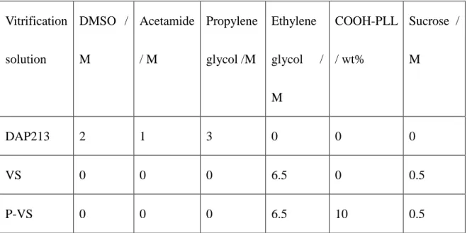
Preparation of VS
The VS developed for cryopreserving human iPS cells14 was used after slight modifications
(Table 1). We prepared a VS based on 6.5 M ethylene glycol (EG, Wako) and 0.75 M sucrose
(Wako) in PBS. COOH-PLL was added to the solution at 10% w/v to evaluate the effects of
the polymer (P-VS). A DAP213 solution (2 M DMSO, 1 M acetamide, and 3 M propylene
glycol [all from Wako] in PBS) was used as a control25. An equilibration solution (ES)
consisting of 15% (v/v) EG in PBS. A rewarming solution (RS) and dilution solution (DS)
9 Tables
Table 1. Composition of various vitrification solutions
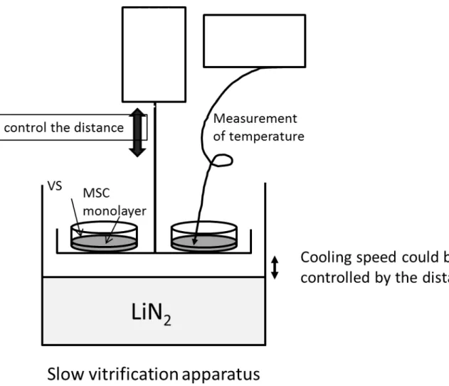
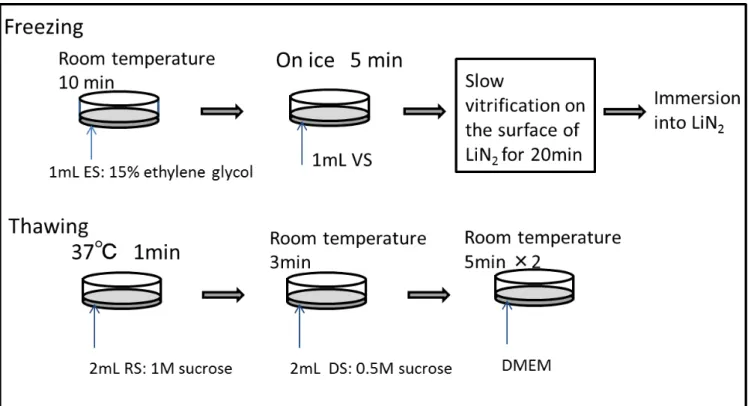
Slow vitrification procedure
First, 1.0 mL of ES was added to the MSC monolayer cultured in a 3.5-cm dish for
pre-equilibration for 10 min. After equilibration, the ES was discarded, and 1.0 mL of three
types of VSs was added to each MSC monolayer on ice. After 5 min, the VS was removed by
aspiration, and the dish was placed on the stainless steel mesh and held various distances
above the surface of liquid nitrogen. The temperature of the MSC monolayer was monitored
with a thermocouple attached on the monolayer. After 20 min, the dish containing the
vitrified MSC monolayer sheet was soaked in liquid nitrogen. An illustration of the practical
procedure of slow vitrification is shown in Figs. S2 and S3. Vitrification solution DMSO / M Acetamide / M Propylene glycol /M Ethylene glycol / M COOH-PLL / wt% Sucrose / M DAP213 2 1 3 0 0 0 VS 0 0 0 6.5 0 0.5 P-VS 0 0 0 6.5 10 0.5
10
The MSC monolayers were warmed on the same day by gently addition of
prewarmed RS (2 mL). After 1 min, the RS was changed to DS. Three minutes later, the
monolayer was washed with DMEM twice for 5 min each.
Evaluation of the survival of MSC monolayers
Two MSC monolayers in the dish were prepared for the same vitrification protocol. One was
used for the evaluation of viability immediately after thawing, and the other was evaluated 1
day after thawing. Thawed MSC monolayers were stained by calcein AM and ethidium
homodimers using a Live/Dead Assay Kit (Life Technologies,Carlsbad, CA, USA). The cells
were then dispersed by trypsin solution, and the viability of the cell monolayers were
determined using trypan blue staining.
Differential scanning calorimetry
To compare the stabilities of the glassy states among the VSs, thermal analysis was
performed using a differential scanning calorimeter (DSC; Q2000; TA Instruments, New
Castle, DE, UAS). Ten microliters of each solution was placed on the aluminum pan. The pan
was then set on the DSC sample chamber, cooled to -170°C at 10°C/min, and then warmed to
11
Induction of differentiation of MSC monolayers
Unfrozen and vitrified MSC monolayers were induced to differentiate into osteoblasts and
adipocytes for 14 days in osteogenic and adipogenic media, respectively26. The osteogenic
medium contained 0.1 mM dexamethasone, 10 mM β-glycerophosphate disodium salt, 0.07 mM L-ascorbic acid 2-phosphate magnesium salt n-hydrate (Sigma-Aldrich), 10% FBS, and
1% antibiotic/antimycotic in DMEM. The adipogenic medium contained 0.5 mM isobutyl
methylxantine, 0.1 mg/mL insulin, 0.2 mM indomethacin, 1 mM dexamethasone, 10% FBS,
and 1% antibiotic/antimycotic in DMEM. The culture medium was changed three times per
week.
Evaluation of differentiation
Monolayer cultures treated with osteogenic differentiation medium were stained with 1%
Alizarin Red S for 15 min after fixation with 4% neutral buffered formalin for mineralized
matrix deposition. Monolayer cultures treated with adipogenic differentiation medium were
stained with oil Red O solution to visualize the formation of fat vacuoles. Briefly, the cells
were fixed with 4% neutral-buffered formalin and incubated for 20 min with 0.2% oil Red O
isopropanol solution.
12
All data are expressed as the means ± standard deviations (SDs). Measurements of post-thaw
viability were collected with three replicates for each sample. All experiments were
conducted in triplicate. To compare data among more than three groups the Tukey-Kramer
test was used. Differences with P values of less than 0.05 were considered statistically
significant.
Results and Discussion
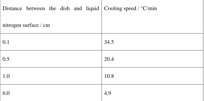
Evaluation of cooling speed
The cooling speed was highly affected by the distance between the dish and liquid nitrogen
surface. Table 2 shows the cooling speed, which was recorded using a thermocouple attached
to the MSC monolayer. The cooling speed was controlled between 4.9 and 34.5°C/min by
changing the distance of the monolayer from the liquid nitrogen surface (Table 2). These
cooling speeds were much slower than that usually used for vitrification8,27. When the dish
containing the MSC monolayer was inserted directly into liquid nitrogen, the cooling speed
13
Table 2. Cooling speed of slow vitrification of MSC monolayers controlled by the distance
between the dish and liquid nitrogen.
Distance between the dish and liquid
nitrogen surface / cm
Cooling speed / °C/min
0.1 34.5
0.5 20.4
1.0 10.8
6.0 4.9
Stabilization of the glassy state and inhibition of devitrification as evaluated by DSC
Thermal analysis of the three VSs was performed using DSC in order to evaluate the
stabilization of the glassy state by COOH-PLL. Fig. 1 illustrates the heat flow curves of the
three VSs. The samples were cooled from 20 to -150°C and warmed from -150 to 0°C at a
rate of 10°C/min on the DSC sample holder. As shown in Fig. 1a, DAP213 crystallized
during cooling at a rate of 10°C/min, while VS and P-VS showed no crystallization. DAP213
has been developed to be optimized for the vitrification of mouse oocytes and embryos25. The
basic principle of the latest high-performance method of vitrifying embryos requires a very
high cooling speed and very small amount of VS to avoid crystallization and
14
During heating, all vitrification solution showed glass transition at around -120°C, and even
DAP213 achieved partial glass transition. VS and P-VS showed recrystallization
(devitrification) during heating at the rate of 10°C/min. Interestingly, the recrystallization
temperature of P-VS (-67.6°C) was higher than that of VS (-98.1°C), and the melting point of
P-VS (-35.6°C) was lower than that of VS (-30.8°C; Fig. 1b). Although the actual rewarming
speed was higher than 10°C/min, we selected this speed in order to compare the risk of
devitrification. These data indicated that COOH-PLL stabilized the glassy state of VS,
leading to a smaller temperature range of the crystalline state of P-VS (from -67.6 to -35.6°C)
than that of VS (from -98.1 to -30.8°C), which may decrease the risk of recrystallization
during thawing. This result was consistent with a previous report in which polyampholytes
inhibited ice recrystallization29.
Toxicity of VSs
MSC monolayers were immersed in DAP213, VS, or P-VS; 10 min later, the monolayers
15
assays are shown in Fig. 2. Many dead cells stained in red were observed in DAP213, while
almost all cells in VS and P-VS were alive. Quantitative analysis by trypan blue staining
showed that the viabilities were 75%, 92%, and 96% for DAP213, VS, and P-VS,
respectively, indicating that DAP213 exhibited the highest cytotoxic effects in MSC
monolayers due to its high osmotic pressure and chemical cytotoxicity. The decrease in
toxicity caused by the addition of COOH-PLL may be the result of improved cell membrane
stability14.
Slow vitrification
Next, we examined the effects of cooling rate on cell viability. Fig. 3 shows images from the
Live/Dead assay at 1 day after thawing of MSC monolayers frozen with the three types of
VSs with various cooling speeds ranging from 4.9 to 34.5°C/min. At cooling speeds of 34.5,
20.4, and 10.8°C/min, almost all cells frozen with P-VS were alive. Some dead cells were
observed in monolayers frozen in VS. However, in contrast, very few live cells were
16
space that was not stained green or red could be observed, particularly in MSC monolayers
frozen with VS. This may be explained by desquamation of the monolayer from the dish due
17
viability. Interestingly, when the cooling speed was decreased to 4.9°C/min, with the distance
between liquid nitrogen and the dish set at 6.0 cm, live cells were observed only when frozen
with P-VS. Fig. 4 shows the quantitative analysis of MSC viability just after thawing (Fig.
4a) and after 1 day of culture (Fig. 4b). The viability of MSCs was higher just after thawing
than at 1 day after thawing. In particular, the viability of cells frozen at a rate of 4.9°C/min in
VS was the lowest due to detachment of a part of the monolayer during thawing. However, it
should be noted that viability by trypan blue staining may be overestimated as some damaged
cells may be considered alive11. These weakened cells may not be able to attach the dish to
grow; thus, viability may be reduced after 1 day of culture. Quantification of viability after 1
day of culture was consistent with the results of the Live/Dead assays (Figs. 3 and 4b). At all
cooling rates, viability with P-VS was significantly higher than those with DAP213 and VS,
18
Ice crystallization was inhibited in VS and P-VS at 10°C/min, as confirmed by DSC (Fig.
19
culture dishes in terms of VS volume, the stabilizing effect of COOH-PLL on the glassy state
was clearly observed in that monolayer sheets frozen with DAP213 or VS turned white when
rewarming. Effective vitrification has been well studied; however, most investigations have
focused on how to obtain rapid cooling using a variety of methods, such as the open pulled
straw method30, cryotop method2,27, and hollow fiber vitrification28. We succeeded in
establishing a new method for obtaining highly viable MSC monolayers via slow (4.9°C/min)
vitrification by adding COOH-PLL to the vitrification solution. This slow vitrification
method was realized for the first time by addition of COOH-PLL; this material had low
toxicity and was effective at stabilizing the vitreous status of the solution during vitrification
and rewarming, thereby resulting in a high rate of survival of MSCs.
Evaluation of differentiation
Fig. 5 shows the results of differentiation of MSCs after slow vitrification with P-VS.
Histological evaluation revealed that MSC monolayers were well differentiated into
osteoblasts (Fig. 5c) and adipocytes (Fig. 5d), similar to nonfrozen cells (Fig. 5a and b).
MSCs have a multipotent capability to differentiate into various functional cell types of
mesodermal tissues31,32. In this study, MSC monolayers were vitrified with our novel method
of slow vitrification with COOH-PLL as a stabilizer of the glassy state, without any other
CPAs and or animal-derived proteins. The results showed that the novel vitrification method
20
differentiation capacity after thawing. In our previous reports, also we found that MSC
differentiation was maintained after slow freezing using COOH-PLL11,12. Thus, these results
confirmed that COOH-PLL did not affect MSC differentiation during preservation.
Conclusion
In this study, we propose a novel slow vitrification method for the cryopreservation of tissue
21
vitrification solution and inhibited recrystallization during rewarming. MSC monolayers
could be cryopreserved, even at a cooling rate of -4.9°C/min, without decreasing cell viability
using P-VS. This novel concept of slow vitrification can be widely applicable for the simple
preservation of tissue-engineered constructs without the requirement for high technical
expertise. Thus, this may facilitate the industrialization of tissue engineering applications by
allowing long-term storage of tissue-engineered constructs.
Supporting Information Available
The following files are available free of charge:
Supplemental synthetic procedures and 1H-NMR of COOH-PLL, schematic illustration of the
slow vitrification apparatus and procedures.
Acknowledgements
This study was supported in part by a Grant-in-Aid, KAKENHI (25242050), for Scientific
Research from the Ministry of Education, Culture, Sports, Science and Technology, Japan,
grant from the Canon Foundation (K11-N-028), and as a Collaborative Research Project
22 References
[1] Rall, W. F.; Fahy, G. M. Ice-free cryopreservation of mouse embryos at -196 degrees C by vitrification. Nature 1985, 313, 573-575.
[2] Kuwayama, M.; Vajta, G.; Kato, O.; Leibo, S. P. Highly efficient vitrification method for cryopreservation of human oocytes. Reprod. Biomed. Online. 2005, 11, 300-308.
[3] Liu, Y.; Xu, X.; Ma, XH.; Liu, J.; Cui, ZF. Effect of various freezing solutions on cryopreservation of mesenchymal stem cells from different animal species. Cryoletters. 2011, 32, 425-435.
[4] Magalhaes, R.; Nugraha, B.; Pervaiz, S.; Yu, H.; Kuleshova, L. L. Influence of cell culture configuration on the post-cryopreservation viability of primary rat hepatocytes. Biomaterials
2012, 33, 829-836.
[5] Mandumpal, J. B.; Kreck, C. A.; Mancera, R. L. A molecular mechanism of solvent cryoprotection in aqueous DMSO solutions. Phys. Chem. Chem. Phys. 2011, 13, 3839-3842. [6] Rall, W. F. Factors affecting the survival of bouse embryos cryopreserved by vitrification. Cryobiology 1987, 24, 387-402.
[7] Reubinoff, B. E.; Pera, M. F.; Vajta, G.; Trounson, A. O. Effective cryopreservation of human embryonic stem cells by the open pulled straw vitrification method. Hum. Reprod.
2001, 16, 2187-2194.
[8] Zhang, X.; Catalano, P. N.; Gurkan, U. A.; Khimji, I.; Demirci, U. Emerging technologies in medical applications of minimum volume vitrification. Nanomedicine 2011, 6, 1115-1129 [9]Steif, P. S.; Noday, D. A.; Rabin, Y. Can thermal expansion differences between cryopreserved tissue and cryoprotective agents alone cause cracking? Cryoletters 2009, 30, 414-421.
[10] Seki, S.; Mazur, P. Effect of warming rate on the survival of vitrified mouse oocytes and on the recrystallization of intracellular ice. Biol. Reprod. 2008, 79, 727-737.
[11] Matsumura, K.; Hyon, S. H. Polyampholytes as low toxic efficient cryoprotective agents with antifreeze protein properties. Biomaterials 2009, 30, 4842-4849.
[12] Matsumura, K.; Hayashi, F.; Nagashima, T.; Hyon, S. H. Long-term cryopreservation of human mesenchymal stem cells using carboxylated poly-L-lysine without the addition of proteins or dimethyl sulfoxide. J. Biomater. Sci. Polym. Ed. 2013, 24, 1484-1497.
[13] Matsumura, K.; Bae, J. Y.; Hyon, S. H. Polyampholytes as cryoprotective agents for mammalian cell cryopreservation. Cell Transplant. 2010, 19, 691–699.
[14] Matsumura, K.; Bae, J. Y.; Kim, H. H.; Hyon, S. H. Effective vitrification of human induced pluripotent stem cells using carboxylated ε-poly-L-lysine. Cryobiology 2011, 63, 76-83
[15] Vorontsov, D. A.; Sazaki, G.; Hyon, S. H.; Matsumura, K.; Furukawa, Y. Antifreeze Effect of Carboxylated ε-Poly-l-lysine on the Growth Kinetics of Ice Crystals. J. Phys. Chem.
23
B. 2014, 118, 10240-10249.
[16] Yamada, N.; Okano, T.; Sakai, H.; Karikusa, F.; Sawasaki, Y.; Sakurai, Y. Thermoresponsive polymeric surfaces; control of attachment and detachment of cultured cells. Macromol. Rapid. Commun. 1990, 11, 571-576.
[17] Yin, H.; Cui, L.; Liu, G.; Cen, L.; Cao, Y. Vitreous cryopreservation of tissue engineered bone composed of bone marrow mesenchymal stem cells and partially demineralized bone matrix. Cryobiology 2009, 59, 180-187.
[18] Spurrier, R.; Speer, A.; Grant, C.; Levin, D.; Grikscheit, T. Vitrification preserves murine and human donor cells for generation of tissue-engineered intestine. J. Surg. Res. 2014, 190, 399-406.
[19] Dahl, SL.; Chen, Z.; Solan, AK.; Brockbank, K.G.; Niklason, L. E.; Song, Y. C. Feasibility of vitrification as a storage method for tissue-engineered blood vessels. Tissue Eng.
2006, 12, 291-300.
[20] Ahmad, H.; Sambanis, A. Cryopreservation effects on recombinant myoblasts encapsulated in adhesive alginate hydrogels. Acta Biomater. 2013, 9, 6814-6822.
[21] Song, Y. C.; Lightfoot, F. G.; Chen, Z.; Taylor, M. J.; Brockbank, K. G. M. Vitreous preservation of rabbit articular cartilage. Cell Preserv. Technol. 2004, 2, 67-74.
[22] Maehara, M.; Sato, M.; Watanabe, M.; Matsunari, H.; Kokubo, M.; Kanai, T.; Sato, M.; Matsumura, K.; Hyon, S. H.; Yokoyama, M.; Mochida, J.; Nagashima, H. Development of a novel vitrification method for chondrocyte sheets. BMC Biotechnology 2013, 13, 58.
[23] Tsutsumi, S.; Shimazu, A.; Miyazaki, K.; Pan, H.; Koike. C.; Yoshida, E.; Takagishi, K.; Kato, Y. Retention of multilineage differentiation potential of mesenchymal cells during proliferation in response to FGF. Biochem. Biophys. Res. Commun. 2001, 288, 413-419. [24] Habeeb, A. F. Determination of free amino groups in proteins by trinitrobenzenesulfonic acid. Anal. Biochem. 1966, 14, 328-336.
[25] Nakagata, N. Survival of mouse morulae and blastocysts derived from in vitro fertilization after ultra rapid freezing. Jikken Dobutsu 1993, 42, 229-231.
[26] Liu, T. M.; Martina, M.; Hutmacher, D. W.; Hui, J. H.; Lee, E. H.; Lim, B. Identification of common pathways mediating differentiation of bone marrow- and adipose tissue-derived human mesenchymal stem cells into three mesenchymal lineages. Stem Cells 2007, 25(3), 750–60.
[27] Hamawaki, A.; Kuwayama, M.; Hamano, S. Minimum volume cooling method for bovine blastocyst vitrification. Theriogenology 2007, 67, 73-80.
[28] Maehara, M.; Matsunari, H.; Honda, K.; Nakano, K.; Takeuchi, Y.; Kanai, T.; Matsuda, T.; Matsumura, Y.; Hagiwara, Y.; Sasayama, N.; Shiraru, A.; Takahashi, M.; Watanabe, M.; Umeyama, K.; Hanazono, Y.; Nagashima, H. Hollow fiber vitrification provides a novel method for cryopreservation in vitro maturation/fertilization-derived porcine embryos. Biol. Reprod. 2012, 87, 133, 1-8.
24
[29]Mitchell, D. E.; Lilliman, M.; Spain, S. G.; Gibson, M. I. Quantitative study on the antifreeze protein mimetic ice growth inhibition properties of poly(ampholytes) derived from vinyl-based polymers. Biomater. Sci. 2014, 2, 1787-1795.
[30] Vajta, G.; Holm, P.; Kuwayama, M.; Booth, P. J.; Jacobsen, H.; Greve, T.; Callesen, H. Open pulled straw (OPS) vitrification: A new way to reduce cryoinjuries of bovine ova and embryos. Mol. Reprod. Dev. 1998, 51, 53-58.
[31] Caplan, A. I.; Bruder, S. P. Mesenchymal stem cells: Building blocks for molecular medicine in the 21st century. Trends. Mol. Med. 2001, 7, 259-264.
[32] Prockop, D. J. Marrow stromal cells as stem cells for non-hematopoietic tissues. Science
1997, 76, 71-74.
25 Figure Legends
Fig. 1. DSC thermograms of various VSs for (a) cooling and (b) heating at 10°C/min.
Fig. 2. Cell viability of MSC monolayers after 10 min treatment at room temperature with (a)
DAP213, (b) VS, and (c) P-VS using a Live/Dead assay kit. Bar, 100 μm.
Fig. 3. Cell viability of MSC monolayers after slow vitrification with DAP213 (a, d, g, and j),
VS (b, e, h, and k), and P-VS (c, f, i, and l). During slow vitrification, the cooling speed was
controlled at 34.5 (a–c), 20.4 (d–f) 10.8 (g–i), and 4.9 (j–l). Bars, 100 μm.
Fig. 4. Quantitative results of viability of MSCs after slow vitrification with various VSs with
different cooling speeds (a) immediately after thawing and (b) after 1 day of culture. ***P <
0.001.
Fig. 5. Histological evaluation of differentiation of MSC monolayers after slow vitrification.
(a, b) Unfrozen control, (c, d) slow vitrification with P-VS. Bars, 100 μm. (a, c) Alizarin Red staining for osteoblast differentiation, (b, d) oil Red O staining for adipocyte differentiation.
26 TOC figure
Supporting Information
Cryopreservation of a two-dimensional monolayer using a slow vitrification method with polyampholyte to inhibit ice crystal formation
Kazuaki Matsumura*, Keiko Kawamoto, Masahiro Takeuchi, Shigehiro Yoshimura, Daisuke Tanaka, Suong-Hyu Hyon
Fig. S1. Synthesis of carboxylated poly-L-lysine. (a) Synthetic scheme and (b) 1H-NMR
chart of COOH-PLL obtained with a Bruker AVANCE III 400 MHz spectrometer
Fig. S2. Schematic illustration of the slow vitrification apparatus for controlling the