INTRODUCTION
Cholecystitis is rare in children. When a patient complains of epigastralgia, pediatricians may con-sider possibilities such as enterocolitis, duodenal ulcer, and Henoch-Schönlein purpura. On the other hand, infectious mononucleosis (IM) associated with primary Epstein-Barr virus (EBV) infection is very familiar, but diagnosis is sometimes difficult due to the unclear symptoms. IM uncommonly causes complications such as hemophagocytic lympho-hystiocytosis, splenic rupture, and encephalitis (1). The present study describes a case of acute acal-culous cholecystitis (AAC) associated with IM.
CASE REPORT
A 6-year-old Japanese girl was admitted to our hospital with a 10-day history of epigastralgia and low-grade fever. She had visited a local hospital twice and had been subscribed antibiotics for en-terocolitis. Her past history was not significant ex-cept for dysphonia, leading to speech therapy at the local hospital. She had not previously experienced repeated abdominal pain. Appetite had remained decreased since disease onset. Physical examination showed that body temperature was 37.3!!, slight facial edema was apparent, the mucous membrane of the pharynx was reddened, and some of the cer-vical lymph nodes were swollen. Localized tender-ness was identified in the right hypochondrium with-out enlargement of the liver or spleen. Jaundice was not observed in the skin or conjunctiva.
Blood examination showed the following : white blood cell (WBC) count, 13.6
!
103/μl (atypicallym-phocytes, 1.0%) ; hemoglobin, 12.2 g/dl ; platelet count, 17
!
104/μl ; C-reactive protein level, 0.6 mg/CASE REPORT
A case of acute acalculous cholecystitis complicated by
primary Epstein-Barr virus infection
Kenichi Suga, Miki Shono, Aya Goji, Sato Matsuura, Miki Inoue, Masami Kawahito,
and Kazuhiro Mori
Department of Pediatrics, Tokushima Prefectural Central Hospital, Tokushima, Japan
Abstract : Acute acalculous cholecystitis (AAC) is a rare complication of infectious mono-nucleosis (IM). An immunocompetent 6-year-old Japanese girl complained of epigastralgia during the course of IM. Ultrasonography (US) revealed a markedly thickened and sono-lucent gallbladder wall. No gallstones were apparent. Antibodies against Epstein-Barr virus (EBV) confirmed primary EBV infection. Cytomegalovirus immunoglobulin M showed a false-positive result in the acute phase, probably due to cross-reaction to EBV nuclear antigen. We diagnosed her as AAC related with primary EBV infection. She recovered com-pletely by conservative treatment. US should be performed in consideration of the pos-sibility of AAC when a patient with IM complains of epigastralgia. J. Med. Invest. 61 : 426-429, August, 2014
Keywords :acute acalculous cholecystitis, Epstein-Barr virus, infectious mononucleosis
Received for publication April 11, 2014 ; accepted June 6, 2014. Address correspondence and reprint requests to Kenichi Suga, Department of Pediatrics, Tokushima Prefectural Central Hos-pital, 1 - 10 - 3 Kuramotocho, Tokushima, Tokushima 770 - 8539, Japan and Fax : +81 - 88 - 637 - 8354.
The Journal of Medical Investigation Vol. 61 2014
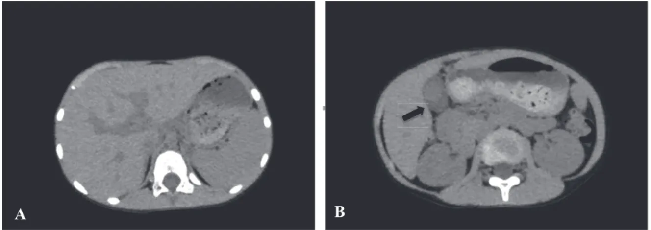
dl ; aspartate aminotransferase (AST), 184 U/l ; alanine aminotransferase (ALT), 139 U/l ; lactate dehydrogenase (LDH), 559 U/l ; total bilirubin, 0.4 mg/dl ; direct bilirubin, 0.1 mg/dl ;γ-glutamyl transferase, 36 U/l ; alkaline phosphatase, 506 U/l ; and leucine aminopeptidase, 86 U/l. Ultrasonogra-phy (US) revealed marked thickening of the gall-bladder wall to 4.6 mm with a sonolucent layer (Fig. 1 A, B). Sludge and pericholecystic fluid was also evident. In addition, localized tenderness was found over the gallbladder, representing a positive sonographic Murphy’s sign. No stones or dilatation of the biliary tract were evident. Computed tomogra-phy suggested acute hepatitis or cholecystitis with collapse of the gallbladder, edematous gallbladder wall, and periportal collar sign ; edematous inflam-mation around the portal veins (Figure 2).
AAC was diagnosed based on the ultrasonography findings. A fat-restricted diet and peripheral infusion were initiated. IM was also suspected based on the clinical presentation with edematous eyelids, pharyn-gitis, lymphadenopathy, and liver injury. On hospi-tal day 3, the ratio of atypical lymphocytes among leukocytes was elevated to 8%. Titers of antibodies
to EBV using an indirect immunofluorescence assay (FA) were increased as follows : EBV-viral capsid antigen (VCA) immunoglobulin (Ig)M 1 : 10 ; and EBV-VCA-IgG 1 : 80. EBV-EBNA (FA) was equivo-cal due to non-specific reaction. Anti-cytomegalovi-rus (CMV) antibody levels by enzyme immunoas-say were also elevated as follows : CMV-IgM, 3.43 index (normal, 0-0.8) and CMV-IgG, 13.2 index (normal, 0-1.9). Abdominal pain and low-grade fe-ver gradually improved. On hospital day 8, abdomi-nal pain became mild and blood results improved as follows : WBC count, 11.9
!
103/μl ; AST, 46 U/l ; ALT, 55 U/l ; ALP, 600 U/l ; LDH, 477 U/l ; and CRP, 0.5 mg/dl. On repeated US, thickening of the gallbladder wall was markedly improved (Fig. 1 C. D). The patient was therefore discharged on day 8. At the 1 month follow-up, the patient appeared healthy and blood results had all normalized. Follow-up after 3 months revealed that EBV-IgG (FA) had significantly increased to 1 : 320 and positive results for EBNA were obtained from enzyme-linked im-munoassay, whereas CMV-IgM was 1.0 index and CMV-IgG had decreased to 11.7 index.
Figure 1. Ultrasonography on admission demonstrates sonolucent thickening of the entire circumference of the gallbladder wall, to 4.6 mm. (A) Horizontal view. B) Sagittal view. ) Thickening of the gall bladder wall appears on ultrasonography to have resolved by hospital day 8. (C) Horizontal view. D) Sagittal view.)
DISCUSSION
Acute cholecystitis is a rare disease in children. Most adult patients with cholecystitis show compli-cations of gallstones and require emergency sur-gery. In children, acute cholecystitis occurs in rela-tion to infecrela-tion rather than to gallstones. Reported associated pathogens include hepatitis A, Brucella, malaria, and Salmonella (2, 3). Some reports have described AAC following systemic diseases, includ-ing systemic lupus erythematosus, nephrotic syn-drome, and Kawasaki disease (4, 5). AAC associ-ated with EBV infection has also been reported, but as far as we know, only eight child patients, includ-ing the present case, have been described (6, 7).
US examination allowed definitive diagnosis of AAC. Our patient had been misdiagnosed with en-terocolitis until US was performed at our hospital. US of AAC showed thickening of the gallbladder wall (!3 mm), sludge, sonographic Murphy’s sign, and pericholecystic fluid (7). None of these features are specific for AAC, combinations of two or more and clinical symptoms are satisfactory for reaching the diagnosis of AAC (7). Gallbladder wall thicken-ing is also familiar in various nonbiliary disorders ; viral hepatitis without cholecystitis, peritonitis, liver cirrhosis, chronic heart failure, and chronic renal failure (8). The exact pathophysiologic mechanism leading to the gallbladder wall thickness in such conditions is uncertain, but it is likely due to venous occlusion of gallbladder wall by elevated portal ve-nous pressure, decreased intravascular osmotic pressure, or subserosal edema, rather than incom-plete gallbladder constriction (8).
The etiology of EBV-associated AAC is unknown.
Dehydration or direct virus invasion to the bile duct is postulated to be related to the pathogenesis (7). Patients with IM sometimes show loss of appetite due to sore throat or malaise. Dehydration may cause cholecystitis due to increased bile viscosity. Two cases of EBV-related AAC with Gilbert’s syn-drome were recently reported from Greece (9). In the present patient, UGTA gene analysis was not performed because jaundice did not appear.
Most children with EBV-associated AAC present with mild clinical symptoms and good prognosis, whereas adult EBV-associated AAC sometimes pro-gresses to serious conditions such as gallbladder perforation, pulmonary edema, or hepatic failure (10).
Co-infection with EBV and CMV was considered due to the positive IgM for both in the acute phase, but follow-up examinations eventually confirmed primary EBV infection, while CMV infection was ruled out because the CMV IgG titer showed no significant increase. Regrettably, we did not perform CMV-DNA analysis using polymerase chain reac-tion. A previous report described false-positive re-sults for CMV IgM during primary EBV infection (11). We thus suggest that EBNA might cross-react with CMV IgM, resulting in EBNA showing a non-specific reaction and CMV IgM showing a false-positive result.
In conclusion, US should be performed when a patient complains of right upper quadrant pain dur-ing the course of IM, in consideration of the possi-bility of AAC. It makes it possible to avoid unnec-essary examinations, treatments, and administration of antibacterial agents.
We declare that conflict of interest is none.
Figure 2. Plain computed tomography on admission suggests acute hepatitis or cholescystitis. A. Low density area is observed around the portal veins. (Periportal collar sign)
B. Edematous swelling of gallbladder wall was shown by arrow. Low echoic lesions surrounded high echoic lesions in gallbladder.
K. Suga, et al. acalculous cholecystitis by EBV infection
REFERENCES
1. Jenson HB. Epstein Barr virus. Nelson text-book of pediatrics. 16 th edition chapter 247, p977-981.Behman RE, Kiegman RM, Jenson HB (eds)
2. Bouyahia O, Khelifi I, Bouafif F, Mazigh Mrad S, Gharsallah L, Boukthir S, Sammoud EI, Gharbi A : Hepatitis A : a rare cause of acute acalculous cholecystitis in children. Med Mal Infect 38(1) : 34-5, 2008
3. Garel L, Lucaya J, Piqueras J : Acute acalculous cholecystitis owing to salmonella sepsis.. Pedi-atr Radiol 33(12) : 905-6, 2003
!
4. Mendonca JA, Marques-Neto JF, Prando P, Appenzeller S : Acute acalculous cholecystitis in juvenile systemic lupus erythematosus. Lupus 18(6) : 561-3, 2009
5. Chen CJ, Huang FC, Tiao MM, Huang YH, Lin LY, Yu HR, Yang KD, Huang YC, Chen CC, Chang WC, Kuo HC : Sonographic gallbladder abnormality is associated with intravenous im-munoglobulin resistance in Kawasaki disease. Scientific World Journal. 2012 ; 2012 : 485758. 6. Iaria C, Arena L, Di Maio G, Fracassi MG,
Leonardi MS, Famulari C, Cascio A : Acute acal-culous cholecystitis as the initial presentation
of primary Epstein-Barr virus infection : a new case and a review of literature. Int J Infect Dis 12(4) : 391-5, 2008
7. Imamoglu M, Sarihan H, Sari A, Ahmetoglu A : Acute acalculous cholecystitis in children ; Diag-nosis and treatment. J Pediatr Surg 37(1) : 36-9, 2002
8. van Breda Vriesman AC, Engelbrecht MR, Smithuis RH, Puylaert JB : Diffuse Gallbladder Wall Thickening : Differential Diagnosis. Am J Roentenol 188 : 495-501, 2007
9. Attilakos A, Prassouli A, Hadjigeorgiou G,
Lagona E, Kitsiou-Tzeli S, Galla A,
Stasinopoulou A, Karpathios T : Acute acalcu-lous cholecystitis in children with Epstein-Barr virus infection : a role for Gilbert’s syndrome? Int J Infect Dis 13(4) : e161-4, 2009
10. O’Donovan N, Fitzgerald E : Gallbladder wall thickening in infectious mono nucleosis : an ominous sign. Postgrad Med J 72 : 299-300, 1996
11. Park JM, Shin JI, Lee JS, Jang YH, Kim SH, Lee KH, Lee CH : False positive immunoglobu-lin M antibody to cytomegalovirus in child with infectious mononucleosis by Epstein-Barr virus infection. Yonsei Med J 31 ; 50(5) : 713-6, 2009