ORIGINAL INVESTIGATION
Impact of individual metabolic risk
components or its clustering on endothelial
and smooth muscle cell function in men
Michio Shimabukuro
1*, Namio Higa
2, Hiroaki Masuzaki
3, Masataka Sata
4and Shinichiro Ueda
5Abstract
Background: Impaired vasoreactivity is often observed in subjects with metabolic syndrome, a condition that
includes the presence of a specific cluster of risk factors for obesity and cardiovascular disease. However, hierarchi-cal causes in the impaired vasoreactivity have not been clarified. We evaluated the impact of individual metabolic risk components or its clustering under the condition of insulin resistance on endothelial and smooth muscle cell function.
Methods: Vascular reactivity to acetylcholine (Ach), with or without nitric oxide synthase (NOS) inhibitor NG
-mon-omethyl-l-arginine (L-NMMA), or sodium nitroprusside (SNP) by forearm venous occlusion plethysmography and
insulin sensitivity index (M mg/kg/min) in euglycemic clamp were measured in men without (n = 18, control group) or with (n = 19, metabolic syndrome group) metabolic syndrome.
Results: (1) Ach-induced maximal forearm blood flow (maxFBF) was impaired in subjects with metabolic syndrome.
In particular, the dependent component of Ach-induced maxFBF was selectively decreased, while the NOS-independent component remained relatively unchanged. (2) Ach-induced maxFBF and ∆Ach-induced maxFBF with L-NMMA were correlated with waist circumference, glucose, and triglycerides, and most strongly correlated with vis-ceral fat area, adiponectin, and M. (3) Multivariate regression analysis indicated that individual metabolic risk compo-nents explained Ach-induced maxFBF by 4–21 %. Clustering of all metabolic risk compocompo-nents increased this to 35 %, and the presence of metabolic syndrome explained 30 %, indicating that defining metabolic syndrome can effectively predict impairment of endothelial dysfunction.
Conclusions: Endothelial dysfunction was correlated with individual metabolic risk components, but more strongly
with clustering of the components under a condition with low insulin sensitivity. We suggest that in subjects with metabolic syndrome, endothelial function is impaired by multiple cardiovascular risk factors exclusively when under the condition of insulin insensitivity and also that defining metabolic syndrome can effectively predict impairment of endothelial dysfunction.
Keywords: Obesity, Metabolic syndrome, Endothelial function, Insulin resistance
© 2016 The Author(s). This article is distributed under the terms of the Creative Commons Attribution 4.0 International License (http://creativecommons.org/licenses/by/4.0/), which permits unrestricted use, distribution, and reproduction in any medium, provided you give appropriate credit to the original author(s) and the source, provide a link to the Creative Commons license, and indicate if changes were made. The Creative Commons Public Domain Dedication waiver (http://creativecommons.org/ publicdomain/zero/1.0/) applies to the data made available in this article, unless otherwise stated.
Background
Metabolic syndrome is a condition that includes the pres-ence of a specific cluster of risk factors for obesity and cardiovascular disease, including abdominal obesity, high
blood pressure, impaired fasting blood glucose, hypertri-glyceridemia and low HDL cholesterol [1]. Of note, the clinical utility of metabolic syndrome has been ques-tioned [2, 3], because different definitions and different clusterings of components of metabolic syndrome may result in variations in cardiovascular risk predictions [4].
Impairment of vasoreactivity has been observed in patients with traditional coronary risk factors, even in the absence of morphological atherosclerotic lesions [5].
Open Access
*Correspondence: mshimabukuro-ur@umin.ac.jp
1 Department of Cardio-Diabetes Medicine, Institute of Biomedical
Sciences, Tokushima University Graduate School, 3-18-15 Kuramoto, Tokushima 770-8503, Japan
Accordingly, the assessment of vasoreactivity can provide pivotal information as a diagnostic and prognostic tool in patients at risk for atherosclerotic cardiovascular disease [6, 7]. The vasoreactivity is often impaired in subjects with metabolic syndrome [8, 9]. Hence, the impaired vas-oreactivity in this syndrome can be provoked by two sce-narios [1]: (1) individual metabolic risk components such as high blood pressure, hyperglycemia, and dyslipidemia, or (2) its clustering under the condition of obesity and/ or insulin resistance. However, the principal scenario for the vascular dysfunction in metabolic syndrome remains obscure.
The current study compared the impact of individ-ual metabolic risk components and its clustering under the condition of obesity and/or insulin resistance on endothelial and smooth muscle cell function in subjects with metabolic syndrome.
Methods Subjects
Male subjects were divided into either the group with-out metabolic syndrome (n = 18, control group) or with metabolic syndrome (n = 19, MS group). A sub-ject was defined as having metabolic syndrome as per the guidelines outlined in the IDF consensus statement [10]. Therefore, the subject had metabolic syndrome if he was obese (according to the Japanese criteria, hav-ing a waist circumference ≥85 cm in men) and had any two of the following four factors: (1) hypertriglyc-eridemia [serum triglyceride concentration ≥150 mg/ dL (1.69 mmol/L)], (2) a low HDL cholesterol level [serum HDL cholesterol concentration of 40 mg/dL (1.04 mmol/L)], (3) an elevated blood pressure (sys-tolic blood pressure ≥130 mmHg and/or dias(sys-tolic blood pressure ≥85 mmHg) or was taking anti-hyper-tensive drugs, and (4) a high fasting plasma glucose level [fasting plasma glucose concentration ≥100 mg/ dL (5.6 mmol/L)]. Participants who were taking insulin regimen or oral anti-diabetic drugs were excluded. Waist circumference was measured in the standing position and subcutaneous fat area (SFA) and intra-abdominal visceral fat area (VFA) were determined at the level of the umbilicus using a standardized method involving computed tomography [11]. Subjects were instructed to refrain from vigorous exercise, anti-hypertensive drugs, anti-hyperlipidemic drugs, non-steroidal anti-inflamma-tory drugs, alcohol, smoking and caffeine for 24 h prior to the study day. The study protocol was approved by the Ethical Committee of the University of the Ryukyus, and obeyed to the standards set by the Declaration of Hel-sinki. Written informed consent was obtained from all subjects.
Biochemical measurements
Venous blood samples were obtained in tubes contain-ing EDTA-sodium (1 mg/mL) and in polystyrene tubes without an anticoagulant. The EDTA-containing tubes were promptly chilled. Plasma was immediately separated by centrifugation at 3000 rpm and 4 °C for 10 min, and serum isolated by centrifugation at 1000 rpm at room temperature for 10 min. Samples were stored at −80 °C until they were assayed. Routine chemical methods were used to determine the serum concentrations of total cho-lesterol, HDL chocho-lesterol, triglycerides, creatinine, glu-cose, and electrolytes. The serum concentration of LDL cholesterol was estimated using Friedewald’s method [12].
Euglycemic hyperinsulinemic clamp
The whole-body insulin sensitivity index (M) was measured using a hyperinsulinemic euglycemic clamp [13] with mod-ifications [14] for 180 min. A primed continuous infusion of insulin (10.8 pmol kg−1 min−1, Novo-Nordisk, Japan)
was administered along with a variable rate infusion of 20 % dextrose (Baxter Health Care, Japan) that was adjusted manually to maintain serum glucose of 5.2 mmol/L. This was determined based on arterialized samples withdrawn every 5 min from an ipsilateral right dorsal hand vein (heated-air blanket was kept at 55 °C). The M value (mg/ kg/min) was calculated during the last 30 min of the study.
Vascular reactivity
Forearm blood flow (FBF) was measured simultaneously in both forearms by bilateral venous occlusion plethysmogra-phy with mercury-in-silicone elastomer strain gauges, as described [14]. All subjects were supine in a quiet, air-con-ditioned room, with both forearms resting slightly above their heart level. Acetylcholine (Ach, 0–400 nmol/min) with or without NG-monomethyl-l-arginine (L-NMMA,
8 µmol/min), a nitric oxide synthase (NOS) inhibitor, or sodium nitroprusside (SNP, 0–30 nmol/min) was infused into a 27-gauge catheter inserted into the brachial artery of the non-dominant arm by volumetric precision pumps. The infusion rate was maintained at 1 mL/min through-out the study unless otherwise indicated. All FBF data were obtained via a Mac Lab Version 4 chart recorder (AD Instruments, Hamstead, London, United Kingdom).
Statistical analysis
Values are expressed as the mean ± SD. Comparisons of vascular responses were analyzed by two-way analysis of variance (ANOVA) for repeated measures on one fac-tor, followed by Holm–Sidak multiple comparison test to compare group means. Simple regression analysis was used to identify significant linear associations of vascular responses with components of the metabolic syndrome
and related variables. Multiple regression analysis for vascular reactivity was performed in standard sequential (hierarchical) models and in a stepwise backward model using variables of individual components in the definition of metabolic syndrome and markers for possible underly-ing mechanisms. All analyses were performed usunderly-ing Jump version 12.1.0 software (SAS Institute Inc., Cary, NC). A
P value of <0.05 was considered statistically significant. Results
General characteristics
Subject characteristics are summarized in Table 1. All subjects with or without metabolic syndrome completed the study. Subjects in the MS group were clinically obese, and displayed a higher body weight, BMI, and waist cir-cumference than subjects without metabolic syndrome. The MS group also had greater SFA and VFA. There were no significant differences in age, heart rate, or smoking status (control 20 %, MS 16 %) between the two groups. The MS group exhibited higher levels of fasting glucose, insulin, HbA1c, total and LDL cholesterol, and
triglycer-ides and lower levels of HDL cholesterol than the control group. The MS group exhibited lower adiponectin than the control group (p < 0.0001). In addition, M values were reduced by half in the MS group.
Vascular reactivity
Vascular responsiveness to Ach
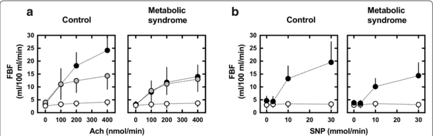
Basal FBF was 3.4 ± 1.3 mL/min/100 mL in the control group and 3.2 ± 1.2 mL/min/100 mL in the MS group
(p = 0.663). As shown in Fig. 1a, Ach-induced maximal FBF (Ach-induced maxFBF) (•) was reduced in the MS group (14.0 ± 4.5 mL/min/100 mL) compared to the con-trol group (24.2 ± 8.0 mL/min/100 mL, p < 0.0001).
NOS‑dependent vasodilation
Ach-induced maximal FBF during co-infusion of L-NMMA was reduced to 14.3 ± 5.2 mL/min/100 mL in the control group, but was not reduced in the MS group (13.0 ± 4.8 mL/min/100 mL) (Fig. 1a). The decline in Ach-induced maximal FBF by L-NMMA (∆maxFBF by L-NMMA), which represents the NOS-dependent vaso-dilation, was 10.2 ± 6.7 and 2.1 ± 2.4 mL/min/100 mL, respectively (p < 0.0001).
Smooth muscle responsiveness to SNP
As shown in Fig. 1b, SNP-induced maximal FBF (SNP-induced maxFBF) was also reduced in the MS group compared to control group: the SNP-induced maxFBF was 19.5 ± 8.0 mL/min/100 mL in the control group and 14.4 ± 5.0 mL/min/100 mL in the MS group (p = 0.021).
Vascular reactivity and components of metabolic syndrome
To explore the contributions of individual metabolic risk components to altered vascular control, the relationships between the components and endothelial and vascular smooth muscle responses were assessed. Significant rela-tionships were observed between Ach-induced maxFBF and waist circumference, glucose, and triglycerides, such that higher levels of these components were associated with lower vasodilation (Fig. 2). SNP-induced maxFBF was correlated only with glucose. In addition, ∆maxFBF by L-NMMA was correlated with SBP, glucose and triglycerides.
Vascular reactivity, abdominal fat distribution, adiponectin, and insulin sensitivity
Next, we evaluated the impact of VFA, SFA, adiponec-tin, and M value, the markers for possible underly-ing mechanisms, on Ach-induced maxFBF (Fig. 3). A strong negative relationship was observed between Ach-induced maxFBF and VFA, but not SFA. Ach-Ach-induced maxFBF was positively correlated with adiponectin, and most strongly correlated with M value. SNP-induced maximal FBF was not correlated with VFA, SFA, or adi-ponectin, but was positively correlated with M value. The ∆maxFBF by L-NMMA was correlated with SBP, glucose and triglycerides. In addition to Ach-induced maxFBF, ∆maxFBF by L-NMMA was negatively correlated with VFA, and positively correlated with adiponectin and M value.
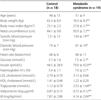
Table 1 General characteristics of the studied patients
Mean ± SD, * p < 0.05, ** p < 0.01 vs control
Control
(n = 18) Metabolic syndrome (n = 19)
Age (years) 48 ± 15 51 ± 9
Body weight (kg) 65.3 ± 8.4 76.4 ± 8.3** Body mass index (kg/m2) 23.1 ± 2.3 27.8 ± 2.7**
Waist circumference (cm) 84.1 ± 9.8 93.9 ± 7.2** Systolic blood pressure
(mm Hg) 121 ± 13 134 ± 19**
Diastolic blood pressure
(mm Hg) 74 ± 7 81 ± 10*
Heart rate (beats/min) 68 ± 8 68 ± 11 Glucose (mmol/L) 5.7 ± 1.6 7.5 ± 2.7* Insulin (pmol/L) 44.5 ± 28.9 79.0 ± 42.0** Haemoglobin A1c (%) 5.4 ± 1.1 7.0 ± 2.1* LDL cholesterol (mmol/L) 2.79 ± 0.70 3.13 ± 0.66 HDL cholesterol (mmol/L) 1.41 ± 0.48 1.22 ± 0.20 Triglyceride (mmol/L) 1.12 ± 0.70 2.53 ± 1.04** Adiponectin (log µg/ml) 0.87 ± 0.15 0.72 ± 0.12** M (mg/kg/min) 7.81 ± 2.86 4.14 ± 2.04**
Multivariate regression analysis of vascular reactivity determinants
Next, we determined the impact of individual meta-bolic risk components or the clustering of components on vascular reactivity by multivariate regression models (Table 2).
For Ach-induced maxFBF, waist circumference (model 1) and triglycerides, but not blood pressure, glu-cose or HDL cholesterol (data not shown), were deter-minants of vascular reactivity, even after correcting for age and smoking status. Clustering of 5 components increased the corrected R2 (model 2), and addition
of M value to the clustering of 5 components further increased the corrected R2, which reached 0.514 (model
3). Meanwhile, the presence of metabolic syndrome, though slightly decreased compared to the clustering of 5 components, was also a determinant of Ach-induced maxFBF (model 4); addition of M value to the pres-ence of metabolic syndrome increased the corrected R2
(model 5).
For SNP FBF, waist circumference (model 1), blood pressure, plasma glucose, triglycerides, and HDL choles-terol (data not shown), were not determinants of vascular reactivity. Clustering of these components with or with-out M value (models 2 and 3), and the presence of meta-bolic syndrome with or without M value (models 4 and 5) were also not determinants of vascular reactivity.
For ∆maxFBF by L-NMMA, waist circumference (model 1) and triglycerides, but not blood pressure, glu-cose or HDL cholesterol (data not shown), were determi-nants of vascular reactivity. Clustering of 5 components and the presence of metabolic syndrome increased the corrected R2 value (models 2 and 3), and addition of M
value further increased the corrected R2 value (models 4
and 5).
In the stepwise backward model (model 6) including the above individual components, defining metabolic syndrome and M value were significant predictors of Ach-induced maxFBF, M value for SNP-induced maxFBF, and defining metabolic syndrome for ∆maxFBF by L-NMMA.
Vascular reactivity and the number of components of metabolic syndrome
Finally, we evaluated the relationship between vascu-lar reactivity and the number of metabolic risk compo-nents in subjects with or without visceral obesity (Fig. 4). In subjects without visceral obesity (waist circumfer-ence <85 cm), subjects with 1 or ≥2 of 4 components, which included a high level of fasting glucose, elevated blood pressure, hypertriglyceridemia, and a low level of HDL cholesterol, did not show a statistical difference in Ach-induced maxFBF, SNP and ∆maxFBF by L-NMMA, compared to the group with 0 component. There were no significant differences in VFA, adiponectin and M value among the subgroups with 0, 1 and ≥2 compo-nents. In subjects with visceral obesity (waist circumfer-ence ≥85 cm), Ach-induced maxFBF was not decreased in the group with 1 component compared to the group with 0 component, but was decreased in the group with ≥2 components. SNP was not different among the subgroups with 0, 1 and ≥2 components. The ∆maxFBF by L-NMMA was decreased in accordance with the num-ber of the components. In the group with ≥3 compo-nents, there were no significant differences in VFA and adiponectin, but M value was significantly decreased.
Fig. 1 Forearm vascular reactivity to a ACh, with or without the nitric oxide synthase (NOS) inhibitor L-NMMA (8 μmol/min), or b SNP, as measured
by bilateral venous occlusion plethysmography in subjects without (n = 18) or with (n = 19) metabolic syndrome. FBF was measured simultane-ously in subjects infused with Ach (closed circles), SNP (closed circles) or Ach plus L-NMMA (gray circles) and non-infused arms (open circles) using bilateral venous occlusion plethysmography. FBF forearm blood flow, ACh acetylcholine, L-NMMA NG-monomethyl-l-arginine, SNP sodium
Discussion
The new findings of this study are: (1) Ach-induced and SNP-induced maximal FBFs were impaired in sub-jects with metabolic syndrome. In particular, we first found that the NOS-dependent component of Ach-induced maxFBF was selectively decreased, while the NOS-independent component remained unchanged. (2) Ach-induced maximal FBF and ∆Ach-induced maximal FBF by L-NMMA, and SNP-induced maxi-mal FBF were correlated strongly with M value than with individual metabolic components. (3) Multivari-ate regression models clearly indicMultivari-ated that defining metabolic syndrome, as compared to individual meta-bolic components, predicts impairment of endothelial dysfunction.
Vascular reactivity
As observed in previous studies of obese subjects [15,
16], Ach-induced maxFBF, a marker of endothelial func-tion, was reduced in MS group. We further investigated vascular NO bioavailability by comparing the dose– response curves of Ach with and without pharmaco-logical NOS inhibition. In the NO clamp technique, the NOS-dependent and NOS-independent components
of FBF can be accurately determined [17]. Co-infusion of L-NMMA and Ach caused a decrease in the level of inhibition of Ach-induced maxFBF in subjects with metabolic syndrome, and equalized the Ach-induced maximum FBF during NOS inhibition between the two groups. This implies that the NOS-dependent compo-nent of Ach-induced maxFBF was selectively decreased in subjects with metabolic syndrome, while the NOS-independent component remained relatively unchanged.
Classically, it has been believed that endothelial func-tion is impaired, even in the first step of atherosclerosis, but smooth muscle cell function is preserved even in the advanced stages of atherosclerosis [5]. Conversely, cur-rent study showed that SNP-induced maximal FBF, a marker of smooth muscle responsiveness, was reduced in the MS group. The data obtained during NO inhibi-tion by L-NMMA, suggest that the whole difference in Ach reactivity between two groups can be mainly due to a defect in the NOS-dependent NO synthesis. How-ever, the impaired smooth muscle vasorelaxation sug-gests an impaired bioavailability and/or responsiveness to endogenous NO [18]. Given that the response to Ach was superimposable at the levels in NO synthesis and/ or its biological activities, the contradictory findings may
Fig. 2 Simple regression analysis between vascular reactivity and components of metabolic syndrome in men without (n = 18, open circles) or
with (n = 19, gray circles) metabolic syndrome. FBF at an Ach infusion of 400 nmol/min, FBF at an SNP infusion of 30 nmol/min, and ∆Ach-induced maximal FBF by co-infusion of L-NMMA at 8 μmol/min. Linear regression analysis was done, and r and p values are shown
be partially explained by a converse compensation for the loss in NO bioavailability [18]. Endothelium-derived hyperpolarizing factor (EDHF) pathway, in which altera-tions was reported under pathological condialtera-tions, might be one such candidate [19]. Fernandes et al. reported that time-to-peak after hyperemia rather than flow mediated dilation (FMD) distinguished metabolic syn-drome from healthy controls [20]. In the time-course analysis of FMD [21], time to peak after hyperemia is not influenced by L-NMMA inhibition, suggesting that other factors, such as differences in vascular compliance and transduction independently of the NOS pathway [20, 22].
Vascular reactivity, abdominal fat distribution, adiponectin and insulin sensitivity
In the current study, Ach-induced maximal FBF, and ∆Ach-induced maximal FBF by L-NMMA were
correlated with waist circumference, glucose and tri-glycerides, and more strongly with M value. This result agrees with a previous study [15] where Steinberg et al. report that obesity/insulin resistance is associ-ated with both blunted endothelium-dependent vaso-dilation and failure of euglycemic hyperinsulinemia to augment endothelium-dependent vasodilation. This suggests that obese/insulin-resistant subjects are char-acterized by endothelial dysfunction and endothe-lial resistance to insulin’s effect on enhancement of endothelium-dependent vasodilation. Our results fur-ther indicated that each of abdominal fat distribution, adiponectin, and insulin sensitivity was correlated with Ach-induced maximal FBF and ∆Ach-induced maxi-mal FBF by L-NMMA more greatly than each of the MS components. Therefore, endothelial function in metabolic syndrome can be explained mostly by mutu-ally dependent insulin resistance, visceral obesity and
Fig. 3 Simple regression analysis between vascular reactivity, abdominal fat distribution, adiponectin and insulin sensitivity index (M) in men
without (n = 18, open circles) or with (n = 19, gray circles) metabolic syndrome. FBF at an Ach infusion of 400 nmol/min, FBF at an SNP infusion of 30 nmol/min, and ∆Ach-induced maximal FBF by co-infusion of L-NMMA at 8 μmol/min. Pearson’s correlation coefficients and p values are shown
hypoadiponectinemia through increased production of reactive oxidative species (ROS) and proinflammatory cytokines [11, 23–29].
Impairment in the SNP-induced maximal FBF was also correlated with M value as well as in Ach-induced
maximal FBF. The observation may be supported by Schinzari et al. showing that the vascular responsiveness to both Ach and SNP was not enhanced during hyperin-sulinemia in patients with metabolic syndrome [30]. They suggests that insulin’s facilitator action on the vasodilator
Table 2 Multivariate regression analysis fo vascular reactivity
a Model 1–5: standard multiple regression analysis b Model 6: stepwise multiple regression analysis
Model 1a Model 2a Model 3a Model 4a Model 5a Model 6b
Ach-induced maximal forearm blood flow (FBF)
Corrected R2 0.228 0.347 0.514 0.297 0.429 0.522
P value 0.009 0.006 0.001 0.002 0.000 0.000
Variables E P E P E P E P E P E P
Age (years) −0.153 0.156 −0.018 0.868 0.085 0.427 −0.109 0.288 0.016 0.883 0 0.712 Current smoking (yes or no) 1.246 0.336 0.532 0.665 −0.770 0.517 0.188 0.876 −0.991 0.419 0 0.562 Waist circumference (cm) ≥85 cm (yes or no) 4.749 0.002 3.387 0.024 2.137 0.116 0 0.134 SBP ≥130 or DBP ≥85 mmHg or use of
antihyper-tensive drugs 1.931 0.146 2.613 0.046 0 0.326
Glucose ≥100 mg/dL (yes or no) 1.947 0.131 −0.374 0.771 0 0.184
Triglycerides ≥150 mg/dL (yes or no) 2.230 0.095 1.305 0.298 0 0.490
HDL-cholesterol ≥150 mg/dL (yes or no) 1.065 0.456 1.493 0.286 0 0.965
Metabolic syndrome (yes or no) 4.675 0.000 −3.120 0.022 −3.560 0.006
M (mg/kg/min) 1.424 0.005 1.258 0.012 1.186 0.005
SNP-induced maximal forearm blood flow (FBF)
Corrected R2 0.123 0.117 0.148 0.173 0.224 0.254
P value 0.063 0.159 0.152 0.026 0.022 0.0017
Variables E P E P E P E P E P
Age (years) −0.233 0.020 −0.153 0.168 −0.114 0.346 −0.213 0.029 −0.151 0.162 0 0.240 Current smoking (yes or no) 1.239 0.293 0.875 0.474 0.229 0.864 0.848 0.448 0.232 0.848 0 0.906 Waist circumference (cm) ≥85 cm (yes or no) 1.669 0.202 1.477 0.304 0.507 0.735 0 0.818 SBP ≥130 or DBP ≥85 mmHg or use of
antihyper-tensive drugs 0.437 0.736 1.123 0.432 0 0.415
Glucose ≥100 mg/dL (yes or no) 2.355 0.069 0.649 0.655 0 0.438
Triglycerides ≥150 mg/dL (yes or no) −0.091 0.944 −0.810 0.563 0 0.726
HDL-cholesterol ≥150 mg/dL (yes or no) 0.792 0.576 1.492 0.343 0 0.519
Metabolic syndrome (yes or no) −2.120 0.061 −1.022 0.430 0 0.336
M (mg/kg/min) 0.872 0.107 0.784 0.103 1.237 0.002
∆Ach-induced maximal forearm blood flow (FBF) by L-NMMA
Corrected R2 0.236 0.460 0.594 0.406 0.533 0.481
P value 0.009 0.001 0.000 0.000 <0.0001 <0.0001
Variables E P E P E P E P E P E P
Age (years) −0.179 0.029 −0.071 0.344 −0.013 0.866 −0.145 0.045 0.074 0.415 0 0.094 Current smoking (yes or no) 0.635 0.512 −0.007 0.993 −0.726 0.397 −0.069 0.935 0.854 0.407 0 0.558 Waist circumference (cm) ≥85 cm (yes or no) 3.048 0.008 1.975 0.068 1.112 0.265 0 0.918 SBP ≥130 or DBP ≥85 mmHg or use of
antihyper-tensive drugs 1.874 0.049 2.161 0.028 0 0.216
Glucose ≥100 mg/dL (yes or no) 1.541 0.085 0.141 0.877 0 0.594
Triglycerides ≥150 mg/dL (yes or no) 1.731 0.073 1.403 0.131 0 0.360
HDL-cholesterol ≥150 mg/dL (yes or no) 1.673 0.100 1.856 0.075 0 0.774
Metabolic syndrome (yes or no) −3.627 0.000 0.950 0.008 −15.01 0.000
machinery was caused by endothelium unresponsiveness (a decrease in NO synthesis and/or a loss of its biologi-cal activities) and by defective sensitization of smooth muscle vasorelaxation (a loss in NO bioavailability) [19]. Aoqui et al. found distinct patterns of microvascular dys-function in metabolic syndrome, with augmented vaso-constriction present in the initial phase of metabolic syndrome independent of endothelial dysfunction [31]. Reportedly, locally produced ROS [25] and/or fat-derived ROS [23] can react with NO, generate peroxynitrite, and impair cyclic GMP-dependent vasodilatation; this mech-anism may partially explain these smooth muscle cell dysfunction [18, 31].
Impact of individual metabolic risk components and its clustering on endothelial and smooth muscle cell function
Since metabolic syndrome is a cluster of relatively het-erogeneous atherosclerotic risk factors, which may solely impair endothelial function, we evaluated the relation-ship between vascular reactivity and individual com-ponents or its clustering. In a simple regression model (Fig. 2), the Ach-induced maxFBF and ∆maxFBF by L-NMMA were lineally correlated with waist circumfer-ence, glucose and triglycerides. However, multivariate regression analysis indicated that individual components
explained only partially Ach-induced maxFBF by 4–21 %, ∆maxFBF by L-NMMA by 6–31 %, and SNP-induced maxFBF by 8–17 % (data not shown). In model 3, includ-ing the 5 components of visceral obesity, high fastinclud-ing glu-cose, elevated blood pressure, hypertriglyceridemia, and a low level of HDL cholesterol, increased the assumption to 35 % in Ach-induced maxFBF and 46 % in ∆maxFBF by L-NMMA, but only 16 % in SNP-induced maxFBF. These results support the notion that clustering of the compo-nents more greatly estimates endothelial dysfunction than individual components, but not smooth muscle cell function. Notably, the presence of metabolic syndrome, while still slightly less than the clustering of 5 compo-nents (model 2), explained 30 % of Ach-induced maxFBF and 41 % of ∆maxFBF by L-NMMA (model 4). These findings support the notion that defining metabolic syn-drome is effective in predicting endothelial dysfunction, which can subsequently predict future cardiovascular events [5, 7]. In the current study, type 2 diabetes mel-litus was prevalent in the MS group (12/18, 67 %) than in the control group (4/19, 21 %) (p < 0.05). When all patients divided into diabetic and non-diabetic groups, Ach-induced maxFBF (21.5 ± 1.7 vs 16.0 ± 2.0 mL/ min/100 mL, p = 0.040), ∆maxFBF by L-NMMA (8.3 ± 6.8 vs 2.8 ± 4.2 mL/min/100 mL, p = 0.008) and
Fig. 4 The relationship between vascular reactivity and number of components of metabolic syndrome in men without visceral obesity (n = 12,
waist circumference <85 cm) or with visceral obesity (n = 25, waist circumference ≥85 cm). Subjects were divided into groups with 0, 1 or 2 ≥of the following metabolic syndrome components: high level of fasting glucose, elevated blood pressure, hypertriglyceridemia, and low level of HDL cholesterol. Data are expressed as mean ± SD. Statistical analysis was done by two-way analysis of variance (ANOVA), followed by Holm–Sidak multiple comparison test to compare group means. P values = * <0.05, *** <0.001 vs. the 0 group
SNP-induced maxFBF (19.4 ± 1.4 vs 13.6 ± 1.7 mL/ min/100 mL, p = 0.011) were all impaired in diabetic groups. Vascular function in type 2 diabetes mellitus is also confounded by cardiovascular risk factors, such as obesity, hypertension and dyslipidemia, suggesting sig-nificance of cardiovascular risk clustering [32].
In our study, endothelial function is not strongly cor-related with individual metabolic risk components, but strongly with insulin resistance. Thus, it is assumed that recovery of vascular function can be obtained less effectively by improvement of individual metabolic parameters [33], but more effectively by improvement of metabolic syndrome or insulin resistance [34]. This notion may be supported by the fact in patients with metabolically healthy obesity (MHO), a medical condi-tion characterized by obesity which does not produce metabolic complications such as dyslipidemia, impaired glucose tolerance or hypertension [35]. The MHO had abnormal vascular reactivity, although their endothelial dysfunction was less pronounced than in patients with metabolic syndrome, indicating that obesity is associated with vascular damage independent of those metabolic abnormalities underlying metabolic syndrome [36].
We also evaluated the relationship between vascular reactivity and the number of metabolic syndrome com-ponents (Fig. 4). In subjects without visceral obesity, there were no differences in Ach-induced maxFBF and ∆maxFBF by L-NMMA among patients with 0, 1 and 2≥ of the 4 selected components. In contrast, in sub-jects with visceral obesity, Ach-induced maxFBF and ∆maxFBF by L-NMMA decreased in subjects with ≥2 components. As such, one or two components cannot be sufficient to cause impairment of endothelial function even in subjects with visceral obesity, but the clustering of ≥2 components can be sufficient. Interestingly, sub-jects without visceral obesity that have ≥2 components showed a subtler endothelial dysfunction than subjects with visceral obesity. This notion agrees with the previous study of Li et al., where they showed impaired endothelial function in subjects with metabolic syndrome as com-pared to individuals with a similar burden of traditional cardiovascular risk factors, but without metabolic syn-drome [37]. Their multivariate regression model found that after adjustment for covariates and 6 traditional car-diovascular risk factors, the presence of metabolic syn-drome had a significant and independent influence on endothelial function (p < 0.01).
In our multivariate model, addition of M value increased the corrected R2 for Ach-induced maxFBF to
51 % in the model including all the components (model 3) and to 43 % in the model including the presence of metabolic syndrome (model 5). Insulin resistance should play a pivotal role in causing endothelial dysfunction by
comorbidity of the metabolic syndrome components. We also found that endothelial function, indicated by Ach-induced maxFBF and ∆maxFBF by L-NMMA, was impaired in the group with ≥2 components, and M value decreased according to the number of components. Col-lectively, our data suggest that in subjects with visceral fat obesity, endothelial function is impaired by multiple car-diovascular risk factors exclusively when under the con-dition of insulin insensitivity.
Study limitations
First, we obtained data from a small number of sub-jects; therefore, there was the risk of type II errors. The strain gauge plethysmography and the euglycemic hyperinsulinemic clamp techniques, used in the study for measurement of vascular function and insulin sen-sitivity, are sensitive and solid, but the time-consuming and invasive characteristics limits number of partici-pants. In contrast, alternative simplified methods such as flow-mediated dilation (FMD) [38] and HOMA-IR [39] are good for recruiting participants, but the reli-ability is limited. Thus, current results should be con-firmed by combinations of multiple techniques and clinical studies with different size. Second, this cross-sectional study has shown only a correlation between vascular dysfunction and insulin resistance in subjects with metabolic syndrome, and has not indicated a cause-effect relationship. In future studies, we need to confirm that a therapeutic approach to improve insulin sensitivity by such as reductions in visceral fat obesity and ectopic fat deposition can recover vascular dys-function in metabolic syndrome. Third, since metabolic syndrome is a cluster of relatively heterogeneous ath-erosclerotic risk factors, which solely affect endothe-lial function, careful consideration should be taken to evaluate underlying mechanisms. Forth, we could not determine the molecular mechanisms by which insu-lin resistance occurs and impairs endothelial function from the current study.
Conclusions
The current study evaluated the impact of individual metabolic risk components or its clustering on endothe-lial and smooth muscle cell function in subjects with metabolic syndrome. The endothelial and smooth muscle cell function were correlated more strongly with cluster-ing of the components under a condition with low insulin sensitivity. Therefore, it may be suggested that in subjects with metabolic syndrome, vascular reactivity is impaired by multiple cardiovascular risk factors exclusively under the condition of insulin insensitivity and also that defin-ing metabolic syndrome can effectively predict impair-ment of vascular reactivity.
Authors’ contributions
MiS researched data and wrote the manuscript. NH researched data and reviewed the manuscript. HM, MaS and SU discussed data and reviewed the manuscript. All authors read and approved the final manuscript.
Author details
1 Department of Cardio-Diabetes Medicine, Institute of Biomedical
Sci-ences, Tokushima University Graduate School, 3-18-15 Kuramoto, Tokush-ima 770-8503, Japan. 2 Cardiovascular Department, Naha City Hospital,
Okinawa, Japan. 3 Division of Endocrinology, Diabetes and Metabolism,
Hema-tology, Rheumatology (Second Department of Internal Medicine), Graduate School of Medicine, University of the Ryukyus, Okinawa, Japan. 4 Department
of Cardiovascular Medicine, Institute of Biomedical Sciences, Tokushima University Graduate School, Tokushima, Japan. 5 Department of Clinical
Phar-macology and Therapeutics, Graduate School of Medicine, University of the Ryukyus, Okinawa, Japan.
Acknowledgements
This study was supported by Grants from the Ministry of Education, Culture, Sports, Science and Technology, Japan, #23591314, #24591338, #24591063. Competing interests
The authors declare that they have no competing interests. Received: 20 March 2016 Accepted: 7 May 2016
References
1. Grundy SM, Brewer HB Jr, Cleeman JI, Smith SC Jr, Lenfant C. Definition of metabolic syndrome: report of the National Heart, Lung, and Blood Insti-tute/American Heart Association conference on scientific issues related to definition. Circulation. 2004;109(3):433–8.
2. Kahn R, Buse J, Ferrannini E, Stern M. The metabolic syndrome: time for a critical appraisal: joint statement from the American Diabetes Association and the European Association for the Study of Diabetes. Diabetes Care. 2005;28(9):2289–304.
3. Simmons RK, Alberti KG, Gale EA, Colagiuri S, Tuomilehto J, Qiao Q, Ramachandran A, Tajima N, Brajkovich Mirchov I, Ben-Nakhi A, et al. The metabolic syndrome: useful concept or clinical tool? Report of a WHO Expert Consultation. Diabetologia. 2010;53(4):600–5.
4. Guize L, Thomas F, Pannier B, Bean K, Jego B, Benetos A. All-cause mortal-ity associated with specific combinations of the metabolic syndrome according to recent definitions. Diabetes Care. 2007;30(9):2381–7. 5. Deanfield JE, Halcox JP, Rabelink TJ. Endothelial function and dysfunction:
testing and clinical relevance. Circulation. 2007;115(10):1285–95. 6. Schachinger V, Britten MB, Zeiher AM. Prognostic impact of coronary
vasodilator dysfunction on adverse long-term outcome of coronary heart disease. Circulation. 2000;101(16):1899–906.
7. Perticone F, Ceravolo R, Pujia A, Ventura G, Iacopino S, Scozzafava A, Ferraro A, Chello M, Mastroroberto P, Verdecchia P, et al. Prognostic sig-nificance of endothelial dysfunction in hypertensive patients. Circulation. 2001;104(2):191–6.
8. Dell’Omo G, Penno G, Pucci L, Mariani M, Del Prato S, Pedrinelli R. Abnor-mal capillary permeability and endothelial dysfunction in hypertension with comorbid Metabolic Syndrome. Atherosclerosis. 2004;172(2):383–9. 9. Walther G, Obert P, Dutheil F, Chapier R, Lesourd B, Naughton G, Courteix
D, Vinet A. Metabolic syndrome individuals with and without type 2 dia-betes mellitus present generalized vascular dysfunction: cross-sectional study. Arterioscler Thromb Vasc Biol. 2015;35(4):1022–9.
10. Alberti KG, Zimmet P, Shaw J. Metabolic syndrome–a new world-wide definition. A consensus statement from the International Diabetes Fed-eration. Diabet Med. 2006;23(5):469–80.
11. Fujita K, Nishizawa H, Funahashi T, Shimomura I, Shimabukuro M. Sys-temic oxidative stress is associated with visceral fat accumulation and the metabolic syndrome. Circ J. 2006;70(11):1437–42.
12. Friedewald WT, Levy RI, Fredrickson DS. Estimation of the concentration of low-density lipoprotein cholesterol in plasma, without use of the preparative ultracentrifuge. Clin Chem. 1972;18(6):499–502.
13. Ueda S, Wada A, Umemura S. Methodological validity and feasibility of the nitric oxide clamp technique for nitric oxide research in human resist-ant vessels. Hypertens Res. 2004;27(5):351–7.
14. Shimabukuro M, Higa N, Tagawa T, Yamakawa K, Sata M, Ueda S. Defects of vascular nitric oxide bioavailability in subjects with impaired glucose tolerance: a potential link to insulin resistance. Int J Cardiol. 2013;167(1):298–300.
15. Steinberg HO, Chaker H, Leaming R, Johnson A, Brechtel G, Baron AD. Obesity/insulin resistance is associated with endothelial dysfunction. Implications for the syndrome of insulin resistance. J Clin Investig. 1996;97(11):2601–10.
16. Weil BR, Stauffer BL, Mestek ML, DeSouza CA. Influence of abdominal obesity on vascular endothelial function in overweight/obese adult men. Obesity (Silver Spring). 2011;19(9):1742–6.
17. Petrie JR, Ueda S, Morris AD, Murray LS, Elliott HL, Connell JM. How reproducible is bilateral forearm plethysmography? Br J Clin Pharmacol. 1998;45(2):131–9.
18. Evgenov OV, Pacher P, Schmidt PM, Hasko G, Schmidt HH, Stasch JP. NO-independent stimulators and activators of soluble guanylate cyclase: discovery and therapeutic potential. Nat Rev Drug Discovery. 2006;5(9):755–68.
19. Félétou M, Vanhoutte Paul M. EDHF: an update. Clin Sci. 2009;117(4):139–55.
20. Fernandes IA, Sales AR, Rocha NG, Silva BM, Vianna LC, da Nobrega AC. Preserved flow-mediated dilation but delayed time-to-peak diameter in individuals with metabolic syndrome. Clin Physiol Funct Imaging. 2014;34(4):270–6.
21. Weissgerber TL. Flow-mediated dilation: can new approaches provide greater mechanistic insight into vascular dysfunction in preeclampsia and other diseases? Curr Hypertens Rep. 2014;16(11):487.
22. Loh KP, Huang SH, Tan BK, Zhu YZ. Cerebral protection of purified Herba Leonuri extract on middle cerebral artery occluded rats. J Ethnopharma-col. 2009;125(2):337–43.
23. Furukawa S, Fujita T, Shimabukuro M, Iwaki M, Yamada Y, Nakajima Y, Nakayama O, Makishima M, Matsuda M, Shimomura I. Increased oxidative stress in obesity and its impact on metabolic syndrome. J Clin Investig. 2004;114(12):1752–61.
24. Shimabukuro M. Cardiac adiposity and global cardiometabolic risk: new concept and clinical implication. Circ J. 2009;73(1):27–34.
25. Chinen I, Shimabukuro M, Yamakawa K, Higa N, Matsuzaki T, Noguchi K, Ueda S, Sakanashi M, Takasu N. Vascular lipotoxicity: endothelial dysfunc-tion via fatty-acid-induced reactive oxygen species overproducdysfunc-tion in obese Zucker diabetic fatty rats. Endocrinology. 2007;148(1):160–5. 26. Fisman EZ, Tenenbaum A. Adiponectin: a manifold therapeutic target
for metabolic syndrome, diabetes, and coronary disease? Cardiovasc Diabetol. 2014;13:103.
27. Brown DI, Griendling KK. Regulation of signal transduction by reactive oxygen species in the cardiovascular system. Circ Res. 2015;116(3):531–49.
28. Libby P, Hansson GK. Inflammation and immunity in diseases of the arte-rial tree: players and layers. Circ Res. 2015;116(2):307–11.
29. Limberg JK, Harrell JW, Johansson RE, Eldridge MW, Proctor LT, Sebranek JJ, Schrage WG. Microvascular function in younger adults with obesity and metabolic syndrome: role of oxidative stress. Am J Physiol Heart Circ Physiol. 2013;305(8):H1230–7.
30. Schinzari F, Tesauro M, Rovella V, Galli A, Mores N, Porzio O, Lauro D, Cardillo C. Generalized impairment of vasodilator reactivity during hyper-insulinemia in patients with obesity-related metabolic syndrome. Am J Physiol Endocrinol Metab. 2010;299(6):E947–52.
31. Aoqui C, Chmielewski S, Scherer E, Eissler R, Sollinger D, Heid I, Braren R, Schmaderer C, Megens RT, Weber C, et al. Microvascular dysfunction in the course of metabolic syndrome induced by high-fat diet. Cardiovasc Diabetol. 2014;13:31.
32. Nevelsteen I, Van den Bergh A, Van der Mieren G, Vanderper A, Mubagwa K, Bult H, Herijgers P. NO-dependent endothelial dysfunction in type II diabetes is aggravated by dyslipidemia and hypertension, but can be restored by angiotensin-converting enzyme inhibition and weight loss. J Vasc Res. 2013;50(6):486–97.
33. Ohno Y, Miyoshi T, Noda Y, Oe H, Toh N, Nakamura K, Kohno K, Morita H, Ito H. Bezafibrate improves postprandial hypertriglyceridemia and
• We accept pre-submission inquiries
• Our selector tool helps you to find the most relevant journal
• We provide round the clock customer support
• Convenient online submission
• Thorough peer review
• Inclusion in PubMed and all major indexing services
• Maximum visibility for your research Submit your manuscript at
www.biomedcentral.com/submit
Submit your next manuscript to BioMed Central
and we will help you at every step:
associated endothelial dysfunction in patients with metabolic syndrome: a randomized crossover study. Cardiovasc Diabetol. 2014;13:71. 34. Kraemer-Aguiar LG, de Miranda ML, Bottino DA, Lima Rde A, de Souza
M, Balarini Mde M, Villela NR, Bouskela E. Increment of body mass index is positively correlated with worsening of endothelium-dependent and independent changes in forearm blood flow. Front Physiol. 2015;6:223. 35. Stefan N, Haring HU, Hu FB, Schulze MB. Metabolically healthy obesity: epidemiology, mechanisms, and clinical implications. Lancet Diabetes Endocrinol. 2013;1(2):152–62.
36. Schinzari F, Iantorno M, Campia U, Mores N, Rovella V, Tesauro M, Di Daniele N, Cardillo C. Vasodilator responses and endothelin-dependent vasoconstriction in metabolically healthy obesity and the metabolic syndrome. Am J Physiol Endocrinol Metab. 2015;309(9):E787–92.
37. Li J, Flammer AJ, Lennon RJ, Nelson RE, Gulati R, Friedman PA, Thomas RJ, Sandhu NP, Hua Q, Lerman LO, et al. Comparison of the effect of the metabolic syndrome and multiple traditional cardiovascular risk factors on vascular function. Mayo Clin Proc. 2012;87(10):968–75.
38. Tomiyama H, Kohro T, Higashi Y, Takase B, Suzuki T, Ishizu T, Ueda S, Yamazaki T, Furumoto T, Kario K, et al. Reliability of measurement of endothelial function across multiple institutions and establishment of reference values in Japanese. Atherosclerosis. 2015;242(2):433–42. 39. Matsuhisa M, Yamasaki Y, Emoto M, Shimabukuro M, Ueda S, Funahashi
T, Matsuzawa Y. A novel index of insulin resistance determined from the homeostasis model assessment index and adiponectin levels in Japanese subjects. Diabetes Res Clin Pract. 2007;77(1):151–4.