(Original)
Matsumoto Shigaku 13 1 329--336, 1987Key words : menomorphic adenoma-microcalcification-ultrastructure-glycosaminoglycans
Light and Electron Microscopic Studies of Microcalcifications
Appearing in Monomorphic Adenomas
of Microcalcifications
Adenomas
CHIHITO NAKAMURA TOSHIYUKI KAWAKAMI
HIROMASA HASEGAWA and SHIGEO EDA
DePartment of Oral Pathology, Matsumoto Dental College (Chief : Prof S. Eda)LaboratoTy
SHOJI AKAHANE
of Electron MicroscoPe, Matsumoto(Chief: S. Ahahane)
Dental College
TADASHI YAMAZAKI
Department of Oral and Maxillofacial Surgery, Shinshu University School of Medicine (Chief : Prof A. Kotani)Summary
Microcalcifications appearing in two cases of monomorphic adenomas were studied
histopathologically, electron microscopically, and electron-microanalytically. One case was
basal cell adenoma that occurred in a 56-year-old man and the other was canalicular
adenoma in a 71-year-old woman. The calcified granules were observed both in the lumina formed by the tumor cells and in the stromal tissues. The surroundings of the granules were
stained by alcian blue and showed a sulfur peak by EPMA. These facts suggest that the
surroundings contain sulfated glycosaminoglycans and that sulfur has a significant role in the mechanisms of pathological calcification as well as physiological calcification.
Presented in part at the 25th Annual Meeting of the Japanese Association for Oral Biology at Fukuoka held on Oct. 2, 1983, and at the 39th Annual Meeting of the Japanese Stomatological Society at Sendai held on May 24, 1985. This study was supported in part by a grant (No. 61570901) from the Ministry of Education, Culture and Science, Japan.
' Present address : Clinic of Oral and Maxillofacial Surgery, Komoro Kosei Hospital. (accepted for publicaton on November 1, 1987)
330 Nakamura, et al : Microcalcifications in Monomorphic Adenomas
Introduction
Microcalcifications are found in both non-neoplastic and neoplastic tissues. The former includes
meninges choroid plexus, pineal boby and thyroid, and the latter is most common in the
menin-giomas, papillary tumors of the thyroid and ovary"}. In addition, they can be seen in tumors derived from the other organs such as the breast, lung, kidney, endometrium, stomach, pituitary gland"), and
salivary gland6}. Although microcalcifications are most common in pleomorphic adenoma, the
calcification is extremely rare in other sarivary gland tumQrs7).
The purpose of the present article is to report the light and electron microscopic findings of the
microcalcifications appearing in monomorphic adenomas and to discuss the role of sulfur in the
mechanisms of pathological calcifications.
Materials and Methods
The specimens used in the present study were obtained from two patients clinically diagnosed
as salivary gland tumors.
Case 1:
A tumor, located in the right sublingual region and in 2.3Å~1.7Å~1.2 cm in size, was surgically
removed from a 56-year-old man. It was examined histopathologically and diagnosed as basal cell adenoma. The patient was discharged from an eventful postoperative course.i')
Case 2:
A globular tumor mass, 1.5 cm in diameter, was enucleated from the right floor of the mouth of a 71-year-old woman and was submitted for histopathological examination. The diagnosis of canalicular adenoma was rendered. The postoperative course was not eventful.
Both specimens, fixed in 10%o formalin, were processed for light microscopy by the following stainings: hematoxylin-eosin (H-E), van Gieson's, alcian blue, toluidine blue (pH 2.5), and von
Kossa's.
For transmission electron microscopy, after removing the paraffin, the remnant materials were washed with O.1 M cacodylate buffer, fixed in Karnovsky's solution, and embedded in Epon 812. The
specimens were not treated with any demineralization. To obtain good orientation and suitable specimens, 1"m thick sections were stained with toluidine blue and checked by light microscopy. Ultrathin sections were cut with a LKB ultratome equipped with a diamond knife (Diatome: Switzerland) and observed under a JEOL JEM 100-B electron microscope operating at 80KV. For scanning electron microscopy and electron probe microanalysis (EPMA), the specimens were processed by critical-point drying and coated with carbon by cathodic sputtering to be examined with a JCXA 733 Super Probe scanning electron microscope.
Results Light Microscopy :
Both tumor masses, covered by a thin fibrous tissue, were composed of cuboidal andlor columnar cells with rather large, deep bosophilic, round to ovoid nuclei. Scant cytoplasm was occasionally found. No mitotic figures were observed in any fields. In case 1, the tumor cells
generally formed solid• nests and partly 1uminal or cystic structures at the center of the nests (Fig. 1). On the other hand, the tumor cells of case 2 were closely packed and formed wide-spread cyst-like 1umina. In both cases, many lumina contained a bosophilic spherical shaped materials (Figs. 1, 2
if}Zgde\ 13(3} 1987 331
arrows). They were positive to von Kossa's stain (Figs. 3, 4 arrows), but their periphery was
partially negative to the stain. Intercellular stroma was scant and inconspicuous. In a part of the intercellular stroma, von Kossa's positive small granules (Figs. 3, 4), which could not be confirmed in the H-E stained sections, were also found (Figs. 3, 4). These areas were stained uniformly red by H-E, blue by Mallory's azan stain and by alcian blue stain (Fig. 5), red by van Gieson's.
Further-more, the areas showed metachromasia by toluidine blue (pH 2.5).
Transmission Electron Microscopy :
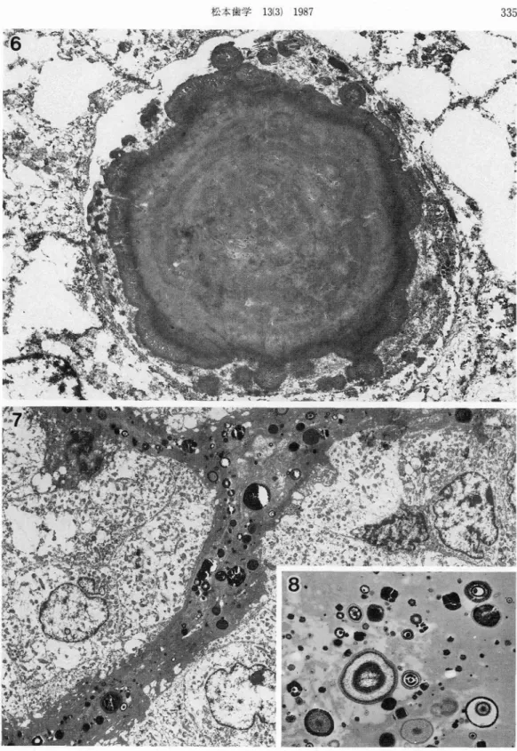
Ultrastructurally, the spherical shaped materials in the lumina of the both cases seemed to be
formed by the fusion of adjacent smaller globules and were sharply delimited, surrounded by
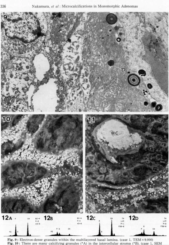
aggregates of electron-moderate fine globular and/or granular materiars (Fig. 6). The calcifications exhibited a relatively electron-lucent center and an electron-dense peripheral end was composed of fine fibriller or needle-like crystallites. No calcified cores could be observed in these structures. In case 1, the light microscopical von Kossa's positive granules in the intercellular stroma were O.2-3#m in size and distributed both in fibrous connective tissues (Figs. 7, 8) and in multilayered
basal laminae (Fig. 9). Many parts of these fibrous connective tissues were electron dense and
partially hyalinized. In case 2, the granules were also located in the relatively electron-dense intercellular stroma, but the distinction between the basal laminae and fibrous connective tissues were not clear.
The small granules were round or ovoid in shape, but a small number were cuboid (Fig. 8), and made of various forms of calcium deposits : amorphous form or crystalline forms of polyheadral or needle shapes, and some deposits formed concentric laminae. The relationship between the calcium
depositions and the membrane of vesicles could not be found.
Scanning Electron Microscopy and Electron Probe Microanalysis :
Under the scanning electron microscope (composition image), relatively large spherules in the 1umina and many small granules in the intercellular connective tissues of both cases were recog-nized as light structures (Figs. 10, 11). EPMA revealed that they mainly consisted of both calcium and phosphorus and that the sulfur peak existed in the contiguous tissues (Figs. 12A-D).
Discussion
Both basal cell adenoma and canalicular adenoma are relatively rare and belongs in a
classification of monomorphic adenoma. Although variety changes, including pathological
calcifica-tion, appear in pleomorphic adenoma (Fukushima 1968`), Hasegawa et al. 19875), Kameyama et al.
i9876)), these two tumors show very little changes in the stroma.
To the best of our knowledge, no research has been done concerning the microcalcifications in monomorphic adenoma. Therefore, both of our cases having calcification are thought to be
extreme-ly rare.
Recently, many facters regarding calcification have been taken up9) : collagen fiber, proteo-glycan (glycosaminoproteo-glycans), lipid, osteocalcin, and matrix vesicle. As the calcification proceeds,
some of them are thought to remain as the structural component in the calcifyjng matrix while others disappear. Chondroitin sulfate, a major constituent of proteoglycans and the most highly acidic substance, has been thought to play a role as calcified nuclei, because of their strong calcium-binding capacity9). Now, it seems more likely that these compounds act as inhibitors of
332 Nakamura, et al : Microcalcifications in Monomorphic Adenomas
calcification and have to be removed before mineralization can occuri-'3,iO•i2,i3•i5,i6).
Regarding the quantitative and qualitative changes of glycosaminoglycans in the calcifying process, many reports have shown the decrease of glycosaminoglycans (particulary sulfated
glycosaminoglycans) with the development of calcification in cartilagei-'3,'O•i2,'3,i5•'6). The fact that
the surroundings of the calcified masses of the intercellular stroma were stained by alcian-blue and metachromatic by toluidine-blue (pH2.5) in our study strongly suggests the existance of sulfated glycosaminoglycans within these areas. The presence of a sulfur peak in the contiguous tissues to the calcified masses may support this suggestion. These may also indicate a significant role of sulfur (chondroitin sulfate, that is sulfated glycosaminoglycans) in the mechanisms of pathological cal-cification as well as a physiological one.
Fukushima (1968)`) has reported the abnormal epithelial secretions in pleomorphic adenoma and adenoid cystic carcinoma. Recentry, Yamazaki, et al. (1987)i') reported a case of basal cel1 adenoma, which is the same as one of our cases (casel), and described the relationship between the abnormal epithelial secretions and the stromal calcifications. Judging from the fact that the cellular basement membrane was positive to PAS reaction, the origin of sulfur responsible for calcification is thought to be the tumor cell.
0n the other hand, a rnembrane of vesicles has been repeatedly demonstrated at the initial
calcification"8). Intracytoplasmic organellae, especially mitochondria, have been considered to be a focus of intracytoplasmic calcification in cartilage, bone, or aortic valveS), with consequent
dis-charge from the cytoplasm and conversion to a matrix vesicle. We were unable to find an obvious
membrane of vesicle despite careful observation. Similar findings, namely the calcification having
no relationship to the vesicles, were observed in duodenal carcinoid tumor by Murayama et al.
(1979)ii). Although there is no denying the matrix vesicle type in our cases because of poor fixation,
we postulate that some of the calcifications will be occurred without concerning membrane of
vesicles.
References
1) Bowness, J.M. and Jacobs, M. (1968) Chondroitin sulfate changes in puppy rib canilage during the
period of calcification. Can. J. Biochem. 46 : 63-67.
2 ) Campo, R. D. (1970) Protein-polysaccharides of cartilage and bone in health and disease. Clin. Orthoped. Rel. Res. 68 : 182-209.
3 ) Dearden, L. C. and Esoinosa, T. (1974) Comparison of Mineralization of the tibial epiphyseal plate in immature rats following treatment with cortisone, propylthiouracil or after fasting. Calcif. Tissue Res.
15 : 93-110.
4) Fukushima, M. (1968) An electron microscopic study of human salivary gland tumorscpleomorphic adenoma and adenoid cystic carcinoma= Bull. Tokyo med. dent. Univ. 15 : 387-408.
5) Hasegawa, H., Kawakami, T., Nakamura, C. and 'Eda, S. (1987) Ultrastructural study of varied calcified materials in the pleomorphic adenoma occurring in the soft palate. Matsumoto Shigaku 13 :
115-121.
6) Kameyama, Y., Sakaki, Y. and Yamada, N. (!987) A case of pleomorphic adenoma with calcified
masses. J. Kyushu Dent. Soc. 41 : 737-741.
7) Kawakami, T., Nakamura, C., Hasegawa, H., Eda, S., Komatsu, M., Furusawa, K. and Akahane, S.
(1986) Ultrastructure of stromal calcification in mucoepidermoid carcinoma. Jpn. J. oral Biol. 28 : 217
-222.
8) Kim, K. M. and Huang, S. (1971) Ultrastructural study of calcification of human aonic valve. Lab.
Invest. 25 : 357-366.
taJztsva"]itL" 13{3) 1987 333
10) Matukas, V. and Krikos, G. A. (1968) Evidence for changes in protein polysaccharide associated with
the onset of calcification in cartilage. J. Cell Biol. 39 : 43-48.
11) Murayama, H., Imai, T., Kikuchi, M.and Kamio, A. (1979) Duodenal carcinoid (apudoma) with
psammoma bodies. Cancer, 43 : 1411-1417.
12) Silbermann, M. and Frommer, J. (1974) Demonstration and distribution of acidic glycosaminoglycans
in mouse secondary cartilage. Histochem. 38:85-93. '
13) Smith, J. W. (1970) The disposition of proteinpolysaccharide in epiphyseal plate canilage of the young rabbit. J. Cell Sci. 6 : 843-864.
14) Takada, K., Yamamoto, Y., Maeyama, I., Nagatani, T. and Watanabe, T. (1974) An elemental analysis
of epiphyseal canilage. Clin. Orthop. Surg. 9 : 742-748.
15) Terashima, Y., Nogami, H., Hirose, J. and Iwata, H. (1976) Zonal analysis of glycosaminoglycans in epiphyseal bone-Biochemical and morphological observatfon in the human fetus and the newborn rat -. Connect. Tissue. 8 : 1-13.
16) Teshima, R. (1977) Studies on calcifiÅëation in normal and osteoarthrotic articular cartilage
structure and elemental analysis-. J. Jap. Orthop. Ass. 52 : 93-100.
17) Yamazaki, T. Kotani, A. and Kawakami, T. (1987) Basal cell adenoma of the sublingual gland. J. oral
,
334 Nakamura, et al ' Microcalcifications in Monomorphic Adenomas
"'//.,$S"k'maec11ge1eeagtw.$11pag,glsi1ee'keegeL///Xime,ge'\ee11ge'/ee'eeX
$,/3-/k-g,ew.•ige,.ab.egitpmtX'\sX"s/t
rc....'/i'pt"'l!'EllgX'di'ec#,eeee,,tsfi•ge•,•/Xl#"X.$t,smp.'tct•tee
.,tw--k.caff.2.;s.ge"."-esg-rw•,•tw-.tkSllh.X.,"$. ee.iigeillis!ee2tsttt'gedet.ksu- s,ebem':"INi:.1.X'ffts
}FEig.1: Tumor cells are arranged m a trabecular or solid pattern Basophilic spherical Shaped materials m the luminal structure (arrow). (case 1, H-E,Å~230)
Fig. 2 : Sphencal shaped mater;als Åqarrows) in some of the 1uminal spaces.(case 2, H-E,Å~150) FEig. 3 : Von Kossa's positive granules (arrowheads) in the intercellular stroma and spherical shaped
matenals m the lumina (arrows) (case 1,Å~150)
lffg. 4 : Spherical shaped materials in the lumina (arrows) are pes!tive to von Kossa's stam (ease
2, Å~ 150)
335
IFYg. 6 : Spherical shaped rnaterial in the 1umen formed by the fusion of adjacent smaller globules.
(case 2, TEM Å~6,500)
Fig. 7 : Electron-dense granules scattered in fibrous connective tissues. (case 1, TEM x3,100) Fig. 8 : The granules are round or ovoid in sliape, and shoving lamelated appearance or solid. (case
336 Nakamura, et al : Microcalcifications in Monomorphic Adenomas
12A,
"E s7es
pt;xs
Åqtr
CAee-A 12B
:Ifss
ps cptec''tv CA ee-A p-4 "-e-`.::d",ti-`..---.-ee
11' .l ,ttt"12D
P s cn Tg Lun P-2 F;G-e-A",.-L...
Fig. 9 : Electron-dense granules within the multilayered basal lainina. (case 1, TEMÅ~9.000) Fig. 10 : There are many calcifying granules ('A) in the intercellular stroma ('B). (case 1, SEM
Composition image Å~1,200)
Fig. 11 : Spherical shaped materials ('C) in the lumen and granules ('D) in the intercellular stroma
are showing calcification. (case 2, SEM Composition image Å~950)
Fig. 12 : Results of EPMA of points A, B (Fig. 10), C and D (Fig. 11). The surroundings of both the
granule and the spherical shaped material show a composition sulfur in addition to