INTRODUCTION
Breast-conserving therapy (BCT) has recently be-come a standard treatment for early breast cancer. The local recurrence rate is decreased using post-operative radiotherapy, which cures minute lesions remaining within the breast (1). In recent years, an increasing number of patients have received breast-conserving surgery. Even locally advanced cancers have received this treatment, following down-staging
with neo-adjuvant chemotherapy. The reported fre-quency of radiation pneumonitis (RP), caused by ra-diotherapy following breast-conserving surgery, var-ies greatly ; cumulative incidence rates of 0.9-85% have been reported (2-9). RP occurs locally, in a limited field of an irradiated lung, at an early time after irradiation(10). Among the pulmonary injuries following radiotherapy of whole breast, the most clinical significant pulmonary disorder is radiation-induced bronchiolitis obliterans organizing pneu-monia (BOOP) syndrome. Radiation-induced BOOP syndrome is characterized by infiltrative shadow expansions outside the irradiation field of lung and migrates ; however, the frequency of radiation- in-duced BOOP is low. Crestani described the follow-ing factors for diagnosfollow-ing radiation-induced BOOP
ORIGINAL
Risk factors for radiation pneumonitis caused by whole
breast irradiation following breast-conserving surgery
Akiko Kubo
1, Kyosuke Osaki
1, Takashi Kawanaka
1, Shunsuke Furutani
1,
Hitoshi Ikushima
2, and Hiromu Nishitani
1 1Department of Radiology and2
Department of Radiation Therapy Technology, Institute of Health Biosciences, the University of Tokushima Graduate School, Tokushima, Japan
Abstract : We evaluated risk factors of radiation pneumonitis (RP) after whole breast ir-radiation following breast-conserving surgery. Four hundred and seventy-two cases un-derwent whole breast irradiation with tangential field following breast-conserving sur-gery in our hospital, between January 2005 and April 2007. Of these cases, we performed statistical analyses for 423 breasts of 413 patients, using a pulmonary dose-volume histo-gram. Patient characteristics, treatment regimens and irradiation methods were included as variables in the analyses on risk factors of RP. As a result, 89 breasts of 84 cases (21%%) were diagnosed with RP. The version 3.0 of the NCI Common Terminology Criteria for Ad-verse Events was used to evaluate the grade of pneumonitis : 77 cases (18.2%%) were diag-nosed as Grade 1 RP, 10 cases (2.3%%) as Grade 2, and 2 cases (0.5%%) as Grade 3. Multivari-ate analysis indicMultivari-ated that the significant risk factors for RP were central lung distance (CLD) ( 1.8 cm) and the short axis length of the radiation field. The incidence of radiation-induced bronchiolitis obliterans organizing pneumonia (BOOP) syndrome significantly correlated only with CLD. The lung volume within the radiation field was shown to be a significant risk factor for RP and radiation-induced BOOP syndrome. J. Med. Invest. 56 : 99-110, August, 2009
Keywords : radiation pneumonitis, radiation-induced BOOP syndrome, breast cancer, breast-conserving therapy
Received for publication December 25, 2008 ; accepted January 26, 2009.
Address correspondence and reprint requests to Akiko Kubo, MD, Department of Radiology, Institute of Health Biosciences, the University of Tokushima Graduate School, Kuramoto - cho, Tokushima 770 - 8503, Japan and Fax +81 - 88 - 633 - 7174.
syndrome (11) : (1) radiotherapy to the breast within the past 12 months, (2) general and/or respiratory symptoms lasting for longer than 2 weeks, (3) ra-diographic lung infiltrations outside the radiation port and (4) no evidence of a specific cause.
Previous clinical studies investigating RP have re-ported on several factors involved in the pathogene-sis of RP, including patient age, lung volume within an irradiated field, chemotherapy before radiother-apy, prescription of tamoxifen during radiotherapy and smoking history (12-15). In the present study, we evaluated the risk factor for RP, including ra-diation-induced BOOP syndrome, induced by whole breast irradiation following breast-conserving sur-gery, with respect to patient background factors and treatment regimens.
MATERIALS AND METHODS
Between January 2005 and April 2007, 472 patient cases were subjected to tangential irradiation follow-ing breast-conservfollow-ing surgery at the Tokushima University Hospital. We conducted follow-up obser-vations for 454 of 472 patients with respect to the occurrence of pulmonary damage using chest radio-graphs. We excluded cases in which the chest wall had been irradiated after mastectomy, and in which irradiation on axilla and supraclavicular nodal re-gions had been simultaneously performed. We in-cluded only those cases in which whole breast tan-gential irradiation had been performed. In these cases, we performed statistical analyses for 423 breasts (413 cases) using a pulmonary dose-volume histogram (DVH). DVH was calculated using a three-dimensional treatment planning device (XiO, ver. 4.33.02, manufactured by CMS). Patient char-acteristics, treatment regimen and irradiation meth-ods were included as variables on the analyses of the risk factors for RP. In 10 cases, tangential irradia-tion was performed simultaneously for both breasts following breast-conserving surgery.
Patients
The patient background of 413 cases is shown in Tables 1 and 2. With respect to chemotherapy, neo-adjuvant chemotherapy was performed in 49 cases, pre-irradiation adjuvant chemotherapy in 5 cases and post-irradiation adjuvant chemotherapy in 100 cases. Of the 313 cases in which endocrine therapy was conducted, 130 concurrently received both endocrine therapy and radiotherapy, whereas
Table 1. Patient characteristics (n = 413)
Age (y), range (median) 24 - 84(52) Side (right/left/bilateral) 212/191/10 Collagen vascular disease (yes/no) 6/407 Allergy disease and /or drug allergy (yes/no) 73/340 Diabetes (yes/no) 12/401 Smoking status (yes/no/unknown) 64/283/76 Usage of other medication unrelated to breast
cancer (yes/no) 126/297 Clinical stage (UICC)
0 54 I 206 II A 101 II B 46 III A 9 III B 3 III C 1 Histologic type
Noninvasive ductal carcinoma 52 Invasive ductal carcinoma 328
other 43 Operation Lumpectomy 316 Quadrantectomy 105 other 2 Chemotherapy (yes/no) 143/283 pre - radiotherapy 54 post- radiotherapy 100 Endocrine therapy ( yes/no ) 313/98 antiestrogen drug 147 aromatase inhibitor 164 Concurrent endocrine therapy( yes/no ) 130/293 antiestrogen drug 47 aromatase inhibitor 87 Abbreviations : UICC = International Union Against Cancer.
Table 2. Radiation therapy details (n = 423 breast) Whole breast irradiation
50 Gy/25 fraction 423 Boost to tumor bed (yes/no)
10 Gy/5 fraction 114/309 Photon energy (4 MVX/6 MVX) 405/18 Wedge filter (yes/no) 422/1 Central lung distance (cm) 0.8 - 3.2 (1.8) Field length, long axis (cm) 14.6 - 21.6 (17.9) Field length, short axis (cm) 3.8 - 11.1 (6.8) Ipsilateral lung V20Gy(mean : 9.6%)
1.8 - 9.6 (%) 213 9.7 - 18.8(%) 210 Ipsilateral lung V10Gy(mean : 12.2%)
2.9 - 12.2(%) 214 12.3 - 22.3 (%) 209 Bilateral lung V20Gy(mean : 4.9%)
0.8 - 4.9 (%) 213 4.9 - 14.2 (%) 210 Bilateral V10Gy(mean : 6.2%)
1.3 - 6.2(%) 213 6.3 - 17.5(%) 210
A
B
C
oral administration was started after radiotherapy in 183 cases. Anti-estrogen agents were administered in 147 cases and aromatase inhibitors (AIs) in 164 cases. In cases where medication was changed dur-ing the follow-up period, the medication that was administered initially was recorded. With regard to patient history, there were 6 cases of collagen dis-ease, 30 cases of double cancer (18 cases of contra-lateral breast cancer), 3 cases of BCT experience for contralateral breast cancer and 73 cases of al-lergic disease. The use of treatment planning CT confirmed the absence of active pulmonary disease in any of these cases at the beginning of radiother-apy. All patients provided written informed consent before radiotherapy.
Radiotherapy
We used 4 or 6 million volt X-rays for the radio-therapy of remaining breasts, and conducted tan-gential irradiation of 50 Gy/25 fraction/5 weeks on all breasts in accordance with the Hinge method (using 4 or 6 million volt X-rays, wedge 15"). A 15" wedge filter was used arbitrarily to equalize the dose distribution. The PRIMUS KD-2 (manufactured by Siemens/Toshiba) was used as a linear accelerator. With regard to 114 cases where the resection mar-gins were positive, boost therapy of 10 Gy/5 frac-tions was added to the tumor beds using electron beams with appropriate energy.
Follow-up
We started the follow up examination with chest radiographs of every 3 months for 1 year after com-pletion of radiotherapy. Based on previous study, radiation pneumonitis tends to occur shortly after the completion of radiotherapy(10). So, after the 1 year follow-up period using chest radiograph, patient’s status were obtained by hospital records or interviews. When the chest radiographs showed an abnormal shadow, we performed the further ex-amination using chest CT in order to evaluate the RP and confirm the grade of RP. The median pe-riod of the follow-up observation after completion of radiotherapy was 11.9 months (range : 2.9-41.6 months). The grade of RP was evaluated in accor-dance with version 3.0 of the NCI Common termi-nology Criteria for Adverse Events (CTC/AE ver. 3.0).
Statistical analysis
We analyzed risk factors for RP using univariate analysis (univariate Cox regression model) and
multivariate analysis (Cox regression model). The variables analyzed were : patient age, history of aller-gic disease, the simultaneous use of systemic medi-cine (except medimedi-cine being used for the treatment of breast cancer), tumor location (every lesion lo-cated at the border between the outside and the in-side of a breast was regarded as being inin-side), op-erative method, presence and timing of chemother-apy, whether the endocrine therapy was used during radiotherapy or in stages, whether the type of drug administered was an anti-estrogen agent or an AI, X-ray energy, total irradiation dose, long and short axis length of the radiation field, central lung distance (CLD) measured from the axial image on the XiO and the volume receiving more than 20 Gy (V20Gy), as well as V10Gy, of ipsilateral and bilateral lungs. Sta-tistical significance was defined as P!0.05 (Figs. 1, 2).
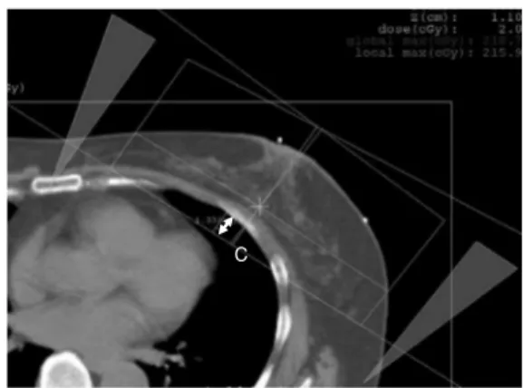
Fig. 2. The furthest distance between the posterior border of the radiation field and the chest wall was measured from the axial image of the treatment planning CT.
C : central lung distance (CLD) (cm).
Fig. 1. Digitally reconstructed radiography image of the three -dimensional treatment planning device in the radiation field A : long axis length of the radiation field (cm), B : short axis length of the radiation field (cm)
At our hospital, the anterior border of the radiation field is set at a distance of 1.5 cm from the skin surface of the nipple base on the CT.
RESULTS
Cumulative incidence of RP
Of the 423 breasts from 413 patients, 89 breasts from 84 cases (21%) were diagnosed with RP, based on the chest radiograph. According to the evaluation of the grade of pneumonitis using CTC/AE ver. 3.0, there were 77 cases (18.2%) of Grade 1 RP, 10 cases (2.3%) of Grade 2, and 2 cases (0.5%) of Grade 3. The age of patients diagnosed with RP ranged from 25 to 80 years (median : 50). Thirty-six cases had RP in their left breasts and 53 in their right breasts. Fifteen cases (16.8%) had a history of allergic dis-ease and 17 (19.1%) had a smoking history. In 32 cases (35.9%), simultaneous medication was done, in addition to that for breast cancer treatment dur-ing radiotherapy. In 26 cases, lesions were found in-side the breasts. In 60 cases, the T factor was T1 or less, while in 60 cases, the N factor was N0. Accord-ing to the stagAccord-ing classification, 49 cases were di-agnosed stage I or below. The operative method of 63 cases was lumpectomy. 72 cases were treated with endocrine therapy, of which 34 were treated during the irradiation period. With respect to fac-tors regarding radiotherapy, 82 cases were treated with 4 MV X-rays, whereas 28 cases underwent boost irradiation. The long axis length of the radia-tion field was 14.6-21.6 cm (median : 17.9 cm) and the short axis length was 3.8-11.1 cm (median : 6.8
cm) ; the CLD was 0.8-3.2 cm (median : 1.8 cm).
Radiation-induced BOOP syndrome
In the present analysis, radiation-induced BOOP syndrome was observed in 12/413 cases (2.9%). All cases of Grade 2 RP or greater developed radiation-induced BOOP syndrome. The age of the patients ranged from 39 to 72 years (median : 50 years old). The clinical symptoms were fever (10 cases), cough-ing (12 cases), decrease in oxygen saturation (8 cases) and sense of dyspnea (8 cases). The timing of the pathogenesis was 2.5-23.1 months after the end of treatment (median : 3.6 months)., Abnormal shadows were observed on the chest radiographs taken 3 months after the completion of radiotherapy in all cases, except for one case, in which RP oc-curred after 23.1 months. Bronchoscopy was per-formed in 8 cases. The total cell number in bron-choalveolar lavage fluid was increased in 7/8 cases. The transbronchial lung biopsy specimens in all 8 cases diagnosed pathologically as organized pneu-monia. Of the 12 cases, steroid administration was implemented in 7 cases, resulting in rapid improve-ment of symptoms. However, recurrence of symp-toms due to the decrease or suspension of the ster-oid administration was observed in 4 cases. The duration of the steroid administration was 5.8-41.4 months (median : 12.3 months) (Table 3).
A 39 year-old woman, in whom RP occurred after
Table 3. Clinical characteristics of 12 patients with radiation - induced BOOP syndrome Patient Age (y) side Endocrine therapy Endocrine therapy (drug) Chemotherapy dose CLD (cm) Short axis (cm) Ipsilateral V20 Gy (%) Steroid Steroid (relapse) Onset, After RT (month) Duration of steroid (month) 1 40 right sequential Tamoxifen - 60 2.4 8.2 12.05 + - 3.2 5.8 2 52 left sequential Anastrozole - 50 2.0 6.6 13.93 + + 4.4 12 3 46 right none - - 50 2.1 7.3 14.00 - - 2.7 -4 53 right concurrent Anastrozole pre + post RT 60 2.0 5.7 7.89 - - 6.0 -5 48 left sequential Tamoxifen - 50 1.7 6.7 8.88 + + 2.5 41.1 6 72 right sequential Anastrozole - 60 2.0 6.4 8.35 + - 6.9 11.1 7 47 right sequential Tamoxifen - 60 1.9 7.3 9.13 - - 3.6 -8 59 left sequential Anastrozole - 50 1.7 6.3 10.56 + + 3.2 35.8 9 69 right sequential Anastrozole - 60 1.8 8.8 10.05 + + 3.0 23.8 10 59 left none - - 50 2.3 6.7 12.14 - - 3.7 -11 44 right concurrent Tamoxifen - 60 2.1 6.6 8.74 - - 3.7 -12 39 left concurrent Tamoxifen - 50 1.7 5.7 9.05 + - 23.1 12.2 Abbreviations : BOOP = bronchiolitis obliterans organizing pneumonia ; RT= radiotherapy ; CLD= central lung distance
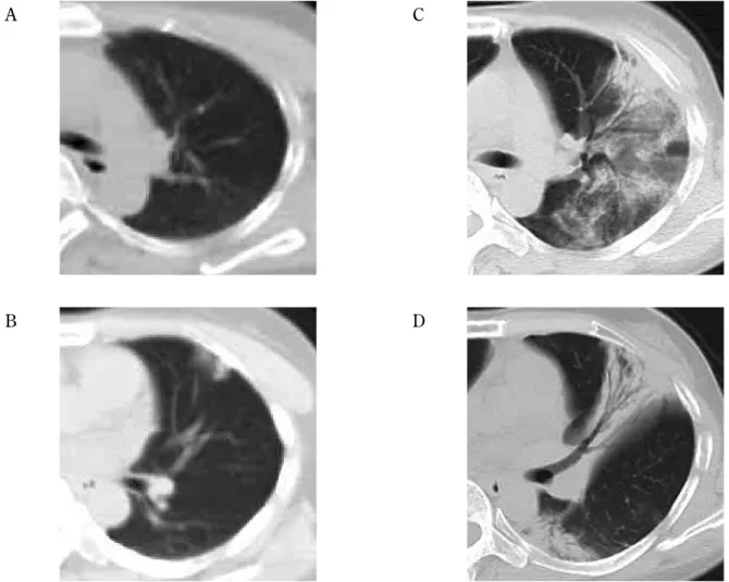
23.1 months, had been taking immunosuppressive agents for a long period due to chronic rheumatoid arthritis. Moreover, radiation treatment planning CT did not show any lesions in her lung fields, and chest radiography taken 3 months after radiotherapy did not reveal any obvious abnormal shadows. Ad-ministration of the immunosuppressive agents was partially suspended 20 months after the completion of the radiotherapy. Moreover, an increased den-sity area that was corresponding to the irradiation field, appeared in the lung field on the CT images at 22 months after radiotherapy. However, she complained of no symptoms at that time. Fever and coughing appeared 23 months after the completion of radiotherapy. Chest radiography revealed an in-filtrative shadow expanding outside the radiation field. She was diagnosed with organized pneumonia using a bronchoscope, and was clinically diagnosed
with radiation-induced BOOP syndrome. Therefore, we started steroid administration and her symptoms improved rapidly. Although this case did not match the criteria proposed by Crestani in terms of the tim-ing of the pathogenesis(11), we extrapolated that RP can occur more than 1 year after irradiation pro-vided that there are special conditions, such as the use of immunosuppressive agents, as in this case (Fig. 3).
Risk factors for RP
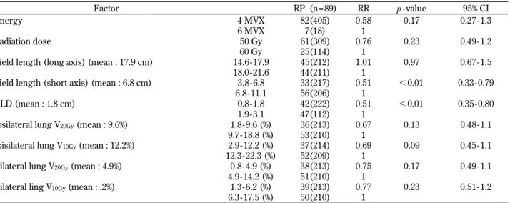
The univariate analysis detected significant dif-ferences in the following factors for incidence of RP of Grade 1 or greater : chemotherapy after ra-diotherapy (p =0.005), chemotherapy in all the pe-riods including before and after radiotherapy (p = 0.001), short axis of the radiation field (!6.8 cm ; p = 0.002), CLD (!1.8 cm ; p =0.002), N factors (!N1 ;
A
A CC
B
B DD
Fig. 3. A 39 - year - old woman had been continuously taking immunosuppressive agents for the treatment of chronic rheumatoid arthri-tis. No abnormalities were recognized from the treatment planning CT before irradiation or from the chest radiograph taken 3 months after irradiation. Partial suspension of immunosuppressive agent administration 20 months after irradiation led to the detection of a localized concentration increasing region by chest CT conducted 22 months after irradiation. No symptoms were indicated at this point. Chest CT conducted 23 months after irradiation, however, revealed a clinical symptom consisting of infiltrative shadows and ground - glass opacity expanding outside the radiation field. They were diagnosed as organized pneumonia by bronchoscopy (A, B : CT performed 20 months after irradiation, C, D : CT performed 23 months after irradiation).
p =0.029), and use of AIs (p =0.028) (Table 4, 5).
The cases of Grade 2 or greater were analyzed in
the same manner. The univariate analysis detected significant differences only in CLD ("1.8 cm ; p =
Table 4. Univariate analysis of patient factors associated with Radiation pneumonitis
Factor RP (n = 89) RR p - value 95% CI Age !50 y 33(191) 0.76 0.20 0.49 - 1.2
"51 y 56(232) 1
Side left 36(201) 0.73 0.15 0.48 - 1.1 right 53(222) 1
Tumor factor T0,Tis,T1 60(301) 0.84 0.43 0.54 - 1.3 T2,T3,T4 29(122) 1
Node factor N0 62(331) 0.60 0.03 0.39 - 0.95 N1,N2,N3 27(92) 1
Clinical stage !I stage 49(263) 0.73 0.14 0.48 - 1.1 !IIA stage 40(160) 1
Location (region) inner side 26(161) 0.65 0.06 0.41 - 1.0 outer side 63(259) 1 unknown 0(3) Operation lumpectomy 63(316) 0.76 0.24 0.48 - 1.2 quadrantectomy 26(105) 1 Allergic disease (+) 15(73) 0.96 0.89 0.55 - 1.7 (-) 74(350) 1
Usage of other medication unrelated to breast cancer (+) 32(126) 1.23 0.34 0.80 - 1.9 (-) 57(297) 1 Smoking status (+) 17(64) 1.14 0.70 0.65 - 1.9 (-) 66(283) 1 unknown 0(76) Chemotherapy (+) 44(143) 1.98 !0.01 1.3 - 3.0 (-) 45(280) 1 chemotherapy (pre - RT) (+) 14(54) 1.27 0.41 0.72 - 2.3 (-) 75(369) 1 chemotherapy (post- RT) (+) 32(100) 1.87 !0.01 1.2 - 2.9 (-) 57(323) 1 Endocrine therapy (+) 72(313) 1.39 0.23 0.82 - 2.4 (-) 17(98) 1 unknown 0(12) antiestrogen drug (+) 26(148) 0.79 0.32 0.50 - 1.3 (-) 63(275) 1 aromatase inhibitor (+) 45(164) 1.59 0.03 1.1 - 2.4 (-) 44(259) 1
(Concurrent endocrine therapy) concurrent 34(130) 1.34 0.20 0.85 - 2.1 sequential 38(183) 1
antiestrogen drug concurrent 10(47) 1.08 0.81 0.56 - 2.1 other 79(376) 1
aromatase inhibitor concurrent 24(83) 1.59 0.05 0.99 - 2.5 other 65(340) 1
Abbreviations : RP = radiation pneumonitis ; RT= radiotherapy ; CLD= central lung distance ; RR= relative risk ; CI = confidence interval.
Table 5. Univariate analysis of radiotherapy factors associated with Radiation pneumonitis
Factor RP (n = 89) RR p - value 95% CI Energy 4 MVX 82(405) 0.58 0.17 0.27 - 1.3
6 MVX 7(18) 1
Radiation dose 50 Gy 61(309) 0.76 0.23 0.49 - 1.2 60 Gy 25(114) 1
Field length (long axis) (mean : 17.9 cm) 14.6 - 17.9 45(212) 1.01 0.97 0.67 - 1.5 18.0 - 21.6 44(211) 1
Field length (short axis) (mean : 6.8 cm) 3.8 - 6.8 33(217) 0.51 !0.01 0.33 - 0.79 6.8 - 11.1 56(206) 1
CLD (mean : 1.8 cm) 0.8 - 1.8 42(222) 0.51 !0.01 0.35 - 0.80 1.9 - 3.1 47(112) 1
Ipsilateral lung V20Gy(mean : 9.6%) 1.8 - 9.6 (%) 36(213) 0.67 0.13 0.48 - 1.1
9.7 - 18.8 (%) 53(210) 1
Ipisilateral lung V10Gy(mean : 12.2%) 2.9 - 12.2 (%) 37(214) 0.69 0.09 0.45 - 1.1
12.3 - 22.3 (%) 52(209) 1
Bilateral lung V20Gy(mean : 4.9%) 0.8 - 4.9 (%) 38(213) 0.75 0.17 0.49 - 1.1
4.9 - 14.2 (%) 51(210) 1
Bilateral ling V10Gy(mean : .2%) 1.3 - 6.2 (%) 39(213) 0.77 0.23 0.51 - 1.2
6.3 - 17.5 (%) 50(210) 1
0.043) and in the operative method (lumpectomy ;
p =0.007). Other than these factors, the patients who
underwent chemotherapy (p =0.090) and the patients
who received radiation dose of 60 Gy (p =0.080) tended to appear RP, but it was not statistically sig-nificant (Table 6, 7).
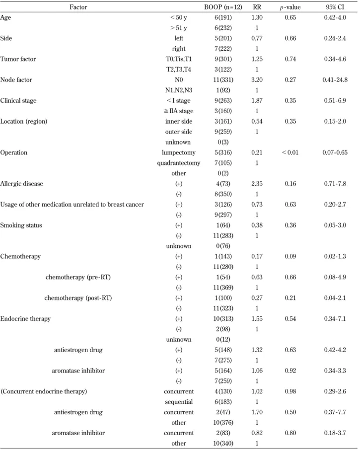
Table 6. Univariate analysis of patient factors associated with RP of Grade 2 or greater, diagnosed as radiation - induced BOOP syndrome
Factor BOOP (n = 12) RR p - value 95% CI Age !50 y 6(191) 1.30 0.65 0.42 - 4.0
"51 y 6(232) 1
Side left 5(201) 0.77 0.66 0.24 - 2.4 right 7(222) 1
Tumor factor T0,Tis,T1 9(301) 1.25 0.74 0.34 - 4.6 T2,T3,T4 3(122) 1
Node factor N0 11(331) 3.20 0.27 0.41 - 24.8 N1,N2,N3 1(92) 1
Clinical stage !I stage 9(263) 1.87 0.35 0.51 - 6.9 !IIA stage 3(160) 1
Location (region) inner side 3(161) 0.54 0.35 0.15 - 2.0 outer side 9(259) 1 unknown 0(3) Operation lumpectomy 5(316) 0.21 !0.01 0.07 - 0.65 quadrantectomy 7(105) 1 other 0(2) Allergic disease (+) 4(73) 2.35 0.16 0.71 - 7.8 (-) 8(350) 1
Usage of other medication unrelated to breast cancer (+) 3(126) 0.73 0.63 0.20 - 2.7 (-) 9(297) 1 Smoking status (+) 1(64) 0.38 0.36 0.05 - 3.0 (-) 11(283) 1 unknown 0(76) Chemotherapy (+) 1(143) 0.17 0.09 0.02 - 1.3 (-) 11(280) 1 chemotherapy (pre - RT) (+) 1(54) 0.63 0.66 0.08 - 4.9 (-) 11(369) 1 chemotherapy (post- RT) (+) 1(100) 0.27 0.21 0.04 - 2.1 (-) 11(323) 1 Endocrine therapy (+) 10(313) 1.55 0.54 0.34 - 7.1 (-) 2(98) 1 unknown 0(12) antiestrogen drug (+) 5(148) 1.32 0.63 0.42 - 4.2 (-) 7(275) 1 aromatase inhibitor (+) 5(164) 1.06 0.92 0.34 - 3.3 (-) 7(259) 1
(Concurrent endocrine therapy) concurrent 4(130) 1.02 0.98 0.29 - 2.6 sequential 6(183) 1
antiestrogen drug concurrent 2(47) 1.70 0.50 0.37 - 7.7 other 10(376) 1
aromatase inhibitor concurrent 2(83) 0.82 0.80 0.18 - 3.7 other 10(340) 1
Abbreviations : BOOP = bronchiolitis obliterans organizing pneumonia ; RT= radiotherapy ; CLD= central lung distance ; RR= relative risk ; CI = confidence interval.
The multivariate analysis detected significant dif-ferences in the following factors for incidence of Grade 1 or greater RP : short axis length (!6.8 cm ;
p =0.024) and CLD (!1.8 cm ; p =0.021) (Table 8).
The multivariate analysis of the RP cases of Grade 2 or greater detected a significant difference only in CLD (!1.8 cm ; p =0.027) (Table 9).
Table 7. Univariate analysis of radiotherapy factors associated with RP of Grade 2 or greater, diagnosed as radiation - induced BOOP syndrome
Factor BOOP (n = 12) RR p - value 95% CI
Energy 4 MVX 12(405)
6 MVX 0(18)
Radiation dose 50 Gy 6(309) 0.36 0.08 0.12 - 1.1 60 Gy 6(114) 1
Field length (long axis) (mean : 17.9 cm) 14.6 - 17.9 6(212) 1.00 0.99 0.32 - 3.1 18.0 - 21.6 6(211) 1
Field length (short axis) (mean : 6.8 cm) 3.8 - 6.8 8(217) 1.76 0.36 0.53 - 5.9 6.8 - 11.1 4(206) 1
CLD (mean : 1.8 cm) 0.8 - 1.8 4(222) 0.29 0.04 0.09 - 0.96 1.9 - 3.1 8(112) 1
Ipsilateral lung V20Gy(mean : 9.6%) 1.8 - 9.6 (%) 7(213) 1.02 0.97 0.32 - 3.2
9.7 - 18.8 (%) 5(210) 1
Ipisilateral lung V10Gy(mean : 12.2%) 2.9 - 12.2 (%) 6(214) 1.01 0.99 0.33 - 3.1
12.3 - 22.3 (%) 6(209) 1
Bilateral lung V20Gy(mean : 4.9%) 0.8 - 4.9 (%) 6(213) 1.04 0.95 0.33 - 3.2
4.9 - 14.2 (%) 6(210) 1
Bilateral ling V10Gy(mean : 6.2%) 1.3 - 6.2 (%) 6(213) 1.04 0.95 0.33 - 3.2
6.3 - 17.5 (%) 6(210) 1
Abbreviations : BOOP = bronchiolitis obliterans organizing pneumonia ; RT= radiotherapy ; CLD= central lung distance ; RR= relative risk ; CI = confidence interval.
Table 8. Multivariate analysis of factors associated with RP
Factor RP (n = 89) RR p - value 95% CI Node factor N0 62(331) 1.08 0.78 0.61 - 1.9
N1,N2,N3 27(92) 1
Location (region) inner side 26(161) 0.76 0.25 0.48 - 1.2 outer side 63(259) 1 unknown 0(3) Chemotherapy (+) 44(143) 1.70 0.16 0.81 - 3.6 (-) 45(280) 1 chemotherapy (post- RT) (+) 32(100) 1.32 0.45 0.65 - 2.7 (-) 57(323) 1 aromatase inhibitor (+) 45(164) 1.39 0.24 0.80 - 2.4 (-) 44(259) 1
aromatase inhibitor concurrent 24(83) 1.31 0.41 0.69 - 2.5 other 65(340) 1
Field length (short axis) (mean : 6.8 cm) 3.8 - 6.8 33(217) 0.60 0.02 0.39 - 0.94 6.8 - 11.1 56(206) 1
CLD (mean : 1.8 cm) 0.8 - 1.8 42(222) 0.58 0.02 0.36 - 0.92 1.9 - 3.1 47(112) 1
Ipsilateral lung V20Gy(mean : 9.6%) 1.8 - 9.6 (%) 36(213) 0.89 0.63 0.56 - 1.4
9.7 - 18.8 (%) 53(210) 1
DISCUSSION
The frequency of pulmonary damage caused by radiotherapy following breast cancer surgery, includ-ing RP occurrinclud-ing at an early time after irradiation and fibrosis occurring at a late time, varies greatly from 0.9% to 85%, depending on reports. Naturally, the detection rates differ between chest radiography and chest CT. According to a previous report, CT evaluation 3 months after radiotherapy showed a frequency of Grade 1 or greater RP of 37% (14). HR-CT evaluation, 4 months after radiotherapy, also de-tected changes in pulmonary parenchyma within ra-diation fields in 85% of cases(7). In general, the fre-quency of RP evaluated by chest radiograph is 0.9-47%. In the present study, 21% of patients showed RP on the chest radiograph at 3 months after comple-tion of radiotherapy. The previous reports, however, also involve local-regional radiotherapy on supracla-vicular and axilla fields, as well as chest-wall irra-diation after mastectomy and differ slightly from our study with respect to the subjects (2, 6, 14, 16, 17). BOOP was reported in 1985 by Epler, et al. It pre-sents symptoms such as coughing, sense of dyspnea, fever and chest pain. On images, it shows patchy consolidation accompanied by ground-glass opaci-ties. Histological characteristics include polypoid masses of granulation tissue in the lumens of small airways, alveolar ducts and some alveoli. Although it can be classified into idiopathic and secondary, the majority of cases are usually idiopathic. The fre-quency of radiation-induced BOOP syndrome fol-lowing breast-conserving surgery of breast cancer has been reported as 2.3% (15), 2.4% (13) and 2.5% (12). In the present study, a similar incidence of
2.9% was obtained as a result. It included one atypi-cal case among the 12 cases in which cliniatypi-cally prob-lematic RP of Grade 2 or greater presented. Apart from this case, however, clinical symptoms requir-ing treatment did not appear before the period of 2.5-6.9 months after radiotherapy. In all cases, slight abnormal shadows can be detected on the chest ra-diographs at the 3-month after radiotherapy. It is considered that RP and radiation-induced BOOP syndrome are two different clinical conditions. RP results from the direct influence of irradiation, whereas radiation-induced BOOP syndrome is be-lieved to be caused by autoimmunization induced indirectly by irradiation (10-13, 18, 19). Neverthe-less, at the onset of radiation-induced BOOP syn-drome, infiltrative shadows resembling ground-glass opacity, in areas corresponding to radiation fields, begin to appear. They then develop into infiltrative shadows expanding outside the radiation field areas. In the present study, there were hardly any clinical symptoms 3 months after radiotherapy. However, the next follow-up observations indicated 7 cases of exacerbated symptoms of Grade 2 or greater, de-spite the fact that the CT taken at 3 months after ra-diotherapy was only able to detect localized shadows within the radiation field. This led us to believe that conducting an evaluation 3 months after radiother-apy was crucial.
There have been several reports analyzing the factors concerning pathogenesis of RP and radia-tion- induced BOOP syndrome caused by irradia-tion following breast cancer surgery. There have been 4 reports investigating radiation-induced BOOP syndrome caused by irradiation after conserving surgery (12, 13, 15, 20). However, no reports have
Table 9. Multivariate analysis of factors associated with Grade2 or greater pneumonitis, diagnosed as radiation - induced BOOP syndrome
Factor BOOP (n = 12) RR p - value 95% CI Chemotherapy (+) 1(143) 0.17 0.09 0.02 - 1.3
(-) 11(280) 1
Radiation dose 50 Gy 6(309) 0.44 0.17 0.14 - 1.4 60 Gy 6(114) 1
Field length (short axis) (mean : 6.8 cm) 3.8 - 6.8 8(217) 1.55 0.49 0.45 - 5.3 6.8 - 11.1 4(206) 1
CLD (mean : 1.8 cm) 0.8 - 1.8 4(222) 0.24 0.03 0.07 - 0.85 1.9 - 3.1 8(112) 1
Ipsilateral lung V20Gy(mean : 9.6%) 1.8 - 9.6 (%) 7(213) 1.64 0.43 0.48 - 5.6
9.7 - 18.8 (%) 5(210) 1
Abbreviations : BOOP = bronchiolitis obliterans organizing pneumonia ; RT= radiotherapy ; CLD= central lung distance ; RR= relative risk ; CI = confidence interval.
been published investigating the effects of patient background and treatment regimens on the patho-genesis of Grade 1 RP. In the present study, we only analyzed cases in which whole breast irradiation had been performed and in which all radiotherapy tech-nical factors had been evaluated. As for the relation-ship between patient characteristics and RP, al-though patient age has often been cited as a risk fac-tor (2, 14, 20-24), no such relationship was estab-lished in the present study. In agreement with pre-vious reports, smoking history and the simultaneous use of other medication were not identified as fac-tors influencing risk of RP (3, 20, 25). The use of radiotherapy and chemotherapy simultaneously creases the risk of RP (9, 17), however, the risk in-duced by the use of chemotherapy in stages varies greatly among reports (15, 20, 26, 27). Our univari-ate analysis also suggested that adjuvant chemother-apy increases the risk of RP.
There are numerous reports indicating the in-volvement of anti-estrogen agents in the pathogene-sis of RP, especially tamoxifen (15, 21, 22, 28, 29). In the present study, however, anti-estrogen agents were not a significant variable for RP, whereas a correlation was found with the use of AIs. Increasing of RP patients who have used AIs might be related to age because AIs are generally used in postmeno-pausal women. With regard to radiotherapy factors, breast and regional irradiation including supracla-vicular and axilla (local-regional radiotherapy) is considered to result in a higher frequency of RP compared with whole breast irradiation (local radio-therapy)(2, 6, 14, 16, 17). Lind, et al. have reported that the risk of RP was significantly higher in local-regional radiotherapy (4.1%) compared with local radiotherapy (0.9%) (16). Kahan (14), likewise, has reported that the incidence of RP and fibrosis were 2.5 times higher in regional irradiation. V20Gywas reported to correlate with lung volume ; the more V20Gyincreased, the higher the incidence of RP (2, 14, 23). So we investigated only cases of breast ir-radiation, the frequency of RP had no significant sta-tistically correlation with V20Gyas well as V10Gyin the present study. Previous reports, on the other hand, took into consideration the regional irradiation, lead-ing to the speculation that significant differences were obtained due to the high influence of the gional irradiation. There have also been several re-ports indicating the involvement of CLD with RP (8, 9). Fernando, et al. (9) have reported the frequency of RP as 4.6% in cases where CLD was 3 cm or more. The present study also showed that CLD of
1.9 cm or more increased the risk of RP and radia-tion-induced BOOP syndrome. Another result of the present study was that the short axis length of the radiation field correlated with RP in the univariate and multivariate analyses. Although there have been reports examining the long axis length of radiation fields (15), it is difficult to compare the short axis with other analyses owing to different operational criteria among institutions. Nevertheless, RP is con-siderably affected by the lung volume to be irradi-ated, in the same way as CLD. The radiation dose exhibited no significant difference, albeit a tendency to correlate with the radiation-induced BOOP syn-drome was observed. There are numerous reports investigating risk factors for the RP and radiation-induced BOOP syndrome. Though, in our study, there are no significant differences with previously suggested factors, such as age and the simultaneous use of hormonal treatment. In multivariate analysis, setting of the radiation field, such as CLD and short axis length of the radiation field, was an important factor. As for the timing of the RP, RP and radiation-induced BOOP syndrome that required treatment were detected on the chest radiography 3 months after the treatment, suggesting that evaluation at this stage is crucial. These results are suggesting that we need to give careful attention to occurrence of RP in around 3 months after completion of RT.
CONCLUSIONS
In the tangential irradiation of whole breast after breast conserving surgery, the significant risk fac-tors of RP are CLD (!1.8 cm) and the short axis length of the radiation field, as determined by multi-variate analysis. The pathogenesis of radiation-in-duced BOOP syndrome significantly correlated with CLD. Therefore, the lung volume within the radia-tion field proved to be a significant risk factor for RP and radiation-induced BOOP syndrome. Regard-ing follow-up after irradiation, the evaluation of chest radiography 3 months after the end of irradiation is postulated to be crucial.
REFERENCES
1. Clarke M, Collins R, Darby S, Davies C, Elphinstone P, Evans E, Godwin J, Gray R, Hicks C, James S, MacKinnon E, McGale P, McHugh T, Peto R, Taylor C, Wang Y : Effects
of radiotherapy and of differences in the extent of surgery for early breast cancer on local re-currence and 15-year survival : an overview of the randomised trials. Lancet 366 : 2087-2106, 2005
2. Lind PA, Wennberg B, Gagliardi G, Fornander T : Pulmonary complications following different radiotherapy techniques for breast cancer, and the association to irradiated lung volume and dose. Breast Cancer Res Treat 68 : 199-210, 2001
3. Holli K, Pitkanen M, Jarvenpaa R, Rajala J, Lahtela S, Hyodynmaa S, Ojala A : Early skin and lung reactions in breast cancer patients after radiotherapy : prospective study. Radio-ther Oncol 64 : 163-169, 2002
4. Zissiadis Y, Langlands AO, Barraclough B, Boyages J : Breast conservation : long-term re-sults from Westmead Hospital. Aust N Z J Surg 67 : 313-319, 1997
5. Fung MC, Schultz DJ, Solin LJ : Early-stage bilateral breast cancer treated with breast-con-serving surgery and definitive irradiation : the University of Pennsylvania experience. Int J Ra-diat Oncol Biol Phys 38 : 959-967, 1997 6. Wennberg B, Gagliardi G, Sundbom L, Svane
G, Lind P : Early response of lung in breast cancer irradiation : radiologic density changes measured by CT and symptomatic radiation pneumonitis. Int J Radiat Oncol Biol Phys 52 : 1196-1206, 2002
7. Ogoh E, Fujimoto K, Meno S, Uchida M, Joh S, Tabuchi E, Toda Y, Onizuka Y, Nishimura H, Hayabuchi N : [HR-CT evaluation of lung parenchymal alterations in patients following breast conservation therapy]. Nippon Igaku Hoshasen Gakkai Zasshi 58 : 331-337, 1998 (in Japanese)
8. Lind PA, Gagliardi G, Wennberg B, Fornander T : A descriptive study of pulmonary complica-tions after postoperative radiation therapy in node-positive stage II breast cancer. Acta Oncol 36 : 509-515, 1997
9. Fernando IN, Powles TJ, Ashley S, Grafton D, Harmer CL, Ford HT : An acute toxicity study on the effects of synchronous chemotherapy and radiotherapy in early stage breast cancer after conservative surgery. Clin Oncol (R Coll Radiol) 8 : 234-238, 1996
10. Abratt RP, Morgan GW : Lung toxicity follow-ing chest irradiation in patients with lung can-cer. Lung Cancer 35 : 103-109, 2002
11. Crestani B, Valeyre D, Roden S, Wallaert B, Dalphin JC, Cordier JF : Bronchiolitis obliter-ans organizing pneumonia syndrome primed by radiation therapy to the breast. The Groupe d’Etudes et de Recherche sur les Maladies Orphelines Pulmonaires (GERM“O”P). Am J Respir Crit Care Med 158 : 1929-1935, 1998 12. Takigawa N, Segawa Y, Saeki T, Kataoka M,
Ida M, Kishino D, Fujiwara K, Ohsumi S, Eguchi K, Takashima S : Bronchiolitis obliter-ans organizing pneumonia syndrome in breast-conserving therapy for early breast cancer : ra-diation-induced lung toxicity. Int J Radiat Oncol Biol Phys 48 : 751-755, 2000
13. Miwa S, Morita S, Suda T, Suzuki K, Hayakawa H, Chida K, Nakamura H : The incidence and clinical characteristics of bronchiolitis obliter-ans organizing pneumonia syndrome after ra-diation therapy for breast cancer. Sarcoidosis Vasc Diffuse Lung Dis 21 : 212-218, 2004 14. Kahan Z, Csenki M, Varga Z, Szil E, Cserhati
A, Balogh A, Gyulai Z, Mandi Y, Boda K, Thurzo L : The risk of early and late lung se-quelae after conformal radiotherapy in breast cancer patients. Int J Radiat Oncol Biol Phys 68 : 673-681, 2007
15. Katayama N, Sato S, Katsui K, Takemoto M, Tsuda T, Yoshida A, Morito T, Nakagawa T, Mizuta A, Waki T, Niiya H, Kanazawa S : Analy-sis of Factors Associated with Radiation-Induced Bronchiolitis Obliterans Organizing Pneumonia Syndrome After Breast-Conserving Therapy. Int J Radiat Oncol Biol Phys : 2008
16. Lind PA, Marks LB, Hardenbergh PH, Clough R, Fan M, Hollis D, Hernando ML, Lucas D, Piepgrass A, Prosnitz LR : Technical factors as-sociated with radiation pneumonitis after local +/- regional radiation therapy for breast cancer. Int J Radiat Oncol Biol Phys 52 : 137-143, 2002 17. Lingos TI, Recht A, Vicini F, Abner A, Silver B, Harris JR : Radiation pneumonitis in breast cancer patients treated with conservative sur-gery and radiation therapy. Int J Radiat Oncol Biol Phys 21 : 355-360, 1991
18. Crestani B, Kambouchner M, Soler P, Crequit J, Brauner M, Battesti JP, Valeyre D : Migra-tory bronchiolitis obliterans organizing pneu-monia after unilateral radiation therapy for breast carcinoma. Eur Respir J 8 : 318-321, 1995
19. Prakash UB : Radiation-induced injury in the “nonirradiated” lung. Eur Respir J 13 : 715-717,
1999
20. Ogo E, Komaki R, Fujimoto K, Uchida M, Abe T, Nakamura K, Mitsumori M, Sekiguchi K, Kaneyasu Y, Hayabuchi N : A survey of radia-tion- induced bronchiolitis obliterans organizing pneumonia syndrome after breast-conserving therapy in Japan. Int J Radiat Oncol Biol Phys 71 : 123-131, 2008
21. Dorr W, Bertmann S, Herrmann T : Radiation induced lung reactions in breast cancer ther-apy. Modulating factors and consequential ef-fects. Strahlenther Onkol 181 : 567-573, 2005 22. Koc M, Polat P, Suma S : Effects of tamoxifen
on pulmonary fibrosis after cobalt-60 radiother-apy in breast cancer patients. Radiother Oncol 64 : 171-175, 2002
23. Lind PA, Wennberg B, Gagliardi G, Rosfors S, Blom-Goldman U, Lidestahl A, Svane G : ROC curves and evaluation of radiation-induced pul-monary toxicity in breast cancer. Int J Radiat Oncol Biol Phys 64 : 765-770, 2006
24. Gagliardi G, Bjohle J, Lax I, Ottolenghi A, Eriksson F, Liedberg A, Lind P, Rutqvist LE : Radiation pneumonitis after breast cancer irra-diation : analysis of the complication probability using the relative seriality model. Int J Radiat Oncol Biol Phys 46 : 373-381, 2000
25. Theuws JC, Kwa SL, Wagenaar AC, Boersma LJ, Damen EM, Muller SH, Baas P, Lebesque JV : Dose-effect relations for early local pulmo-nary injury after irradiation for malignant lym-phoma and breast cancer. Radiother Oncol 48 : 33-43, 1998
26. Van Haecke P, Vansteenkiste J, Paridaens R, Van der Schueren E, Demedts M : Chronic lymphocytic alveolitis with migrating pulmo-nary infiltrates after localized chest wall irra-diation. Acta Clin Belg 53 : 39-43, 1998
27. Taghian AG, Assaad SI, Niemierko A, Floyd SR, Powell SN : Is a reduction in radiation lung volume and dose necessary with paclitaxel che-motherapy for node-positive breast cancer? Int J Radiat Oncol Biol Phys 62 : 386-391, 2005 28. Huang EY, Wang CJ, Chen HC, Sun LM, Fang
FM, Yeh SA, Hsu HC, Hsiung CY, Wu JM : Multivariate analysis of pulmonary fibrosis after electron beam irradiation for postmastectomy chest wall and regional lymphatics : evidence for non-dosimetric factors. Radiother Oncol 57 : 91-96, 2000
29. Bentzen SM, Skoczylas JZ, Overgaard M, Overgaard J : Radiotherapy-related lung fibro-sis enhanced by tamoxifen. J Natl Cancer Inst 88 : 918-922, 1996