Mycobacterium abscessus 肺感染症の臨床的検討
小橋 吉博 岡 三喜男
要旨:〔目的〕Mycobacterium abscessus による肺感染症の臨床像を検討した。〔対象と方法〕対象は, 当院および関連施設において最近 5 年間に M.abscessus肺感染症と診断した 4 例とし,2008年の日本 結核病学会の診断基準を満たしていた。これらの臨床所見に関して過去の報告例と比較しながら検討 した。〔結果〕対象の平均年齢は 56歳(男性 1 例,女性 3 例),基礎疾患は全例にみられ,悪性腫瘍 2 例,陳旧性肺結核 1 例,膠原病(免疫抑制剤投与中)1 例であった。発見動機は,他疾患で経過観 察中が 1 例で,他の3例は自覚症状の出現であった。検査所見では 4 例中 3 例に中等度の炎症所見を 認め,細菌学的には 4 例中 3 例が塗抹陽性(気管支鏡下検体 2 例)であった。画像所見は空洞を有す る結核類似型が 1 例,小結節・気管支拡張型が 3 例で病変の拡がりは全例片側肺以内であった。治療 は 4 例中 3 例に IPM/cs,AMK,CAM,LVFX等を用いた多剤併用療法が行われ,いずれも有効であっ たが,他の 1例は治療開始前に原疾患の増悪で死亡した。〔結論〕M.abscessus肺感染症は基礎疾患を 有する症例に併発することが多かったが,気管支鏡を用いて積極的に診断しえた 2 例では早期に適切 な抗菌薬投与ができ,改善が得られていた。 キーワーズ:Mycobacterium abscessus 肺感染症 Ⅰ. 緒 言Mycobacterium abscessus(M. abscessus)は迅速発育型の 非結核性抗酸菌で土壌や水道水などに常在し,皮膚軟部 組織や骨の感染症の原因菌として知られている。また, M. abscessus などの迅速発育菌群は気管支鏡の汚染で気 管支洗浄液から検出されることもあり,肺に感染するこ とは比較的稀である1)。近年われわれは,M. abscessus に よる肺感染症を経験するようになってきたため,臨床像 を過去の報告例とも比較しながら検討したので報告する。 Ⅱ. 対象と方法 対象は,当院および関連施設において M. abscessus 肺 感染症と診断した 4 例である。これらの全例は 2008 年 に日本結核病学会が提唱した肺非結核性抗酸菌症の診断 基準2)を満たしていた。これら 4 例に対して,背景因子, 発見動機,細菌学的所見を含めた検査所見,画像所見, 治療および予後に関して,retrospectiveに検討した。また, 過去 15年間に本邦で報告されてきた M.abscessus肺感染 症 24 例に対しても同様に検討し,当院および関連施設 において経験した 4 例と比較検討した。 Ⅲ. 結 果 当院および関連施設で経験した 4 例の M.abscessus肺 感染症の主な臨床所見を Table 1に示した。平均年齢は 56 歳,性別は男性 1 例に対し,女性 3 例であった。喫煙 歴は 1 例にみられ,基礎疾患は全例が有しており,その 内訳は悪性腫瘍 2 例,陳旧性肺結核 1 例,膠原病で免疫 抑制剤投与中が 1 例であった。発見動機は,4 例中 3 例 が自覚症状の出現,残りの 1 例は他疾患の経過観察中に 発見されていた。炎症所見では,4 例中 3 例が軽度から 中等度陽性を示していた。また,細菌学的検査では 4 例 中 3 例において抗酸菌塗抹陽性であったが,うち 2 例は 気管支肺胞洗浄液からの検体を用いてであった。画像所 見については,結核類似型で大きな空洞を有する症例が 1 例(Fig. 1A,1B),小結節・気管支拡張型が 3 例(Fig.
川崎医科大学呼吸器内科 連絡先 : 小橋吉博,川崎医科大学呼吸器内科,〒701_0192 岡
山県倉敷市松島 577(E-mail: yoshihiro@med.kawasaki-m.ac.jp) (Received 31 Jul. 2009/Accepted 30 Sep. 2009)
Table 1 Pulmonary Mycobacterium abscessus disease experienced in our hospital
Tbc : Tuberculosis, BALF : Bronchoalveolar lavage fluid, RUL : Right upper lobe, RML : Right middle lobe, RLL : Right lower lobe, LLL : Left lower lobe, CAM : clarithromycin, AMK : amikacin, LVFX : levofloxacin, IPM/cs : imipenem/cilastatin,
Extension of lesion : 1 (within one-third of unilateral lung field), 2 (within unilateral lung field), 3 (over unilateral lung field)
Case Age Sex Smokinghistory Underlyingdisease Clinical symptom
Laboratory findings Clinical specimen Radiological findings
Treat-
ment Prognosis WBC
(/μl) (mm/br)T2SR (mg/dl)CRP Smear Culture Loca- tion Exten- sion Cavity Bronchi- ectasis 1 2 3 4 58 M 58 F 36 F 72 F 20/day (40years) (−) (−) (−) Lung cancer (Adenoca) Old Tbc Breast Ca (ope) ANCA associated vasculitis (Steroid (+)) Fever Cough Hemo-sputum (−) Fever Cough 11120 7850 4680 15960 96 19 58 103 12.25 0.17 2.70 2.41 (+) (+) (Sputum) (−) (+) (Sputum) (+) (+) (BALF) (+) (+) (BALF) RUL RUL RML RML LLL RML RLL 1 2 2 2 (+) (−) (+) (−) (−) (+) (+) (+) (−) CAM AMK IPM/cs AMK IPM/cs LVFX CAM AMK IPM/cs Death Improved Improved Improved
Fig. 1 Chest X-ray (A) and chest CT (B) revealed cavity lesion due to Mycobacterium abscessus in the right upper lobe and mass shadow due to lung adenocarcinoma in the left upper lobe.
(B)
Fig. 2 Chest X-ray revealed infiltration shadows in the middle and lower field of the right lung (A). Chest CT showed infiltration and nodular lesions with bronchiectasis in the right middle lobe (B).
(A)
Fig. 3 Chest X-ray revealed infiltration shadow in the right lower lung field before treatment (A). Chest CT showed small nodular shadows with bronchiectasis in the right lung and lingula seg- ment of the left lung before treatment (B) and its shadow has been improved after treatment (C).
(C)
Fig. 4 Chest X-ray revealed infiltration shadow in the right lower lung field (A). Chest CT re- vealed small nodular shadows with bronchiecta- sis in the middle lobe of the right lung before treatment (B) and its shadow has been improved after treatment (C). (B) (C) (A) (B) (A) 2A,2B,3A,3B,4A,4B)あり,病変の拡がりは全例片 側肺以内にとどまっていた。治療に関しては,症例 1 は 肺癌に対する化学療法中に合併した症例であり,原疾患 の増悪に診断の遅れも加わって治療開始前に死亡した。 他の 3例は改善が得られ,うち 2 例は気管支鏡検査を施 行し,早期に診断治療ができたため,カルバぺネム系抗 菌薬(イミぺネム,IPM/cs),アミノ配糖体抗菌薬(アミ カシン,AMK),マクロライド系抗菌薬(クラリスロマ イシン,CAM)もしくはニューキノロン系抗菌薬等(レ ボフロキサシン,LVFX)による多剤併用療法により, 症状もしくは炎症所見および画像所見(Fig. 3C,4C)と もに改善が得られていた。 次に,1994年以降に本邦で報告された M.abscessus肺 感染症 24例をまとめて臨床所見を Table 2に示した。対
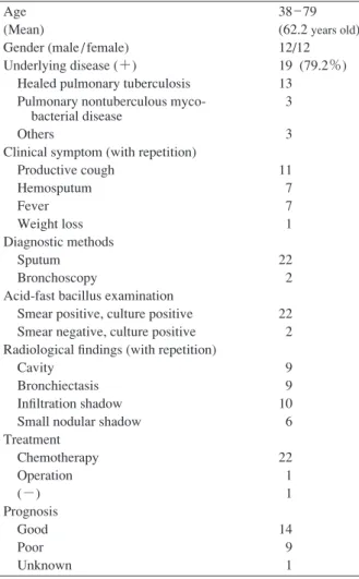
Table 2 Pulmonary Mycobacterium abscessus disease reported previously in Japan (24 cases)
Age (Mean)
Gender (male / female) Underlying disease (+) Healed pulmonary tuberculosis Pulmonary nontuberculous myco- bacterial disease
Others
Clinical symptom (with repetition) Productive cough Hemosputum Fever Weight loss Diagnostic methods Sputum Bronchoscopy
Acid-fast bacillus examination Smear positive, culture positive Smear negative, culture positive Radiological findings (with repetition) Cavity
Bronchiectasis Infiltration shadow Small nodular shadow Treatment Chemotherapy Operation (−) Prognosis Good Poor Unknown 38 _ 79 (62.2 years old) 12/12 19 (79.2%) 13 3 3 11 7 7 1 22 2 22 2 9 9 10 6 22 1 1 14 9 1 象患者の平均年齢は 62.2歳,男女比は同率であった。基 礎疾患は 79% と大半の症例が有しており,陳旧性肺結 核が最も多くみられ,呼吸器疾患が大半を占めており, われわれの症例とは異なっていた。発見動機は,咳嗽や 血痰などの自覚症状を主訴として受診してからが多かっ た。診断は,喀痰から頻回に M. abscessus が分離され, 塗抹陽性であった症例が大半を占めていた。治療は,1 例に施行できなかったのと,1 例には外科的手術が施行 された以外は,大半の症例に複数の抗菌薬による多剤併 用療法が施行されていた。しかしながら,予後に関して は治療への反応が悪い症例が多く,14例(58%)の有効 率にとどまっていた。 Ⅳ. 考 察 M. abscessus は迅速発育菌群の中では検出される頻度 は高く,非結核性抗酸菌の中でも Mycobacterium avium complex(MAC),M. kansasii に次いで多く,病原性が強 いため肺感染症と診断された場合の臨床的意義は大きい と考えられている。 M. abscessus 肺感染症は,1993年に Griffithらが米国に おける迅速発育菌群による肺感染症 154例の中でまとめ て報告している3)。全体の 82% が M.abscessus によるも ので,平均年齢は 54 歳,女性のほうが多かったが,基 礎疾患のない症例は 32% で一次感染型のほうが少な かった。基礎疾患としては,陳旧性肺結核や肺 MAC症 といった抗酸菌感染症が最も多く,発見動機も咳嗽,発 熱,血痰といった自覚症状出現を契機としていた。画像 所見では,実質影,間質影,粒状網状影がほぼ同等にみ られたが,空洞は少なく,病変は上葉に優位にみられて いた。しかし,最近の海外からの報告4) 5)では上葉優位 の空洞性病変,多発する結節影,中葉・舌区の気管支拡 張性変化といった肺 MAC症に類似した所見を呈する症 例が多いと述べられている。本邦における最近の M. abscessus 肺感染症の症例では,50 歳以上の中高年女性 に多く,肺に基礎疾患を有しない一次感染型が多く,肺 MAC 症に類似した上葉に空洞性病変を有するものや中 葉・舌区に小結節や気管支拡張性変化を有する症例の比 率がほぼ同じと報告されている6) ∼ 18)。われわれの施設 において経験した 4 例では,全例が何らかの基礎疾患を 有し,うち 3 例が Fig. 2,3,4 に示したように中葉・舌 区を中心に小結節・気管支拡張性変化を呈し,画像的に は肺 MAC症に類似した所見であった。今回の 4 症例で は,既往歴としての肺結核の合併による肺病変は 1 例の みであったが,肺 MAC症に類似する所見をとりやすい ことから,細菌学的検査を正確にかつ積極的に行うこと は早期診断・治療に重要と思われた。 M. abscessus 肺感染症に対する治療に関しては,従来か らM. abscessus は抗結核薬に対して耐性であるが,CAM, AMK,セフォキシチン(CXT),IPM/csに対しては in vitro で感受性を示すとされている。2007 年の ATS によるガ イドライン19)でも,CAMに加えて AMK,CXTもしくは IPM/cs のうちのいくつかの薬剤を数カ月間は併用して いくことを推奨しているが,これらの薬剤に耐性を示す 菌にはリネゾリドも考慮する必要があること,しかし, 根治的な治療は病巣部を外科的に切除するしかないこと も記載されている。われわれが経験した 4 例では,うち 1 例が他科で肺腺癌治療中に胸部異常陰影が出現してい たにもかかわらず,診断治療が遅れたことと原疾患の増 悪も加わり,呼吸不全で死亡したが,治療ができた 3 例 中 2 例は気管支鏡検査を積極的に行い,早期に正確な診 断が得られたために,CAM,AMK,IPM/cs,LVFX 等に よる多剤併用療法を適切に施行できた結果,治療効果は 有効で改善が得られていた。 以上,最近 5 年間でわれわれは当院および関連施設に おいて M. abscessus による肺感染症 4 例を経験した。肺 非結核性抗酸菌症の中で M. abscessus を原因菌として発
症する症例も増加してきており,肺 MAC症との臨床像 も類似している一方で,特効薬はないもののガイドライ ン上も治療法が異なることから,気管支鏡を用いた積極 的な診断法も試みながら,早期診断治療を試みることが 重要と考えられる。 文 献 1 ) 冨岡治明, 齋藤 肇, 江崎孝行, 他:「結核」, 泉孝英 編, 医学書院, 東京, 1998, 273_293. 2 ) 日本結核病学会非結核性抗酸菌症対策委員会,日本呼 吸器学会感染症・結核学術部会:肺非結核性抗酸菌症 診断に関する指針─2008年. 結核. 2008 ; 83 : 525_526. 3 ) Griffith DE, Girard WM, Wallace RJ Jr : Clinical features of
pulmonary disease caused by rapidly growing mycobacteria. Am Rev Respir Dis. 1993 ; 147 : 1271 _ 1278.
4 ) Han D, Lee RS, Koh WJ, et al. : Radiographic and CT find- ings of nontuberculous mycobacterial pulmonary infection caused by Mycobacterium abscessus. AJR. 2003 ; 181 : 513 _ 517.
5 ) Jeong YJ, Lee KS, Koh WJ, et al.: Nontuberculous mycobac- terial pulmonary infection in immunocompetent patients : Comparison of thin-section CT and histopathologic findings. Radiology. 2004 ; 231 : 880 _ 886.
6 ) 川島 崇, 来生 哲, 荒川正昭:Mycobacterium chelonae subsp. abscessus による肺感染症の 2 症例. 感染症学雑 誌. 1994 ; 68 : 416_420.
7 ) 猶 木 克 彦, 大 角 光 彦, 高 杉 知 明, 他:Mycobacterium
chelonae subsp. abscessus による肺感染症 2 例. 日胸疾 会誌. 1996 ; 34 : 1264_1270.
8 ) 竹村佳純, 岩崎吉伸, 駒谷暢代, 他:糖尿病を基礎疾 患に持つ Mycobacterium abscessus による非定型抗酸菌 症の 1 例. 日呼吸会誌. 2002 ; 40 : 61_65.
9 ) Tanaka E, Kimoto T, Tsuyuguchi K, et al. : Successful treatment with faropenem and clarithromycin of pulmonary
Mycobacterium abscessus infection. J Infect Chemother. 2002 ; 8 : 252 _ 255. 10) 田川曉大, 池原邦彦, 西山晴美, 他:Mycobacterium abscessus 肺感染症の1 例. 日呼吸会誌. 2003 ; 41 : 546 _ 550. 11) 松澤邦明, 塚口勝彦, 岡村英生, 他:原発性マクロアミ ラーゼ血症に合併した Mycobacterium abscessusによる 非結核性抗酸菌症の 1 症例. 日呼吸会誌. 2004 ; 42 : 519 _ 522. 12) 笠井昭吾, 徳田 均, 吉川充浩, 他:大量排菌が持続 し, 気管内潰瘍が唯一の病巣であった Mycobacterium abscessus 感染症の1 例. 日呼吸会誌. 2004 ; 42 : 919 _ 923. 13) 小林信之, 鈴木 学:M. abscessus, M. szulgai による肺 感染症. 日胸. 2007 ; 66 : 558_566. 14) 小泉晴美:Mycobacterium abscessus. 日胸. 2004 ; 63 : S185 _ S187. 15) 西澤依小, 藤村政樹, 田上敦朗, 他:薬剤感受性検査結 果と臨床経過に乖離を認めた Mycobacterium abscessus 肺感染症の 2 例. 日呼吸会誌. 2005 ; 43 : 241_246. 16) 鹿 間 裕 介, 神 尾 義 人, 栗 生 和 幸, 他:Clarithromycin, amikacin, imipenem/cilastatin による化学療法が有効と考 えられた Mycobacterium abscessus 肺感染症の 1 例. 日 呼吸会誌. 2006 ; 44 : 800_806.
17) Sugino K, Kobayashi M, Iwata M, et al.: Successful treat- ment with pneumonectomy for pulmonary abscessus infec- tion. Intern Med. 2008 ; 48 ; 459 _ 463.
18) Fujita K, Tanaka E, Hatta K, et al.: An autopsy case of Myco-
bacterium abscessus pulmonary infection complicated with rheumatoid arthritis. Intern Med. 2008 ; 47 : 1273 _ 1276. 19) Griffith DE, Aksamit T, Brown-Elliot BA, et al. : An official
ATS/IDSA statement : diagnosis, treatment, and prevention of nontuberculous mycobacterial diseases. Am J Respir Crit Care Med. 2007 ; 175 : 357 _ 416.
−−−−−−−− Original Article −−−−−−−−
CLINICAL ANALYSIS OF MYCOBACTERIUM ABSCESSUS
PULMONARY INFECTION IN OUR HOSPITAL
Yoshihiro KOBASHI and Mikio OKA Abstract [Objective] We analyzed the clinical characteris-
tics of pulmonary infection due to Mycobacterium abscessus. [Materials and Methods] Four cases diagnosed with M.
abscessus pulmonary infection encountered at Kawasaki Medical School Hospital and affiliated hospitals over the last five years were enrolled in this study. They all satisfied the diagnostic criteria of the Japanese Society for Tuberculosis. The clinical findings in this study were also compared to those of previously reported cases in Japan.
[Results] The average age of the four cases was 56 years (one male and three females). All four cases showed underly- ing diseases, comprising two cases with malignancy, one with old pulmonary tuberculosis and one with collagen vascular disease receiving immunosuppressive treatment. Three cases were detected based on clinical symptoms, and one was inci- dentally identified during follow-up for another underlying disease. Laboratory examinations revealed mild or moderate inflammatory responses in three of the four cases, and three of the four were smear-positive for acid-fast bacilli in the clinical specimens (sputum in one and bronchial alveolar lavage fluid in two) microbiologically. The radiological exam- ination revealed that one case showed tuberculosis resembling a cavitary lesion and three showed the small nodular and
bronchiectatic type. The extent of lesions was within the uni- lateral lung in all cases. Concerning treatment for M. absces-
sus pulmonary infection, combined multi-drug chemotherapy using IPM/cs, AMK, CAM, and LVFX was carried out in three of the four cases, achieving a satisfactory clinical effect. However, one case died due to progression of the underlying disease before the initiation of treatment.
[Conclusion] Although M.abscessus pulmonary infection was more frequent in cases with underlying disease, the early, appropriate administration of antibiotics was performed in two of the four cases correctly diagnosed using bronchoscopic pro- cedures, resulting in clinical improvement.
Key words : Mycobacterium abscessus pulmonary infection Division of Respiratory Diseases, Department of Medicine, Kawasaki Medical School
Correspondence to : Yoshihiro Kobashi, Division of Respira- tory Diseases, Department of Medicine, Kawasaki Medical School, 577 Matsushima, Kurashiki-shi, Okayama 701_ 0192 Japan. (E-mail : yoshihiro@med.kawasaki-m.ac.jp)