熊本大学学術リポジトリ
Structure and organization of the human TRKA gene encoding a high affinity receptor for nerve growth factor
journal or
publication title
Japanese Journal of Human Genetics
volume 42
number 2
page range 343‑351
year 1997‑06
URL http://hdl.handle.net/2298/9081
Structure and organization of the human TRKA gene encoding a high affinity receptor for nerve growth factor
Yasuhiro Indo*, Sek Mardy, Motoko Tsuruta, Mohammad Azharul Karim**, and Ichiro Matsuda
Department of Pediatrics, Kumamoto University School of Medicine, 1-1-1 Honjo, Kumamoto 860, Japan
* To whom correspondence should be addressed.
** Present address: Department of Medical Genetics and Pediatrics, University of Wisconsin- Madison Medical School, Madison, Wisconsin, U.S.A.
(Received January 28, 1997; Revised version accepted April 8, 1997)
Summary
Nerve growth factor (NGF) induces neurite outgrowth and promotes survival of embryonic sensory and sympathetic neurons. TRKA, a receptor tyrosine kinase cloned from a human colon cancer was later found to be expressed in the nervous system and phosphorylated in response to NGF. Somatic rearrangement(s) of the TRKA gene (also designated NTRK1) are responsible for formation of some oncogenes. Genetic defects in TRKA are responsible for a human disorder, congenital insensitivity to pain with anhidrosis (CIPA). We report here isolation and characterization of the TRKA gene which spans at least 23 kb and is split into 17 exons. Exon sizes range from 18 to 394 bp and intron sizes range from 170 bp to at least 3.3 kb. Sizes and boundaries of the exons were determined, and all the splice donor and acceptor sites conformed to the GT/AG rule. Approximately 1.2 kb of the 5’-flanking regions was sequenced, and putative regulatory elements were identified. These results will be useful for studies on the developmental and biological regulation of the TRKA gene and for further
characterization of mutations in CIPA patients as well as elucidation of mechanisms responsible for rearrangement(s) observed in human tumors.
Key Words
TRKA, nerve growth factor, nerve growth factor receptor, receptor tyrosine kinase, congenital insensitivity to pain with anhidrosis
Cell survival, growth and differentiation in nervous systems are mediated by numerous growth factors, including neurotrophic factors (neurotrophins). Nerve growth factor (NGF), the first neurotrophic factor to be discovered, supports the survival of sympathetic ganglion neurons and subpopulations of mainly nociceptive sensory neurons in dorsal root ganglia derived from the neural crest as well as ascending cholinergic neurons of the basal forebrain (Levi-Montalcini, 1987; Thoenen and Barde, 1980). The TRKA (also named NTRK1) was isolated from a colon carcinoma as a potential new member of the tyrosine kinase gene family (Martin-Zanca et al., 1986) and was later found to be expressed in the nervous system (Martin-Zanca et al., 1990). TRKA is a receptor tyrosine kinase and is
phosphorylated in response to NGF (Kaplan et al., 1991; Klein et al., 1991).
Congenital insensitivity to pain with anhidrosis (CIPA; McKusick: 256800, also known as congenital sensory neuropathy with anhidrosis, hereditary sensory and autonomic neuropathy type IV) is an autosomal-recessive genetic disease characterized by recurrent episodes of unexplained fever, anhidrosis (absence of sweating) and absence of reaction to noxious stimuli, self-mutilating behavior, and mental retardation (Swanson, 1963; Dyck, 1984; McKusick, 1994). Recently, we have reported that the gene responsible for CIPA is TRKA, suggesting that the NGF-TRKA system plays a crucial role in development and function of the nociceptive reception as well as establishment of thermoregulation via sweating systems in humans (Indo et al., 1996).
The TRKA gene is located on the q arm of chromosome 1 (Miozzo et al., 1990; Morris et al., 1991). The TRKA -derived oncogenes are also detected in human breast tumor cells (Kozma et al., 1988) or in papillary thyroid carcinoma (Butti et al., 1995; Greco et al., 1995).
These oncogenes are activated by somatic rearrangements juxtaposing their tyrosine kinase domain to the 5’-end sequences derived from unrelated loci and producing chimeric
oncogenes whose products display a constitutive and ectopic tyrosine kinase activity.
Breakpoints producing some oncogenes often involve a specific region of the TRKA gene and part of its sequence has been described (Greco et al., 1993). However, the exact location of this region in the whole TRKA gene is unknown, as structure of this gene has not been documented.
We have now defined the structural and genomic organization of TRKA. This knowledge will be useful for studies in developmental and biological regulation of the TRKA gene and further characterization of mutation(s) in CIPA patients and as well as elucidation of mechanisms responsible for rearrangement(s) of TRKA in human tumors.
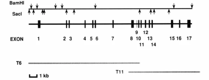
A phage library constructed from human leukocytes (Clontech, Palo Alto, CA) was screened to obtain DNA fragments from the TRKA gene. We used a human TRKA cDNA (pLM6) (Martin-Zanca et al., 1989) as a radioactively labeled probe and isolated two clones (T6 and T11), covering the entire genomic region of TRKA, as shown in Fig. 1. We
previously characterized the TRKA gene encoding the intracellular domain (Indo et al., 1996).
In the present study, all the exon/intron splice junctions were determined by comparing the human TRKA genomic sequences with the human TRKA cDNA sequences. Size of introns was estimated by the sequence of restriction fragments or polymerase chain reaction. The human TRKA gene divided into 17 exons ranged in size from 18 bases (exon 9) to 394 bases (exon 17), and 16 introns ranged in size from 170 bases (intron 9) to at least 3.3 kb (intron 1).
The entire human TRKA gene was estimated to span at least 23 kb. The sequences of exon- intron boundaries are presented in Table 1. All of the splice donor and acceptor sites
conformed to the GT/AG rule for nucleotides immediately flanking the exon border (Shapiro and Senapathy, 1987).
A single transmembrane domain divides the TRKA protein into an extracellular and an intracellular domain (Snider, 1994; Barbacid, 1995). The extracellular domain is important for specific NGF binding and includes a signal peptide, three tandem leucine-rich motifs flanked by two cysteine clusters, and two immunoglobulin-like domains (or motifs). The intracellular domain includes a juxtamembrane region, a tyrosine kinase domain, and a very short carboxy-terminal tail (Barbacid, 1995). We found a general correlation between the genomic organization of the TRKA gene and the functional organization of TRKA protein.
Exon 1 contains the signal peptide and the first cysteine cluster. Three leucine-rich motifs are encoded by exons 2, 3 and 4, respectively. This suggests that simple duplication can account for the variable numbers of the motif. Exon 5 contains the second cysteine cluster. The first immunoglobulin-like motif is encoded by exons 6 and 7, while the second immunoglobulin-
like motif is encoded by the single exon 8. Thus, the splice sites of the TRKA gene encoding the extracellular domain separate the functional domains so that each domain is encoded by separate exons. Exon 9 is a small (18-bp) one incorporated into mRNA by alternative splicing and six amino acid residues encoded by this exon are present in the extracellular domain of the neuronal-specific TRKA receptor (Barker et al., 1993). The transmembrane domain is encoded by exons 10 and 11 and the intracellular domain of TRKA is encoded by exons 11-17. The juxtamembrane domain is encoded by exons 11 and 12. The domain contains an IXNPXpY motif where p indicates phosphorylation at Tyr-490 residue of the activated TRKA (Dikic et al., 1995). This motif is encoded by exon 12 and is recognized by an Shc adaptor protein required for activation of the Ras-MAPK pathway (Obermeier et al., 1994; Stephens et al., 1994). The tyrosine kinase domain which is phosphorylated in
response to NGF and is critical for the intercellular signaling is encoded by exons 13-17. A consensus sequence motif YXXM which interacts with phosphatidylinositol-3’ kinase, is located at the end of the kinase catalytic domain (Tyr-751 residue in TRKA) (Obermeier et al., 1993a; Soltoff et al., 1992) and encoded by exon 17. The short carboxy-terminal tail of 15 amino acids is also encoded by exon 17 and includes a conserved Tyr residue (Tyr-785 in TRKA) which is responsible for binding of phospholipase Cγ (Obermeier et al., 1993b; Loeb
et al., 1994). Thus, the functional domains or motifs are generally encoded by different exons, except for exons 10 and 11 which contain a small portion of the transmembrane region and a portion of the juxtamembrane region, respectively.
In addition, there were discrepancies between the nucleotide sequences in exons 7 and 8 of the genomic clone (T6) and those noted in the cDNA (Martin-Zanca et al., 1989). A single base substitution of T-871 to G in exon 7 and substitutions of dinucleotide CG (983 and 984) to GC in exon 8 changed codons as follows: Leu-263 to Val and Ser-300 to Cys, respectively.
We also found these two substitutions in cDNAs from two normal controls and four CIPA subjects (data not shown). These amino acid changes were located in the immunoglobulin- like motifs 1 and 2, respectively. In rat TRKA cDNA, two amino acids corresponding to these residues are also Val and Cys, respectively (Meakin et al., 1992). It is noteworthy because this immunoglobulin-like motif-2 is the structural element that determines the interaction of
neurotrophins with their receptors (Urfer et al., 1995). If our data are accurate, two Cys residues should be conserved in the second immunoglobulin-like motif of all human TRK family members as in the first immunoglobulin-like motif (Nakagawara et al., 1995).
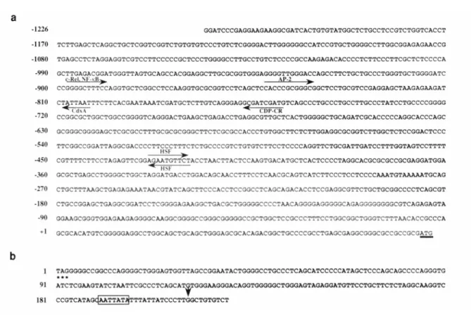
The nucleotide sequence of 1226 bp upstream from the base number 1 of the TRKA cDNA (Martin-Zanca, et al., 1989) is shown in Fig. 2a. A consensus CAAT or TATA elements were not present upstream of the putative region for transcription initiation. To determine regulatory sites in the TRKA gene, we used a computer program “TFSEARCH”
based on a database, “TRANSFAC”, which compiled eukaryotic cis-acting regulatory DNA elements and trans-acting factors (Wingender, 1994). Sequences from vertebrates were selected on the threshold of 95.0. Sequences similar to the binding site for several
transcription factors located between -420 and -990. Sequences homologous to the binding site for the following proteins were seen in this region: c-Rel /NF-κB (Kunsch et al., 1992;
Baeuerle and Henkel, 1994; Schmidt et al., 1996) at position -898, AP-2 (Mitchell et al., 1991; Faisst and Meyer, 1992) at position -852, CdxA (Margalit et al., 1993; Frumkin et al., 1993) at position -809, CCAAT displacement protein (CDP-CR) (Neufeld et al., 1992;
Harada et al., 1995) at position -767, and heat shock transcription factor (HSF) at position - 429 (both directions) (Kroeger and Morimoto, 1994). Determination of the precise putative promoter sites in transcriptional regulation of the TRKA gene is the subject of ongoing study.
The nucleotide sequence of the 3’ exon region of the gene is shown in Fig. 2b. The site of the polyadenylation signal was inferred from the cDNA. A noncanonical polyadenylation signal was present 21 bases upstream of the polyadenine tail.
Oncogenic rearrangements often involve the same region of the TRKA gene, resulting in the same junction. Our study indicates that the region frequently involved in the
rearrangements is located in exons 8 through 12 of the TRKA. The structural and genomic organization of the whole human TRKA gene will provide a basis for elucidation of mechanisms responsible for such rearrangement(s). In addition, we found a microsatellite region (GT or CA repeat) located in intron 12. The nucleotide sequence flanking this locus was reported (Greco et al., 1993) and discussed (Butti et al., 1995). Position of this locus (AFMa127wh9) and data on the heterozygosity were described (Dib et al., 1996).
Clinically, CIPA is a serious illness that might be fatal in the first years of life if the hyperpyrexia is not properly overcome. In older children, osteomyelitis and bone and/or joint deformities demand surgical procedures sometimes involving extensive amputations (Dyck, 1984; McKusick, 1994). To data, three different mutations in four families have been identified. A deletion-, a splice- and a missense-mutations all in the region encoding the tyrosine kinase domain were detected in these families. The present study revealed that a single base deletion and missense mutations are located in exon 14 of this gene. Splice mutation is located in the 5’-splice donor site of intron 15. This study will facilitate analyses of CIPA mutations in other regions of TRKA. Identification of such mutations will further genetic diagnoses of this painless but serious disease.
Acknowledgements
We thank L.F. Parada for providing the pLM6 plasmid and M. Ohara for assistance with the manuscript. This work was funded in part by a Grant-in-Aid for Scientific Research (C) from the Ministry of Education, Science, Sports and Culture of Japan and the Research Grant (8A-7) for Nervous and Mental Disorders from the Ministry of Health and Welfare of Japan.
References
Baeuerle PA, Henkel T (1994): Function and activation of NF-κB in the immune system.
Annu Rev Immunol 12: 141-179
Barbacid M (1995): Structural and functional properties of the TRK family of neurotrophin receptors. Ann N Y Acad Sci 766: 442-458
Barker PA, Lomen HC, Gensch EM, Meakin SO, Glass DJ, Shooter EM (1993): Tissue- specific alternative splicing generates two isoforms of the trkA receptor. J Biol Chem 268: 15150-15157
Butti MG, Bongarzone I, Ferraresi G, Mondellini P, Borrello MG, Pierotti MA (1995): A sequence analysis of the genomic regions involved in the rearrangements between TPM3 and NTRK1 genes producing TRK oncogenes in papillary thyroid carcinomas. Genomics 28: 15-24
Dib C, Faure S, Fizames C, Samson D, Drouot N, Vignal A, Millasseau P, Marc S, Hazan J, Seboun E, Lathrop M, Gyapay G, Morissette J, Weissenbach J (1996): A comprehensive genetic map of the human genome based on 5,264 microsatellites. Nature 380: 152-154 Dikic I, Batzer AG, Blaikie P, Obermeier A, Ullrich A, Schlessinger J, Margolis B (1995):
Shc binding to nerve growth factor receptor is mediated by the phosphotyrosine interaction domain. J Biol Chem 270: 15125-15129
Dyck PJ (1984): Neuronal atrophy and degeneration predominantly affecting sensory and autonomic neurons. In: Dyck PJ, Thomas PK, Lambert EH, Bunge R (eds). Peripheral neuropathy. W. B. Saunders Company, Philadelphia. pp 1557-1599
Faisst S, Meyer S (1992): Compilation of vertebrate-encoded transcription factors. Nucleic Acids Res 20: 23-26
Frumkin A, Haffner R, Shapira E, Tarcic N, Gruenbaum Y, Fainsod A (1993): The chicken CdxA homeobox gene and axial positioning during gastrulation. Development 118: 553-562
Greco A, Mariani C, Miranda C, Pagliardini S, Pierotti MA (1993): Characterization of the NTRK1 genomic region involved in chromosomal rearrangements generating TRK oncogenes. Genomics 18: 397-400
Greco A, Mariani C, Miranda C, Lupas A, Pagliardini S, Pomati M, Pierotti MA (1995): The DNA rearrangement that generates the TRK-T3 oncogene involves a novel gene on chromosome 3 whose product has a potential coiled-coil domain. Mol Cell Biol 15: 6118- 6127
Harada R, Berube G, Tamplin OJ, Denis LC, Nepveu A (1995): DNA-binding specificity of the cut repeats from the human cut-like protein. Mol Cell Biol 15: 129-140
Indo Y, Tsuruta M, Hayashida Y, Karim MA, Ohta K, Kawano T, Mitsubuchi H, Tonoki H, Awaya Y, Matsuda I (1996): Mutations in the TRKA/NGF receptor gene in patients with congenital insensitivity to pain with anhidrosis. Nature Genet 13: 485-488
Kaplan DR, Hempstead BL, Martin ZD, Chao MV, Parada LF (1991): The trk proto- oncogene product: a signal transducing receptor for nerve growth factor. Science 252:
554-558
Klein R, Jing SQ, Nanduri V, O'Rourke E, Barbacid M (1991): The trk proto-oncogene encodes a receptor for nerve growth factor. Cell 65: 189-197
Kozma SC, Redmond SM, Fu XC, Saurer SM, Groner B, Hynes NE (1988): Activation of the receptor kinase domain of the trk oncogene by recombination with two different cellular sequences. EMBO J 7: 147-154
Kroeger PE, Morimoto RI (1994): Selection of new HSF1 and HSF2 DNA-binding sites reveals difference in trimer cooperativity. Mol Cell Biol 14: 7592-7603
Kunsch C, Ruben SM, Rosen CA (1992): Selection of optimal κB/Rel DNA-binding motifs:
interaction of both subunits of NF-κB with DNA is required for transcriptional activation.
Mol Cell Biol 12: 4412-4421
Levi-Montalcini R (1987): The nerve growth factor: thirty-five years later. EMBO J 6: 1145- 1154
Loeb DM, Stephens RM, Copeland T, Kaplan DR, Greene LA (1994): A Trk nerve growth factor (NGF) receptor point mutation affecting interaction with phospholipase C-γ 1 abolishes NGF-promoted peripherin induction but not neurite outgrowth. J Biol Chem 269: 8901-8910
Margalit Y, Yarus S, Shapira E, Gruenbaum Y, Fainsod A (1993): Isolation and
characterization of target sequences of the chicken CdxA homeobox gene. Nucleic Acids Res 21: 4915-4922
Martin-Zanca D, Hughes SH, Barbacid M (1986): A human oncogene formed by the fusion of truncated tropomyosin and protein tyrosine kinase sequences. Nature 319: 743-748 Martin-Zanca D, Oskam R, Mitra G, Copeland T, Barbacid M (1989): Molecular and
biochemical characterization of the human trk proto-oncogene. Mol Cell Biol 9: 24-33 Martin-Zanca D, Barbacid M, Parada LF (1990): Expression of the trk proto-oncogene is
restricted to the sensory cranial and spinal ganglia of neural crest origin in mouse development. Genes Dev 4: 683-694
McKusick VA (1994): Mendelian inheritance in man. The Johns Hopkins University Press, Baltimore
Meakin SO, Suter U, Drinkwater CC, Welcher AA, Shooter EM (1992): The rat trk
protooncogene product exhibits properties characteristic of the slow nerve growth factor receptor. Proc Natl Acad Sci USA 89: 2374-2378
Miozzo M, Pierotti MA, Sozzi G, Radice P, Bongarzone I, Spurr NK, Della PG (1990):
Human TRK proto-oncogene maps to chromosome 1q32-q41. Oncogene 5: 1411-1414 Mitchell PJ, Timmons PM, Hebert JM, Rigby PW, Tjian R (1991): Transcription factor AP-2
is expressed in neural crest cell lineages during mouse embryogenesis. Genes Dev 5: 105- 119
Morris CM, Hao QL, Heisterkamp N, Fitzgerald PH, Groffen J (1991): Localization of the TRK proto-oncogene to human chromosome bands 1q23-1q24. Oncogene 6: 1093-1095 Nakagawara A, Liu XG, Ikegaki N, White PS, Yamashiro DJ, Nycum LM, Biegel JA,
Brodeur GM (1995): Cloning and chromosomal localization of the human TRK-B tyrosine kinase receptor gene (NTRK2). Genomics 25: 538-546
Neufeld EJ, Skalnik DG, Lievens PM, Orkin SH (1992): Human CCAAT displacement protein is homologous to the Drosophila homeoprotein, cut. Nature Genetics 1: 50-55
Obermeier A, Lammers R, Wiesmuller KH, Jung G, Schlessinger J, Ullrich A (1993a):
Identification of Trk binding sites for SHC and phosphatidylinositol 3'-kinase and formation of a multimeric signaling complex. J Biol Chem 268: 22963-22966 Obermeier A, Halfter H, Wiesmuller KH, Jung G, Schlessinger J, Ullrich A (1993b):
Tyrosine 785 is a major determinant of Trk-substrate interaction. EMBO J 12: 933-941 Obermeier A, Bradshaw RA, Seedorf K, Choidas A, Schlessinger J, Ullrich A (1994):
Neuronal differentiation signals are controlled by nerve growth factor receptor/Trk binding sites for SHC and PLC γ. EMBO J 13: 1585-1590
Schmidt UR, Memet S, Lilienbaum A, Feuillard J, Raphael M, Israel A (1996): NF-κB
activity in transgenic mice: developmental regulation and tissue specificity. Development 122: 2117-2128
Shapiro MB, Senapathy P (1987): RNA splice junctions of different classes of eukaryotes:
sequence statistics and functional implications in gene expression. Nucleic Acids Res 15:
7155-7174
Snider WD (1994): Functions of the neurotrophins during nervous system development:
what the knockouts are teaching us. Cell 77: 627-638
Soltoff SP, Rabin SL, Cantley LC, Kaplan DR (1992): Nerve growth factor promotes the activation of phosphatidylinositol 3-kinase and its association with the trk tyrosine kinase.
J Biol Chem 267: 17472-7
Stephens RM, Loeb DM, Copeland TD, Pawson T, Greene LA, Kaplan DR (1994): Trk receptors use redundant signal transduction pathways involving SHC and PLC-γ 1 to
mediate NGF responses. Neuron 12: 691-705
Swanson AG (1963): Congenital insensitivity to pain with anhidrosis. Arch Neurol 8: 299-306 Thoenen H, Barde Y-A (1980): Physiology of nerve growth factor. Physiol Rev 60: 1284-1335 Urfer R, Tsoulfas P, O'Connell L, Shelton DL, Parada LF, Presta LG (1995): An
immunoglobulin-like domain determines the specificity of neurotrophin receptors.
EMBO J 14: 2795-2805
Wingender E (1994): Recognition of regulatory regions in genomic sequences. J Biotechnol 35: 273-280
Figures
Fig. 1. Physical map of the human TRKA gene.
Fig. 1. Physical map of the human TRKA gene.
The structure of the gene is shown by a thick line. Exons 1-17 are shown as vertical lines and numbered. Below the gene structure, the genomic DNA fragments from the phage clones are shown by dotted lines. BamHI and SacI sites are shown above.
Fig. 2. Nucleotide sequences of the 5’ and 3’ regions of the human TRKA gene.
Fig. 2. Nucleotide sequences of the 5’ and 3’ regions of the human TRKA gene.
(a) Nucleotide sequence of the 5’ region of the gene.
The sequence is numbered according to Martin-Zanca et al (Martin-Zanca et al., 1989), starting from guanine at base number 1 of the cDNA. Sequences with a horizontal arrow indicate the putative region resembling binding sites for specific transcription factors. The directions of the sequence are indicated by the directions of the arrows. The translation start codon ATG is underlined.
(b) Nucleotide sequences of the 3’ region of the gene.
The termination codon, TAG, is marked (***). The sequence is numbered starting from thymidine of the termination codon. A vertical arrowhead indicates the poly(A) addition site.
The noncanonical poly(A) addition signal is boxed.
Table 1
Exon-Intron Organization of the human TRKA gene
Size cDNA Size
Exon (bp) positiona Intron (kb) Exon Intron Exon
1 296
1-296
1 3.3 GAG CT
Glu Leu gtgagtgtccggcgggcggt ... cccatccgctctccccacag C TAC Leu Tyr 2 75 297-371 2 0.3 AAC CT
Asn Leu gtgagggaaacggggactgt ... cctcctgcacccctccccag C ACC Leu Thr 3 72 372-443 3 2.1 CGC CT
Arg Leu gtgagtgtggccagtgctgg ... ctgtgtctccacgcccgcag G AAT Leu Asn 4 69 444-512 4 1.2 GAA CT
Glu Leu gtgagtgggggcgcttccag ... gtgtcccccatgccccccag G GTC Leu Val
5 146 513-658 5 0.3 TGT G
Cys Gly gtaggtgccgggtgagggag ... cgggcgtcctgggtggccag GT GTG Gly GTG 6 143 659-801 6 2.3 GTG ATG
Val Met gtgagaagaccttcgctggc ... ccctctctttcctgatctag AAA TCT Lys Ser
7 133 802-934 7 2.1 TCC T
Ser Phe gtgagtctcagtggcagctc ... ttgctctttctggcccacag TC CCG Phe Pro 8 327 935-1261 8 0.4 CCT G
Pro Val gtgcgagggccatcctgaac ... tctcctccctcctgctgcag TC TCC Val Ser
9b 18 - 9 0.2 GTG G
Val Asp gtgagtagcccaaggtggag ... cctgccctgtgtccctacag AC ACT Asp Thr 10 56 1262-1317 10 0.3 TTT GGG
Phe Gly gtgagataggaagtagaagc ... ctacccctgtcccccaccag GTC TCG Val Ser 11 103 1318-1420 11 0.5 AAC C
Asn Arg gtgagtcggggctgcagaggg ... cggctgtgtctcctctctag GC CCG Arg Pro 12 147 1421-1567 12 0.4 GCC T
Ala Cys gtgaggggctatgctgggtc ... gaccctgcaagccccctcag GT GTT Cys Val 13 131 1568-1698 13 0.2 GTC AAG
Val Lys gtgagaccctgccccggggg ... tccctgccgcttccatccag GCA CTG Ala Leu 14 173 1699-1871 14 2.5 CTC CG
Leu Arg gtaccagcacctggcctcag ... tctccttttcttgttcacag A TCC Arg Ser 15 241 1872-2112 15 0.7 TAC CGT
Tyr Arg gtaagggtcctttgtcccca ... gcagtgtccgcccgtggcag GTG GGA Val Gly 16 159 2113-2271 16 1.2 ACG GAG
Thr Glu gtcagccccggcccatggtc ... tgtctctccggtggccccag GCA ATC Ala Ile
17 394 2272-2665 CTT G
a Numbered according to Martin-Zanca et al. (1986).
b A small exon incorporated into mRNA of neural tissues by alternative splicing (Barker et al., 1993).