Fukushima Medical University
This document is downloaded at: 2021-11-07T23:17:35Z
Title Baseline serum syndecan-4 predicts prognosis after the onset of acute exacerbation of idiopathic interstitial pneumonia( 本文 )
Author(s) 佐藤, 佑樹
Citation
Issue Date 2018-03-21
URL http://ir.fmu.ac.jp/dspace/handle/123456789/757
Rights © The Author(s). This is the peer reviewed version. Published version is "PLoS One. 2017 May 3;12(5):e0176789. doi:
10.1371/journal.pone.0176789", used under CC BY 4.0 DOI
Text Version ETD
Baseline Serum Syndecan-4 Predicts Prognosis after the Onset
1
of Acute Exacerbation of Idiopathic Interstitial Pneumonia
2
3 4
Yuki Sato
5 6 7
Department of Pulmonary Medicine, Fukushima Medical University School of
8
Medicine, Fukushima, Fukushima, Japan
9 10 11 12 13 14 15 16 17 18 19 20
1
Abstract
1 2
Background: Patients with idiopathic interstitial pneumonia can experience
3
acute respiratory worsening, also known as acute exacerbation, with a large
4
deterioration on prognosis. The precise mechanism remains unclear; however,
5
syndecan-4 may be involved. Syndecan-4, a transmembrane heparan sulfate
6
proteoglycan expressed in a variety of cells (e.g., epithelial cells, macrophages,
7
fibroblasts, etc.), performs various biological roles by binding to several proteins
8
through its heparan sulfate glycosaminoglycan side chains. The goal of this study
9
was to clarify the role of syndecan-4 in acute exacerbation of idiopathic interstitial
10
pneumonia.
11
Methods: Patients with idiopathic interstitial pneumonia who had been
12
sequentially admitted to our hospital due to acute exacerbation were
13
retrospectively analyzed. First, serum syndecan-4 levels in the acute
14
exacerbation and clinically stable phases were compared. Second, the
15
relationship between serum syndecan-4 levels and clinical parameters was
16
analyzed. Third, the relationship between serum syndecan-4 levels and
17
prognosis was evaluated.
18
2
Results: Serum syndecan-4 levels were significantly lower in patients with acute
1
exacerbation of idiopathic interstitial pneumonia than in patients in the clinically
2
stable phase. Serum syndecan-4 levels also showed a significant positive
3
correlation with white blood cell count and a weak positive tendency with KL-6
4
and baseline %VC. Prognosis was significantly worse in patients with idiopathic
5
interstitial pneumonia with high baseline serum syndecan-4 levels than with low
6
baseline levels. Multiple logistic analysis indicated baseline serum syndecan-4
7
level as the only prognostic predictor following acute exacerbation.
8
Conclusions: Baseline serum syndecan-4 is a possible prognostic biomarker
9
after the onset of acute exacerbation of idiopathic interstitial pneumonia.
10 11
12 13 14 15 16 17 18
3
Introduction
1
Idiopathic pulmonary fibrosis (IPF) is a chronic, progressive, and intractable
2
fibrosing lung disease that can progress to rapid respiratory failure during the
3
disease course. IPF of unknown cause and without symptoms of left heart failure,
4
pulmonary embolism, or lung infection is referred to as acute exacerbation (AE)
5
of IPF [1-8]. The reported frequency of AE is 1, 2, or 3 years, at rates of 8.6%,
6
12.6%, and 23.9%, respectively [9]. The post-onset mortality rate is
7
approximately 50%, so IPF has an extremely poor prognosis [10,11]. AE
8
accounts for 40% of all IPF-related deaths [12], making it a critical event during
9
the clinical course of IPF. However, the pathogenesis of AE is poorly understood,
10
and no standard treatment is yet available.
11
One potential factor that may contribute to the pathogenesis of AE of interstitial
12
pneumonia is the proteoglycan syndecan-4. Proteoglycans are glycoproteins
13
composed of a core protein associated with one or more side chain
14
glycosaminoglycans, which are highly negatively charged sulfated
15
polysaccharides. The syndecans are a family of proteoglycans associated with
16
three to five heparan sulfate or chondroitin sulfate side chains. They are
17
transmembrane proteins and are expressed on the surfaces of many cells. The
18
4
syndecan family in humans has four members—syndecan-1 to syndecan-4—that
1
have reported involvement in lung development, wound healing, and
2
inflammatory reactions [13]. Syndecans exist in cell surface forms, as well as in
3
soluble forms that are shed from the cell membrane by matrix metalloproteinases
4
(MMPs) [14-16]. The heparan sulfate side chains are thought to bind to various
5
cytokines, chemokines, and growth factors, thereby regulating biological activity
6
[17].
7
Our previous comparison of syndecan-4-deficient mice and wild-type mice
8
indicated that neutrophil migration and injury are increased in the lungs of
9
syndecan-4-deficient mice after intratracheal instillation of lipopolysaccharide
10
(LPS) [18]. We also found higher serum syndecan-4 levels in hospitalized
11
patients with bacterial pneumonia than in healthy subjects, and the syndecan-4
12
levels tended to increase over the course of pneumonia in those patients with a
13
favorable prognosis [19]. Another effect of syndecan-4 may involve its interaction
14
with the C-X-C motif chemokine ligand 10 (CXCL10), which shows an anti-fibrotic
15
effect upon binding to syndecan-4 in a bleomycin-induced mouse model of
16
pulmonary fibrosis [20]. These results suggest an important role for syndecan-4
17
in limiting lung inflammation and fibrosis. The aim of the present study was to
18
5
clarify the role of syndecan-4 in AE of idiopathic interstitial pneumonia (IIP).
1
2
Materials and Methods
3
Subjects
4
The ethics committee of Fukushima Medical University approved this work
5
(approval number: 1105), and all clinical investigation were conducted according
6
to the principles of the Declaration of Helsinki. Informed consent was not obtained
7
because the data were analyzed anonymously. Patients who were sequentially
8
hospitalized for AE of IIP (AE-IIP) at our department between 2007 and 2014,
9
patients with clinically stable IIP (SD-IIP) without subjective symptoms of dyspnea
10
or rapid deterioration on image findings for at least 3 months, and healthy
11
volunteers (HV) were included in this retrospective analysis. The diagnostic
12
criteria for AE were set according to a definition of AE in a previous report [21].
13
The criteria for inclusion were as follows: progression of dyspnea within the past
14
month; new bilateral infiltrative shadows or ground glass opacities on high-
15
resolution computed tomography (HRCT); and a decrease in partial pressure of
16
arterial oxygen (PaO2) of at least 10 torr or a PaO2/fraction of inspiratory oxygen
17
(FiO2) (P/F) ratio of < 300 mmHg. Patients with pneumonia, heart failure,
18
6
pulmonary embolism, and pneumothorax were excluded. Patients whose
1
progression of pulmonary fibrosis was clearly associated with another disease
2
(collagen disease, drug-induced lung injury, pneumoconiosis, hypersensitivity
3
pneumonitis, sarcoidosis, pulmonary Langerhans cell histiocytosis,
4
lymphangioleiomyomatosis, etc.) were also excluded. IPF was diagnosed using
5
the definition in the 2011 ATS/ERS/JRS/ALAT joint statement [22], and all patients
6
with IIP met the 2013 ATS/ERS Update of the International Multidisciplinary
7
Classification of the IIP [23]. Diagnosis of systemic inflammatory response
8
syndrome (SIRS) [24] and APACHE II classifications [25] were made according
9
to past diagnostic criteria at the time of admission. All patients underwent steroid
10
pulse therapy as treatment.
11
12
Measurement of serum syndecan-4
13
We first compared serum syndecan-4 levels in patients with AE-IIP and SD-IIP,
14
as well as in HV. Syndecan-4 levels were measured as described previously [19]
15
with a commercially available ELISA kit (IBL, Takasaki, Japan) according to the
16
manufacturer’s protocol.
17 18
7
The relationship between serum syndecan-4 and clinical
1
parameters
2
We also examined the relationship between serum syndecan-4 levels and
3
clinical characteristics, including blood laboratory findings and HRCT findings
4
upon admission.
5
6
The relationship between serum syndecan-4 and
7
prognosis
8
We compared the clinical data of a survival group and a non-survival group, as
9
defined at 60 days after admission for AE. We also used univariate and
10
multivariate analysis to evaluate prognostic factors, and we used Kaplan-Meier
11
analysis to examine the relationship between syndecan-4 level and prognosis.
12
13
Statistical analysis
14
Data were expressed as means ± the standard error of the mean (SEM). Two
15
unpaired groups were compared with the Mann-Whitney U test or Fisher’s exact
16
test, while multiple groups were compared with ANOVA with the Tukey HSD.
17
Serum syndecan-4 levels in each IIP patient during AE and during the stable
18
8
phases (baseline) were analyzed with Wilcoxon’s signed-rank test. Correlations
1
between serum syndecan-4 levels and clinical parameters were analyzed using
2
Spearman’s correlation coefficient and survival predictors 60 days after
3
admission were subjected to logistic regression analysis in the univariate and
4
multivariate analyses. Prognoses according to syndecan-4 levels were
5
compared with a log-rank test. The analysis was performed using IBM SPSS
6
Statistics 17.0 software (IBM Japan; Tokyo, Japan), and a p value of < 0.05 was
7
considered statistically significant.
8
9
Results
10
Clinical characteristics
11
Fifty-six AE-IIP patients were hospitalized during the study period; 21 were
12
patients with IPF and 35 were patients with IIP other than IPF (non-IPF). The
13
patients had a mean age of 70 ± 1 years, and included 47 men and nine women.
14
The AE-IIP patients had a reduced P/F ratio of 206 ± 16 mmHg, as well as
15
elevated KL-6, surfactant protein (SP)-A, and SP-D levels of 1524 ± 127 U/mL
16
(reference < 500 U/mL), 119 ± 7 ng/mL (reference < 43.8 ng/mL), and 344 ± 53
17
ng/mL (reference < 110 ng/mL), respectively (Table 1). A comparison of the AE-
18
9
IIP and SD-IIP patients revealed a significantly higher white blood cell (WBC)
1
count, significantly higher lactate dehydrogenase (LDH), SP-A, and C-reactive
2
protein levels, and a higher erythrocyte sedimentation rate in the AE-IIP patients.
3
The levels of SP-D also tended to be higher in the AE-IIP patients.
4
5
Table 1. Clinical Characteristics of Healthy Volunteers, Patients with AE-IIP,
6
and Patients with SD-IIP
7
Healthy Volunteers
Patients With SD-IIP
Patients With AE-IIP
P Value
Subjects (n) 45 62 56
Age (years) 43±2*† 68±1 70±1 <0.01
Gender (M/F) 29/16 47/15 47/9 0.078
IPF/non IPF (n) NA 33/29 21/35 0.063
Duration from diagnosis to hospitalization for AE (months)
NA NA 24±4
Duration of symptoms before admission (days)
NA NA 9±1
Survivor /Non-survivor at 60 days after admission (n)
NA NA 25/31
Glucocorticoid pulse therapy (n) NA NA 56
Cyclophosphamide pulse therapy (n) NA NA 7
Sivelestat (n) NA NA 23
10
WBC (/μl) NA 7907±315 10439±618 0.001
LDH (IU/L) NA 260±8 450±56 <0.001
KL-6 (U/ml) NA 1463±144 1524±127 0.485
SP-A (ng/ml) NA 98±8 119±7 0.012
SP-D (ng/ml) NA 250±24 344±53 0.084
CRP (mg/dl) NA 1.36±0.48 7.97±0.88 <0.001
ESR (mm/h) NA 28±3 39±4 0.012
PCT (ng/ml) NA NA 0.65±0.27
BNP (pg/ml) NA NA 90.1±21.2
FBG (mg/dl) NA NA 453±16
FDP (μg/ml) NA NA 13.7±3.5
D-dimer (μg/ml) NA NA 6.6±1.6
P/F on admission (mmHg) NA NA 206±16
SIRS (+/-) NA NA 34/22
SIRS score NA NA 1.9±0.1
APACHE II score NA NA 13.7±0.7
VC (L) NA 2.55±0.18 NA
%VC (%) NA 79.8±4.8 NA
Serum syndecan-4 (ng/ml) 16.05±0.77 25.22±3.72*ǂ 10.65±0.73 <0.001
SD: stable disease, AE: acute exacerbation, IPF: idiopathic pulmonary fibrosis, WBC: white blood cell count, LDH: lactate
1
dehydrogenase, KL-6: Krebs von den Lungen-6, SP-A: surfactant protein-A, SP-D: surfactant protein-D, CRP: c-reactive
2
protein, ESR: erythrocyte sedimentation rate, PCT: procalcitonin, BNP: brain natriuretic peptide, FBG: fibrinogen, FDP:
3
fibrin and fibrinogen degradation product, P/F: arterial partial pressure of carbon dioxide/Fraction of inspiratory oxygen,
4
SIRS: systemic inflammatory response syndrome, VC: vital capacity. APACHE II score: acute physiology and chronic
5
health evaluation II score. * Healthy Volunteers vs Patients with SD-IIP. †Healthy Volunteers vs Patients with AE-IIP. ǂ
6
Patients with SD-IIP vs Patients with AE-IIP. Mean ± SEM.
7
11
Serum syndecan-4 levels in AE-IIP patients
1
A comparison of serum syndecan-4 levels among the HV, SD-IIP, and AE-IIP
2
groups revealed significantly higher serum syndecan-4 levels in the SD-IIP
3
patients than in the HV and AE-IIP groups (25.22 ± 3.72 ng/mL vs. 16.05 ± 0.77
4
ng/mL vs. 10.65 ± 0.73 ng/mL; Fig 1). A comparison of the serum syndecan-4
5
levels between baseline and AE for 34 of the 56 AE-IIP patients for whom analysis
6
was possible during stability revealed significantly lower serum syndecan-4 levels
7
during AE than at baseline for the same patients (14.06 ± 0.78 ng/mL vs. 10.47 ±
8
0.69 ng/mL, p = 0.01 for baseline and AE, respectively; Fig 2). The mean time
9
between blood sampling and AE was 556 ± 104 days.
10
12 1
Fig 1. Comparison of serum syndecan-4 levels in healthy volunteers and in
2
patients with SD-IIP and AE-IIP. Serum syndecan-4 levels were significantly
3
higher in the SD-IIP group than in the HV and AE-IIP groups. Serum syndecan-4
4
levels did not differ between the HV and AE-IIP groups. HV: healthy volunteer
5
group, SD-IIP: patient group with stable idiopathic interstitial pneumonia, AE-IIP:
6
patient group with acute exacerbation of idiopathic interstitial pneumonia. ANOVA
7
with Tukey HSD was used for statistical analysis. *: p < 0.05 vs HV. †: p < 0.001
8
vs AE-IIP. Data are expressed as means ± the standard error of the mean (SEM).
9
13 1
Fig 2. Changes in serum syndecan-4 levels of IIP patients before and upon
2
AE. Serum syndecan-4 levels were compared in the same patients for whom
3
analysis was possible during stability and upon acute exacerbation (n = 34).
4
Serum syndecan-4 levels were significantly lower during AE than in the stable
5
phase. AE: acute exacerbation. Wilcoxon’s signed-rank test was used for
6
statistical analysis.
7
8
The relationship between serum syndecan-4 levels and
9
clinical parameters
10
We also examined the correlation between the serum syndecan-4 levels of the
11
14
AE-IIP patients upon admission for AE and their clinical parameters, including
1
blood laboratory results, SIRS score, and APACHE II score. Serum syndecan-4
2
levels during AE were significantly positively correlated with WBC count and
3
showed a weakly positive tendency to correlation with KL-6 and baseline %vital
4
capacity (VC); however, no correlations were observed with SP-A or SP-D (Table
5
2). The possible correlation between serum syndecan-4 levels and clinical
6
parameters was also analyzed separately in the IPF and non-IPF groups by first
7
analyzing the correlation between serum syndecan-4 levels on admission and the
8
clinical parameters evaluated in Table 2. The IPF patients (n = 21) showed results
9
consistent with those of all AE-IIP patients, i.e., a significant positive correlation
10
was observed between serum syndecan-4 levels on admission and WBC count.
11
By contrast, analysis of non-IPF patients (n = 35) revealed a significant positive
12
correlation between syndecan-4 levels on admission and KL-6 and baseline %VC
13
and a significant negative correlation with PaO2. Division of the 34 AE-IIP patients
14
with baseline syndecan-4 levels into a low baseline serum syndecan-4 group and
15
a high baseline serum syndecan-4 group (based on a median baseline syndecan-
16
4 level of 12.8 ng/mL) revealed no differences in the two groups for time from
17
baseline to AE (423 ± 148 days vs. 688 ± 142 days) or frequency of use of
18
15
pirfenidone (7/17 vs. 4/17), prednisolone (9/17 vs. 9/17), or immunosuppressants
1
(2/17 vs. 6/17) during stability. The two groups also showed no differences in LDH
2
(274 ± 11 IU/L vs. 282 ± 20 IU/L), KL-6 (1327 ± 171 U/mL vs. 1166 ± 167 U/mL),
3
SP-A (106 ± 17 ng/mL vs. 84 ± 13 ng/mL), SP-D (269 ± 51 ng/mL vs. 217 ± 33
4
ng/mL), VC (2.02 ± 0.26 L vs. 2.04 ± 0.27 L), or %VC (64.3 ± 6.8% vs. 91.0 ±
5
30.1%; Table 3) during stability. No correlations were observed between the
6
baseline serum syndecan-4 levels and the time to onset of AE. The baseline
7
syndecan-4 levels also showed no correlation with LDH, KL-6, SP-A, SP-D,
8
baseline VC, or %VC. The syndecan-4 levels also did not differ between IPF and
9
non-IPF patients, in either the stability period (13.69 ± 4.63 ng/mL vs. 14.34 ±
10
4.62 ng/mL) or the exacerbation period (9.19 ± 3.86 ng/mL vs. 11.53 ± 6.17
11
ng/mL).
12
13
Table 2: Correlations of Serum Syndecan-4 Levels on Admission with
14
Clinical Parameters
15
Correlation coefficients P value
WBC 0.426 0.001
LDH -0.074 0.587
KL-6 0.256 0.057
SP-A 0.079 0.569
SP-D 0.142 0.307
16
CRP -0.157 0.249
ESR -0.174 0.263
PCT -0.104 0.573
BNP -0.067 0.644
FDP -0.161 0.285
D-dimer -0.208 0.135
PaO2 -0.215 0.112
P/F 0.135 0.323
SIRS score -0.209 0.123
Baseline VC 0.104 0.491
Baseline %VC 0.271 0.069
APACHE II score -0.132 0.336
WBC: white blood cell count, LDH: lactate dehydrogenase, KL-6: Krebs von den Lungen-6, SP-A: surfactant protein-A,
1
SP-D: surfactant protein-D, CRP: c-reactive protein, ESR: erythrocyte sedimentation rate, PCT: procalcitonin, BNP: brain
2
natriuretic peptide, FBG: fibrinogen, FDP: fibrin and fibrinogen degradation product, PaO2: arterial partial pressure of
3
carbon dioxide, P/F: arterial partial pressure of carbon dioxide/Fraction of inspiratory oxygen, SIRS: systemic inflammatory
4
response syndrome, VC: vital capacity. APACHE II score: acute physiology and chronic health evaluation II score. Mean
5
± SEM
6 7
Table 3: Clinical Characteristics of Idiopathic Interstitial Pneumonia
8
Patients with High and Low Baseline Serum Syndecan-4 levels
9
High baseline
serum syndecan-4
group
Low baseline
serum syndecan-4
group
P value
Subjects (n) 17 17
Age (years) 71.3±1.6 67.9±1.9 0.120
Gender (M/F) 13/4 16/1 0.335
Duration until acute exacerbation from initial stable analysis (days)
423±148 688±142 0.352
17
Use of pirfenidone (+/-) 7/10 4/13 0.465
Use of oral immunosuppressive agents (+/-) 2/15 6/11 0.225
LDH (IU/L) 274±11 282±20 0.787
KL-6 (U/ml) 1327±171 1166±167 0.517
SP-A (ng/ml) 106±17 84±13 0.327
SP-D (ng/ml) 269±51 217±33 0.885
Baseline VC (L) 2.02±0.26 2.04±0.27 0.664
Baseline %VC (%) 64.3±6.8 91.0±30.1 0.748
KL-6: Krebs von den Lungen-6, SP-A: surfactant protein-A, SP-D: surfactant protein-D, VC: vital capacity. Patients with
1
patients with acute exacerbation of idiopathic interstitial pneumonia were stratified according to median serum syndecan-
2
4 concentration (Low baseline serum syndecan-4 group; < 12.8 ng/ml. High baseline serum syndecan-4 group; > 12.8
3
ng/ml). All blood laboratory parameters were analyzed at the time of blood sampling during stability. Mean ± SEM.
4 5
The relationship between serum syndecan-4 levels and
6
prognosis
7
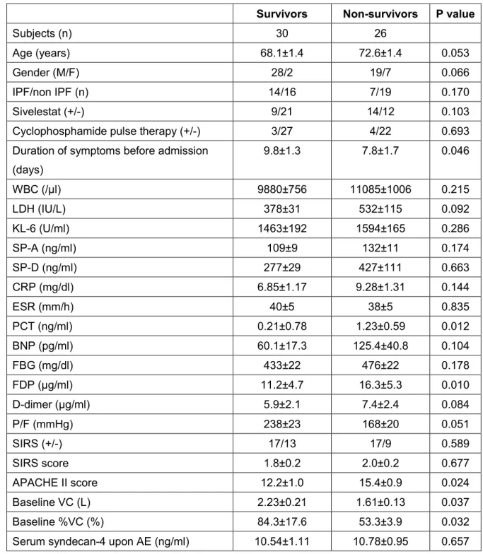
We compared prognoses 60 days after admission for AE in the 56 patients with
8
AE-IIP. After 60 days, the survival rate was 53.6% (30 patients). Comparison of
9
the clinical characteristics of the survival (n = 30) and non-survival (n = 26) groups
10
revealed higher FDP and APACHE II levels, lower baseline VC and %VC, and
11
significantly shorter duration between initial stable analysis and AE for the non-
12
survival than for the survival group (Table 4). The serum syndecan-4 levels upon
13
admission for AE did not differ between the two groups, but the non-survival group
14
had a significantly higher baseline serum syndecan-4 level (16.13 ± 1.12 ng/mL
15
18
vs. 12.61 ± 0.97 ng/mL).
1
2
Table 4: Comparison of the Clinical Characteristics of Survivors and Non-
3
survivors of AE-IIP
4
Survivors Non-survivors P value
Subjects (n) 30 26
Age (years) 68.1±1.4 72.6±1.4 0.053
Gender (M/F) 28/2 19/7 0.066
IPF/non IPF (n) 14/16 7/19 0.170
Sivelestat (+/-) 9/21 14/12 0.103
Cyclophosphamide pulse therapy (+/-) 3/27 4/22 0.693
Duration of symptoms before admission (days)
9.8±1.3 7.8±1.7 0.046
WBC (/μl) 9880±756 11085±1006 0.215
LDH (IU/L) 378±31 532±115 0.092
KL-6 (U/ml) 1463±192 1594±165 0.286
SP-A (ng/ml) 109±9 132±11 0.174
SP-D (ng/ml) 277±29 427±111 0.663
CRP (mg/dl) 6.85±1.17 9.28±1.31 0.144
ESR (mm/h) 40±5 38±5 0.835
PCT (ng/ml) 0.21±0.78 1.23±0.59 0.012
BNP (pg/ml) 60.1±17.3 125.4±40.8 0.104
FBG (mg/dl) 433±22 476±22 0.178
FDP (μg/ml) 11.2±4.7 16.3±5.3 0.010
D-dimer (μg/ml) 5.9±2.1 7.4±2.4 0.084
P/F (mmHg) 238±23 168±20 0.051
SIRS (+/-) 17/13 17/9 0.589
SIRS score 1.8±0.2 2.0±0.2 0.677
APACHE II score 12.2±1.0 15.4±0.9 0.024
Baseline VC (L) 2.23±0.21 1.61±0.13 0.037
Baseline %VC (%) 84.3±17.6 53.3±3.9 0.032
Serum syndecan-4 upon AE (ng/ml) 10.54±1.11 10.78±0.95 0.657
19
Change in serum syndecan-4* (ng/ml) 2.30±1.01 5.43±1.11 0.142
AE: acute exacerbation, IPF: idiopathic pulmonary fibrosis, WBC: white blood cell count, LDH: lactate dehydrogenase,
1
KL-6: Krebs von den Lungen-6, SP-A: surfactant protein-A, SP-D: surfactant protein-D, CRP: c-reactive protein, ESR:
2
erythrocyte sedimentation rate, PCT: procalcitonin, BNP: brain natriuretic peptide, FBG: fibrinogen, FDP: fibrin and
3
fibrinogen degradation product, P/F: arterial partial pressure of carbon dioxide/Fraction of inspiratory oxygen, SIRS:
4
systemic inflammatory response syndrome, VC: vital capacity. APACHE II score: acute physiology and chronic health
5
evaluation II score. * Difference in serum syndecan-4 between before and upon AE in the identical patients. Survival was
6
evaluated at 60 days after admission. All blood laboratory parameters were analyzed on admission. Mean ± SEM.
7 8
A univariate analysis of prognostic factors for survival 60 days after admission
9
for AE revealed that age, P/F ratio, APACHE II scores, baseline VC, and baseline
10
serum syndecan-4 levels were predictive factors for death. A significant multi-
11
collinearity was observed between P/F ratio and APACHE II scores, so we chose
12
four variables (age, APACHE II scores, baseline VC, and baseline serum
13
syndecan-4 levels) for multivariate analysis. This analysis revealed baseline
14
serum syndecan-4 level alone as a significant prognostic factor (hazard ratio
15
[HR]: 1.286, 95% confidence interval [CI]: 1.044–1.584; p < 0.05; Table 5).
16
17
Table 5: Univariate Analysis of Survival Prediction
18
Univariate analysis
19
Variable HR 95%CI P value
Age 1.085 1.005–1.171 0.037
Duration of symptoms before admission (days)
0.968 0.903–1.037 0.352
20
PCT 4.673 0.622–35.102 0.134
FDP 1.010 0.983–1.037 0.480
D-dimer 1.012 0.964–1.061 0.640
P/F 0.994 0.989–1.000 0.032
APACHE II score 1.142 1.017–1.283 0.025
Baseline VC 0.376 0.149–0.948 0.038
Baseline %VC 0.975 0.948–1.002 0.074
Baseline serum syndecan-4 1.216 1.015–1.458 0.034
PCT: procalcitonin, FDP: fibrin and fibrinogen degradation product, P/F: arterial partial pressure of carbon dioxide/Fraction
1
of inspiratory oxygen, APACHE II score: acute physiology and chronic health evaluation II score, VC: vital capacity.
2 3
Multivariate analysis
4
Variable HR 95%CI P value
Age 0.983 0.854–1.130 0.804
APACHE IIscore 1.123 0.911–1.385 0.278
Baseline VC 0.273 0.058–1.283 0.100
Baseline serum syndecan-4 1.286 1.044–1.584 0.018
APACHE II score: acute physiology and chronic health evaluation II score, VC: vital capacity.
5 6
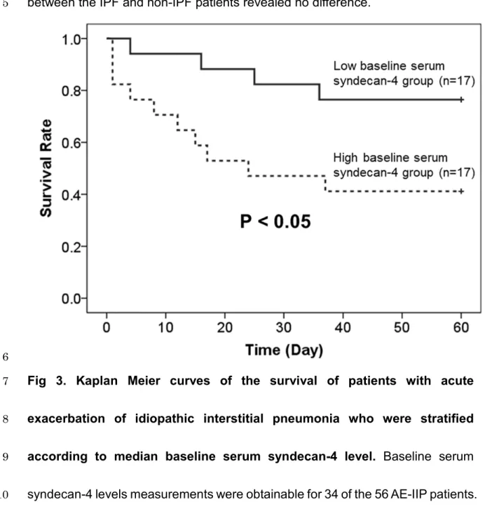
Baseline serum syndecan-4 levels were available for 34 of the 56 AE-IIP
7
patients; therefore, we divided these patients into high (n = 17) and low (n = 17)
8
baseline serum syndecan-4 groups, using the median baseline serum syndecan-
9
4 level (12.8 ng/mL) as a reference. As shown in Fig 3, prognosis was poorer for
10
the high baseline serum syndecan-4 group. A further division of these 34 AE-IIP
11
patients into groups with a high (n = 17) and low (n = 17) serum syndecan-4 upon
12
AE, using the median serum syndecan-4 level (11.3 ng/mL) during AE as a
13
21
reference, revealed no difference in prognosis. Analysis of all 56 AE-IIP patients
1
also revealed no difference in prognosis between the groups with high (n = 28)
2
and low (n = 28) serum syndecan-4 upon AE, using the median serum syndecan-
3
4 level (10.6 ng/mL) during AE as a reference. Comparison of the prognosis
4
between the IPF and non-IPF patients revealed no difference.
5
6
Fig 3. Kaplan Meier curves of the survival of patients with acute
7
exacerbation of idiopathic interstitial pneumonia who were stratified
8
according to median baseline serum syndecan-4 level. Baseline serum
9
syndecan-4 levels measurements were obtainable for 34 of the 56 AE-IIP patients.
10
22
The survival rate was significantly better for patients with low baseline serum
1
syndecan-4 levels than with high baseline serum syndecan-4 levels. (Low
2
baseline serum syndecan-4 group, < 12.8 ng/ml; High baseline serum syndecan-
3
4 group, > 12.8 ng/ml.) A log-rank test was used for statistical analysis.
4
5
Discussion
6
In the present study, we demonstrated that: (1) baseline serum syndecan-4
7
levels were higher in patients with IIP than in the HV group, and the levels
8
decreased during AE; (2) serum syndecan-4 levels during AE were positively
9
correlated with WBC count and showed weak positive tendencies with KL-6 and
10
baseline %VC; (3) after AE, significantly higher baseline serum syndecan-4 levels
11
were observed in the non-survival group than in the survival group; and (4)
12
baseline serum syndecan-4 levels were the sole factor capable of predicting
13
prognosis after AE in a multivariate analysis.
14
The AE of interstitial pneumonia is a clinically important event that influences
15
the prognosis of patients with interstitial pneumonia. In IPF, AE occurs at
16
frequencies of 8.6% and 23.9% at 1 and 3 years after diagnosis, respectively,
17
with a reported post-onset mortality rate of approximately 50%. AE therefore
18
23
accounts for 40% of all IPF-related deaths. AE also occurs in forms of interstitial
1
pneumonia other than IPF [26, 27], with suspected involvement of viral infection,
2
aspiration, exposure to air pollution, progression of abnormal fibroproliferation
3
related to acute stress on the lungs, and other factors [28-30]. Increased
4
neutrophil counts in bronchoalveolar lavage fluid [31], and histopathological
5
findings of diffuse alveolar damage characterized by the formation of a hyaline
6
membrane, similar to those of acute respiratory distress syndrome are observed
7
[32-36]. However, organizing pneumonia and extensive fibroblastic foci can also
8
be present, in addition to diffuse alveolar damage [37]. The presence of
9
considerable epithelial injury has also been suggested due to higher levels of KL-
10
6 and SP-D during AE of IPF than during stability [38, 39]. The levels of pro-
11
inflammatory cytokines, such as CXCL1 and CXCL8/IL-8, may increase, together
12
with the levels of M2 cytokines such as CCL18. M2 cytokines are considered to
13
play an important role in wound healing, so these increases suggest strong
14
upregulation of both lung damage and the wound healing process [40].
15
Much remains unclear about the pathogenesis of AE of IPF, although Kondoh
16
et al. reported risk factors that included a decrease of at least 10% in a
17
patient’s %VC value within 6 months, and an increased AaDO2 level at baseline
18
24
[41]. Louis et al. reported a rise in plasma CXCL13 levels in IPF patients, higher
1
CXCL13 levels in IPF patients with AE or in those who develop AE within 6
2
months, and poor prognosis when the CXCL13 level increases by 50% or more
3
during the course of IPF, irrespective of the initial level, to cause subsequent AE-
4
related respiratory failure [42]. Jenkins et al. also reported an association
5
between increased degradation of extracellular matrix proteins by matrix
6
metalloproteinases and disease progression, and that the rate of increase of
7
degradation products was a predictor of survival [43]. However, no predictors of
8
onset of AE of IPF or prognostic biomarkers after onset have been established
9
thus far.
10
The members of the syndecan family of proteoglycans typically have three to
11
five heparan sulfate and chondroitin sulfate chains, and exist as transmembrane
12
proteins on epithelial cells, endothelial cells, macrophages, and fibroblasts in the
13
lungs. Humans have four isoforms of syndecan (syndecan-1 to syndecan-4).
14
Syndecan-4-deficient mice have a high mortality, as determined using mouse
15
models of sepsis induced by intraperitoneal administration of LPS [44], and they
16
show severe liver failure in response to concanavalin A [45], suggesting an
17
important role of syndecan-4 in limiting inflammation. We also reported an anti-
18
25
inflammatory effect of syndecan-4 in mice treated intratracheally with LPS [18]
1
and with viable bacteria [19]. Arif et al. found that intravenous administration of
2
recombinant syndecan-4 to mice increased bronchiolar progenitor cells and
3
markedly decreased lung inflammation caused by naphthalene and bleomycin
4
[46]. Jiang et al., using syndecan-4-deficient mice and a mouse model of
5
bleomycin-induced pulmonary fibrosis, reported that syndecan-4 decreased
6
fibrosis in the lungs by binding to CXCL10, a chemokine with an anti-fibrotic effect
7
[20]. These findings suggest an important role for syndecan-4 in the pathogenesis
8
of inflammation and fibrosis of the lung. In the present study, serum syndecan-4
9
levels during AE of IIP had a positive correlation with WBC count and weak
10
positive tendencies with KL-6 and baseline %VC. The study population was
11
divided into two groups, an IPF group and non-IPF group, and analysis of the
12
correlations of serum syndecan-4 levels during AE and clinical parameters
13
revealed differences between the two groups. In the IPF group, serum syndecan-
14
4 level during AE was positively correlated with WBC, whereas in the non-IPF
15
group, serum syndecan-4 level during AE had significant positive correlations
16
with KL-6 and baseline %VC, and a significant negative correlation with PaO2.
17
The precise cause(s) of these results is not evident, but the difference in the
18
26
pathogenesis of acute exacerbation of IPF and non-IPF, if it exists, might explain
1
the inconsistent results found between the IPF and non-IPF groups. The results
2
presented here suggest that serum syndecan-4 levels reflect the degree of
3
inflammation and fibrosis in AE of IIP. Although serum syndecan-4 levels
4
decrease during AE, the precise cause is unknown because the source and
5
kinetics of serum syndecan-4 in the human body have not yet been clarified.
6
Excess secretion from the kidneys may be one cause of decrease in serum
7
syndecan-4 during AE; Schmidt et al. have recently reported elevated levels of
8
urinary glycosaminoglycans in patients with acute respiratory distress syndrome
9
[47].
10
In the present study, the univariate analysis comparing factors between the
11
survival and non-survival groups after the onset of AE revealed that the patients
12
in the non-survival group were older and had significantly lower P/F ratio and
13
baseline VC, and significantly higher APACHE II and baseline serum syndecan-
14
4 levels. A multivariate analysis of survival based on the univariate analysis
15
results also revealed that the baseline serum syndecan-4 level was the sole factor
16
yielding a prognostic prediction after AE. The factors related to survival in AE of
17
IPF reported thus far (SIRS score, LDH, KL-6, procalcitonin, severity of
18
27
hypoxemia and degree of respiratory impairment prior to AE) [48–52] did not
1
reveal associations with death; only baseline serum syndecan-4 level was a
2
prognostic factor, which has interesting ramifications in terms of the pathogenesis
3
of this disease. Patients with an elevated baseline serum syndecan-4 level likely
4
had a poor prognosis during AE because, at the time of blood sampling during
5
stability, syndecan-4 had increased in their lungs in response to a higher level of
6
inflammatory and fibrotic activity of interstitial pneumonia. The resulting damage
7
to the lung tissue would then prompt processes to relieve inflammation and
8
promote repair in the lungs. However, our results showing an association
9
between low levels of baseline syndecan-4 and better prognosis in AE-IIP
10
patients seem to be inconsistent with this hypothesis. Syndecan-4 exists on the
11
cell surface and is shed by proteases such as MMPs, resulting in two forms of
12
syndecan; cell surface and soluble forms [14-16]. The exact kinetics of syndecan-
13
4 protein in the human body have not been clarified, and we only analyzed serum
14
syndecan-4 which is a soluble form. Inflammatory and fibrotic responses increase
15
the expression of cell surface form of syndecan-4. We have previously reported
16
that intratracheal instillation of LPS and viable bacteria into mice increases mRNA
17
expression of syndecan-4 in the lungs [18, 19], and immunohistochemical
18
28
analysis showed increased expression of proteoglycans in patients with
1
pulmonary fibrosis including IPF [53]. Moreover, more shedding of membrane
2
syndecan-4 must be induced in fibrotic lungs, because MMP levels are reported
3
to be increased in pulmonary fibrosis [54, 55]. IPF patients with a high MMP-7
4
level are reported to have a poor prognosis [56], and our results are consistent
5
with this. On the other hand, from the viewpoint of the role of syndecan-4 in IIP,
6
elevated levels of syndecan-4, which possibly functions to inhibit excess
7
pulmonary inflammatory and fibrotic processes, might not be sufficient to
8
suppress excess inflammatory and fibrotic processes in IIP lungs. To clarify the
9
exact role of syndecan-4 in the pathogenesis of pulmonary fibrosis, further
10
investigations are necessary.
11
This study has some limitations. First, it was a retrospective study of IIP
12
patients who had developed AE, so we did not analyze predictors of onset of AE
13
following stability. Although we compared serum syndecan-4 levels in the same
14
patients for whom analysis was possible during stability and upon acute
15
exacerbation, the number of patients was limited. Our analysis of IIP patients did
16
not show a relationship between the baseline syndecan-4 level and time to AE
17
onset, so further study is required to determine if analysis of baseline serum
18
29
syndecan-4 can predict the subsequent risk of onset of AE. If serum syndecan-4
1
proves to be a prognostic indicator of both the risk of AE onset as well as
2
prognosis after onset, this would facilitate the development of treatment
3
strategies, such as the consideration of aggressive therapy from an early stage
4
in IIP patients who show an elevated baseline syndecan-4 level. This possibility
5
should be evaluated by a prospective study. Second, we also need to consider
6
the possible participation of bacterial pneumonia, as our previous study showed
7
increased serum syndecan-4 levels under this condition [19]. We ensured an
8
accurate diagnosis of AE by careful evaluation of various clinical findings,
9
including sputum culture, urinary antigens such as Streptococcus pneumoniae
10
and Legionella pneumophilia, serum β-D-glucan, and cytomegalovirus antigen.
11
The results in our patients did not suggest the presence of respiratory infection
12
and HRCT, conducted on all patients, did not show the typical radiological findings
13
of bacterial pneumonia, such as segmental consolidations. However, exclusion
14
of respiratory infection, or distinguishing respiratory infection from AE, can
15
sometimes be difficult. In addition, some events, including respiratory infection,
16
cause acute deterioration of interstitial pneumonia, and complete exclusion of
17
triggered events is often difficult in the clinical setting.
18
30
A revised definition of AE was recently published by the international working
1
group, which introduced a completely new term of “triggered AE” [8]. The report
2
defined two types of AE: “idiopathic” and “triggered.” In the present study,
3
procalcitonin levels, which have been reported to be increased in patients with
4
AE-IIP [50, 57], were higher in the high baseline serum syndecan-4 group than in
5
the low baseline serum syndecan-4 group. We cannot exclude the possibility that
6
some of our AE-IIP patients may have had “triggered” AE. The potential
7
differences in clinical characteristics, such as prognosis, have not been
8
established for “triggered” and “idiopathic” AE, so further studies are required to
9
identify these. Third, the mean age was also significantly lower in our HV group
10
compared to the SD-IIP and AE-IIP groups. The relationship between age and
11
serum syndecan-4 levels has not yet been investigated in healthy individuals, so
12
final conclusions must await further studies.
13
Taken together, however, the results of the present study indicate that baseline
14
serum syndecan-4 levels could be clinically useful as a prognostic indicator after
15
the onset of AE.
16
17
Acknowledgements
18
31
The authors would like to thank Dr. Yoshinori Tanino, Dr. Xintao Wang, Dr.
1
Takefumi Nikaido, Dr. Suguru Sato, Dr. Kenichi Misa, Dr. Ryuichi Togawa, Dr.
2
Charles Frevert, Dr. Mitsuru Munakata, and Dr. Masatsugu Orui.
3
4
References
5
1. Kondo A, Saiki S. Acute exacerbation in idiopathic interstitial pneumonia. In:
6
Harasaka M, Fukuchi Y, Morinari H, editors. Interstitial pneumonia of
7
unknown etiology. Tokyo: University of Tokyo Press; 1989. pp. 33-42.
8
2. Kondoh Y, Taniguchi H, Kawabata Y, Yokoi T, Suzuki K, Takagi K. Acute
9
exacerbation in idiopathic pulmonary fibrosis: analysis of clinical and
10
pathologic findings in three cases. Chest. 1993; 103: 1808–1812.
11
3. Akira M, Hamada H, Sakatani M, Kobayashi C, Nishioka M, Yamamoto S. CT
12
findings during phase of accelerated deterioration in patients with idiopathic
13
pulmonary fibrosis. AJR Am J Roentgenol. 1997; 168: 79–83.
14
4. Ambrosini V, Cancellieri A, Chilosi M, Zompatori M, Trisolini R, Saragoni L, et
15
al. Acute exacerbation of idiopathic pulmonary fibrosis: report of a series. Eur
16
Respir J. 2003; 22: 821–826.
17
5. Kim DS, Park JH, Park BK, Lee JS, Nicholson AG, Colby T. Acute
18
32
exacerbation of idiopathic pulmonary fibrosis: frequency and clinical features.
1
Eur Respir J. 2006; 27: 143–150.
2
6. Kondoh Y, Taniguchi H, Yokoi T, Nishiyama O, Ohishi T, Kato T, et al.
3
Cyclophosphamide and low-dose prednisolone in idiopathic pulmonary
4
fibrosis and fibrosing nonspecific interstitial pneumonia. Eur Respir J. 2005;
5
25: 528–533.
6
7. Kubo H, Nakayama K, Yanai M, Suzuki T, Yamaya M, Watanabe M, et al.
7
Anticoagulant therapy for idiopathic pulmonary fibrosis. Chest. 2005; 128:
8
1475–1482.
9
8. Collard HR, Ryerson CJ, Corte TJ, Jenkins G, Kondoh Y, Lederer DJ, et al.
10
Acute Exacerbation of Idiopathic Pulmonary Fibrosis: An International
11
Working Group Report. Am J Respir Crit Care Med. 2016; 194: 265-275.
12
9. Kondoh Y, Taniguchi H, Katsuta T, Kataoka K, Kimura T, Nishiyama O, et al.
13
Risk factors of acute exacerbation of idiopathic pulmonary fibrosis.
14
Sarcoidosis Vasc Diffuse Lung Dis. 2010; 27: 103-110.
15
10. Ley B, Collard Hr, King TE. Clinical course and prediction of survival in
16
idiopathic pulmonary fibrosis. Am J Respir Crit Care Med. 2011; 37: 356-363.
17
11. Luca Richeldi. Time for Prevention of Idiopathic Pulmonary Fibrosis
18
33
Exacerbation. Ann Am Thorac Soc. 2015; 12: S181-185.
1
12. Natsuizaka M, Chiba H, Kuronuma K, Otsuka M, Kudo K, Mori M, et al.
2
Epidemiologic survey of Japanese patients with idiopathic pulmonary fibrosis
3
and investigation of ethnic differences. Am J Respir Crit Care Med. 2014; 190:
4
773-779.
5
13. Götte M. Syndecans in inflammation. FASEB J. 2003; 17: 575-591.
6
14. Papraeger AC. Syndecan-regulated receptor signaling. J Cell Biol. 2000; 149:
7
995-998.
8
15. Fitzgerald ML, Wang Z, Park PW, Murphy G, Bernfield M. Shedding of
9
syndecan-1 and-4 ectodomains is regulated by multiple signaling pathways
10
and mediated by a TIMP-3-sensitive metalloproteinase. J Cell Biol. 2000;
11
148: 811-824.
12
16. Pruessmeyer J, Martin C, Hess FM, Schwarz N, Schmidt S, Kogel T, et al. A
13
disintegrin and metalloproteinase 17 (ADAM17) mediates inflammation-
14
induced shedding of syndecan-1 and-4 by lung epithelial cells. J Biol Chem.
15
2010; 285: 555-564.
16
17. Manon-Jensen T, Itoh Y, Couchman JR. Proteoglycans in health and disease:
17
the multiple roles of syndecan shedding. FEBS J. 2010; 277: 3876-3889.
18
34
18. Tanino Y, Chang MY, Wang X, Gill SE, Skerrett S, McGuire JK, et al.
1
Syndecan-4 regulates early neutrophil migration and pulmonary inflammation
2
in response to lipopolysaccharide. Am J Respir Cell Mol Biol. 2012; 47: 196-
3
202.
4
19. Nikaido T, Tanino Y, Wang X, Sato S, Misa K, Fukuhara N, et al. Serum
5
Syndecan-4 as a Possible Biomarker in Patients With Acute Pneumonia. J
6
Infect Dis. 2015; 212: 1500-1508.
7
20. Jiang D, Liang J, Campanella GS, Guo R, Yu S, Xie T, et al. Inhibition of
8
pulmonary fibrosis in mice by CXCL10 requires glycosaminoglycan binding
9
and syndecan-4. J Clin Invest. 2010; 120: 2049-2057.
10
21. Inokoshi Y, Tanino Y, Wang X, Sato S, Fukuhara N, Nikaido T, et al. Clinical
11
significance of serum hyaluronan in chronic fibrotic interstitial pneumonia.
12
Respirology. 2013; 18:1236-1243.
13
22. Raghu G, Collard HR, Egan JJ, Martinez FJ, Behr J, Brown KK, et al. An
14
official ATS/ERS/JRS/ALAT statement: idiopathic pulmonary fibrosis:
15
evidence-based guidelines for diagnosis and management. Am J Respir Crit
16
Care Med. 2011; 183: 788-824.
17
23. Travis WD, Costabel U, Hansell DM, King TE Jr, Lynch DA, Nicholson AG, et
18
35
al. ATS/ERS Committee on Idiopathic Interstitial Pneumonias. An official
1
American Thoracic Society/European Respiratory Society statement: Update
2
of the international multidisciplinary classification of the idiopathic interstitial
3
pneumonias. Am J Respir Crit Care Med. 2013; 188: 733-748.
4
24. Bone RC, Balk RA, Cerra FB, Dellinger RP, Fein AM, Knaus WA, et al.
5
Definitions for sepsis and organ failure and guidelines for the use of
6
innovative therapies in sepsis. Chest. 1992; 101: 1644-1655.
7
25. Knaus WA, Draper EA, Wagner DP, Zimmerman JE. APACHE II: a severity
8
of disease classification system. Crit Care Med. 1985; 13: 818-829.
9
26. Rice AJ, Wells AU, Bouros D, du Bois RM, Hansell DM, Polychronopoulous
10
V, et al. Terminal diffuse alveolar damage in relation to interstitial pneumonias.
11
An autopsy study. Am J Clin Pathol. 2003; 119: 709-714.
12
27. Park IN, Kim DS, Shim TS, Lim CM, Lee SD, Koh Y, et al. Acute exacerbations
13
of interstitial pneumonia other than idiopathic pulmonary fibrosis. Chest.
14
2007; 132: 214-220.
15
28. Song JW, Hong SB, Lim CM, Koh Y, Kim DS. Acute exacerbation of idiopathic
16
pulmonary fibrosis: incidence, risk factors and outcome. Eur Respir J. 2011;
17
37: 356-363.
18
36
29. Johannson KA, Vittinghoff E, Lee K, Balmes JR, Ji W, Kaplan GG, et al: Acute
1
exacerbation of idiopathic pulmonary fibrosis associated with air pollution
2
exposure. Eur Respir J. 2014; 43: 1124-1131.
3
30. Collard HR, Yow E, Richeldi L, Anstrom KJ, Glazer C; IPFnet investigators.
4
Suspected acute exacerbation of idiopathic pulmonary fibrosis as an outcome
5
measure in clinical trials. Respir Res. 2013; 14: 73.
6
31. Kurosu K, Takiguchi Y, Okada O, Yumoto N, Sakao S, Tada Y, et al.
7
Identification of annexin 1 as a novel autoantigen in acute exacerbation of
8
idiopathic pulmonary fibrosis. J Immunol. 2008; 181: 756-767.
9
32. Parambil JG, Myers JL, Ryu JH. Histopathologic features and outcome of
10
patients with acute exacerbation of idiopathic pulmonary fibrosis undergoing
11
surgical lung biopsy. Chest. 2005; 128: 3310-3315.
12
33. Tiitto L, Bloigu R, Heiskanen U, Paakko P, Kinnula VL, Kaarteenaho-Wiik R.
13
Relationship between histopathological features and the course of idiopathic
14
pulmonary fibrosis/usual interstitial pneumonia. Thorax. 2006; 61: 1091-1095.
15
34. Ambrosini V, Cancellieri A, Chilosi M, Zompatori M, Trisolini R, Saragoni L, et
16
al. Acute exacerbation of idiopathic pulmonary fibrosis: report of a series. Eur
17
Respir J. 2003; 22: 821-826.
18
37
35. Papiris SA, Kagouridis K, Kolilekas L, Bouros D, Manali ED. Idiopathic
1
pulmonary fibrosis acute exacerbations: where are we now? Expert Rev
2
Respir Med. 2014; 8: 271-273.
3
36. Collard HR, Moore BB, Flaherty KR, Brown KK, Kaner RJ, King TE Jr, et al.
4
Acute exacerbations of idiopathic pulmonary fibrosis. Am J Respir Crit Care
5
Med. 2007; 176: 636-643.
6
37. Churg A, Müller NL, Silva CI, Wright JL. Acute exacerbation (acute lung injury
7
of unknown cause) in UIP and other forms of fibrotic interstitial pneumonias.
8
Am J Surg Pathol. 2007; 31: 277-284.
9
38. Collard HR, Calfee CS, Wolters PJ, Song JW, Hong SB, Brady S, et al.
10
Plasma biomarker profiles in acute exacerbation of idiopathic pulmonary
11
fibrosis. Am J Physiol Lung Cell Mol Physiol. 2010; 299: L3-7.
12
39. Antoniou KM, Wells AU. Acute exacerbations of idiopathic pulmonary fibrosis.
13
Respiration. 2013; 86: 265-274.
14
40. Schupp JC, Binder H, Jäger B, Cillis G, Zissel G, Müller-Quernheim J, et al.
15
Macrophage activation in acute exacerbation of idiopathic pulmonary fibrosis.
16
PLoS One. 2015; 10: e0116775.
17
41. Kondoh Y, Taniguchi H, Ebina M, Azuma A, Ogura T, Taguchi Y, et al. Risk
18