1
Diversity and properties of
aerobic anoxygenic photosynthetic bacteria in epilithic biofilms of a river
A doctoral dissertation
Department of Biological Sciences
Graduated School of Science and Engineering Tokyo Metropolitan University
Setsuko Hirose
March 2015
Abstract
Aerobic anoxygenic photosynthetic bacteria grow heterotrophically by respiration.
They produce bacteriochlorophyll but do not grow anaerobically in the light. Aerobic anoxygenic photosynthetic bacteria were found to be a considerable fraction of marine bacterioplankton; however their distribution in freshwater environments has not been well-understood. Biofilm on stones under water is an important microbial habitat in the ecosystem of rivers. In this study, I explored aerobic anoxygenic photosynthetic bacteria in the streambed biofilms of the upper reaches of Tama River, Japan using culture-dependent and culture-independent approaches. The physiological properties of isolates were also characterized.
Total genomic DNAs were directly extracted from the biofilm collected in Ohme-city. Diversity of the PCR-amplified pufM gene coding for a subunit of the photochemical reaction center was analyzed. Thirty seven sequences were obtained and phyolgenetically divided into 18 operational taxonomic units (OTUs). Each OTU was defined as a group having amino acid sequence identities above 90%. One of the OTUs was closely related to anaerobic anoxygenic photosynthetic bacteria Rhodoferax fermentans. Using culture-dependent approach, isolates closely related to R. fermentans and anaerobic anoxygenic photosynthetic bacteria Rhodopseudomonas palustris were isolated from the same sample. Seventeen other OTUs showed less than 85 % identities to the PufM amino acid sequences of any known photosynthetic bacteria. Nine of them made clades with the sequences obtained from aerobic anoxygenic photosynthetic bacteria.
3
cultivation under aerobic conditions in the dark was conducted. Cells of biofilms collected from Ohme-city were suspended in sterilized distilled water and spread on agar plates. Spectroscopic analyses of the colonies indicated that about 20 % of the total number of colonies contained bacteriochlorophyll. Twenty eight strains were isolated and all isolates were confirmed to be aerobic anoxygenic photosynthetic bacteria because of the absence of growth under anaerobic photoheterotrophic conditions.
Bacteriochlorophyll production was also confirmed for all the isolates. The bacteriochlorophyll contents varied by strains from 0.13 to 3.58 nmol per mg of dry cell weight. The isolated strains were phylogenetically divided into 8 groups belonged to alpha-1, alpha-3 and alpha-4 subclass of the class Alphaproteobacteria and the class Betaproteobacteria with 16S rRNA gene sequence similarities of 96 % or more. Among them, 4 groups were distantly related to the previously known bacteria.
The effect of organic nutrient concentrations on growth of the isolates was evaluated. Two strains grew in a low-nutrient medium (containing 0.5 g of organic compounds per liter) but did not grow in a high-nutrient medium (containing 2.5 g of organic compounds per liter). Other strains grew in both media.
Sampling was also conducted in Okutama-machi, 20 km upstream of the sampling site in Ohme-city. Aerobically grown randomly selected 13 strains of bacteriochlorophyll-containing bacteria were isolated. They were phylogenetically divided into 10 groups belonging alpha-1 and alpha-4 subclass of the class Alphaproteobacteria and the class Betaproteobacteria. Among them 5 groups were common to that in Ohme-city. Other than the groups obtained in Ohme-city, 4 additional groups which distantly related to the previously known bacteria were isolated.
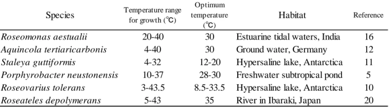
Physiological properties of several phylogenetically and metabolically novel isolates were studied and compared to those of their neighbors. They showed different properties from their phylogenetic neighbors. Distinct temperature dependence was shown by the strains. They did not grow at a moderately high temperature at 40ºC.
Some strains did not grow even at 35ºC.
This study showed phylogenetically divergent aerobic anoxygenic photosynthetic bacteria in addition to anaerobic anoxygenic photosynthetic bacteria inhabited biofilms of the river. Phylogenetic analyses indicated that 8 groups representing novel taxa severally were present in the isolates. It seems that the river biofilms contain a variety of habitats for aerobic anoxygenic photosynthetic bacteria as well as anaerobic ones.
Differences in growth properties and bacteriochlorophyll contents may reflect environmental diversity of the habitats.
5
CONTENTS
Acknowledgements --- 1
General Introduction --- 2
CHAPTER I ---9
Diversity of Purple Photosynthetic Bacteria, Inferred from pufM Gene, within Epilithic Biofilm in Tama River in Ohme-city, Japan Abstract --- 10
Introduction --- 11
Materials and Methods --- 13
Results --- 15
Discussion --- 17
References --- 19
CHAPTER II --- 26
Phylogentically Diverse and Novel Aerobic Anoxygenic Photosynthetic Bacteria were Isolated from Epilithic Biofilms in Tama River in Ohme-city, Japan Abstract --- 27
Introduction --- 28
Materials and Methods --- 29
Results --- 34
Discussion --- 38
References --- 40
CHAPTER III --- 56
Physiological Properties of Novel Aerobic Anoxygneic Photosynthetic Bacteria Isolated from Epilithic Biofilms in Tama River in Ohme-city, Japan Abstract --- 57
Introduction --- 59
Materials and Methods --- 61
Results --- 64
Discussion --- 67
References --- 73
CHAPTER IV --- 96
Phylogentic Diversity of Anoxygenic Photosynthetic Bacteria Isolated from Epilithic Biofilms in Tama River in Okutama-machi, Upstream of Ohme-city Abstract --- 97
Introduction --- 98
Materials and Methods --- 99
Results --- 101
Discussion --- 104
7
Concluding Remarks --- 118
Acknowledgements
I would like to express my gratitude to my supervisors, Dr. Katsumi Matsuura and Dr. Shin Haruta for introducing me to the field of bacterial ecology.
I thank to Dr. Satoshi Hanada for his valuable advices for bacterial isolation and characterization. I also thank Dr. Keizo Shimada for his helpful suggestions in the studies on aerobic anoxygenic photosynthetic bacteria. Special thanks are due to all the members of the Environmental Microbiology Laboratory for their help and kindness.
I thank the Tokyo Metropolitan Board of Education and Mr. Shigeru Nonaka, the principal of Tokyo Metropolitan Sunagawa High School, and my colleagues for allowing me to study at Tokyo Metropolitan University.
Finally, I wish to thank my husband for his encouragement.
This work was supported in part by a Grant-in-Aid for Scientific Research from the Ministry of Education, Culture, Sports, Science and Technology of Japan to K. M.
Chapter IV of this work was joint research with Ms. Chizuru Kato as her graduation work.
2
General Introduction
Phylogenetic positions of photosynthetic bacteria
Photosynthetic bacteria (phototrophic bacteria) comprise several independent phyla in bacterial phylogenetic tree, i.e., filamentous anoxygenic photosynthetic bacteria (Chloroflexi), green sulfur bacteria (Chlorobi), Cyanobacteria, heliobacteria (Firmicutes) and purple bacteria (Proteobacteria) (10). Photosynthetic bacteria were also recently found in the phyla Acidobacteria (3) and Gemmatimonadetes (22).
Among them, bacterial groups other than Cyanobacteria conduct anoxygenic photosynthesis, therefore they are called to be anoxygenic photosynthetic bacteria (anoxygenic phototrophic bacteria), whereas, Cyanobacteria conduct oxygenic photosynthesis and are called to be oxygenic photosynthetic bacteria (oxygenic phototrophic bacteria).
Energy metabolism of anoxygenic photosynthetic bacteria
Anoxygenic photosynthetic bacteria make bacteriochlorophyll as a photosynthetic pigment. Anoxygenic photosynthetic bacteria such as purple nonsulfur bacteria and filamentous anoxygenic photosynthetic bacteria are facultative anaerobes and use organic compounds as carbon and energy sources. They grow under anaerobic conditions in the light (photoheterotrophic growth) and aerobic conditions in the dark (chemoheterotrophic growth).
Aerobic anoxygenic photosynthetic bacteria
Anoxygenic photosynthetic bacteria had been known to conduct photosynthesis under anaerobic conditions. In 1978, bacteriochlorophyll-containing bacteria which grow only under aerobic conditions were reported at the first time (4, 13). As they do not grow under anaerobic conditions even in light, they were called aerobic anoxygenic photosynthetic bacteria (aerobic anoxygenic phototrophic bacteria) (14). Until now, more than 60 species of aerobic anoxygenic photosynthetic bacteria have been described in Alpha-, Beta- and Gammaproteobacteria from various environments (21).
Phylogenetically, aerobic anoxygenic photosynthetic bacteria and anaerobic purple photosynthetic bacteria are related to each other (11). Photosynthetic genes and consequently photosynthetic apparatus of both bacteria are closely similar.
Proteobacteria are thought to have diverged from a photosynthetic ancestor (19).
Aerobic anoxygenic photosynthetic bacteria and anaerobic purple photosynthetic bacteria are direct descendants of the ancestor (2).
Most of aerobic anoxygenic photosynthetic bacteria have significantly lower amounts of bacteriochlorophyll than anaerobic purple photosynthetic bacteria (5, 14), but some species have similar amounts of bacteriochlorophyll to those of the anaerobic ones (14).
Phylogeny of aerobic anoxygenic photosynthetic bacteria
Phylogenetically, aerobic anoxygenic photosynthetic bacteria have wide distribution in the phylum Proteobacteria. The majority of these bacteria has been found in the class Alphaproteobacteria (21).
4
anoxygenic photosynthetic bacteria have been reported in the alpha-1, alpha-2 and alpha-3 subclass of Alphaproteobacteria, however in the alpha-4 subclass, only members of aerobic anoxygneic photosynthetic bacteria were found and no anaerobic anoxygnic photosynthetic species was contained in the subclass. All the aerobic anoxygenic photosynthetic members those belong to the alpha-3 subclass of Alphaproteobacteria were derived from saline or oceanic environments and they all belonged to the Roseobacter clade in the alpha-3 subclass of the class Alphaproteobacteria (20).
Anoxygneic photosynthetic bacteria have been rarely found in the class Betaproteobacteria. Roseateles depolymerans (16) was the first reported aerobic anoxygneic photosynthetic member in Betaproteobacteria. Next to this discovery, Aquincola tertiaricarbonis (9, 12) have been described as aerobic anoxygneic photosynthetic bacteria in the class Betaproteobacteria. Methyloversatilis universalis (6) which belongs to Betaproteobacteria have been also shown to have genes coding subunits of photosynthetic reaction center. Several strains of aerobic anoxygneic photosynthetic bacteria belonging to the class Gammaproteobacteria, such as marine bacterium Congregibacter litoralis (15) have been known.
Recently, an aerobic anoxygenic photosynthetic member was also found in the phylum Gemmatimonadetes (22).
Ecological importance of aerobic anoxygenic photosynthetic bacteria
Kolber et al. reported that aerobic anoxygenic bacterial photosynthesis is widespread and accounts for 2-5% of the photosynthetic electron transport in the upper ocean (6). They also reported that photosynthetically competent anoxygenic bacteria are
abundant in the upper open ocean and comprise at least 11% of the total microbial community (7). After these reports, many intensive studies have revealed the presence, abundance and diversity of aerobic anoxygenic photosynthetic bacteria in various regions of the oceans.
Biofilm on submerged stones of rivers (epilithic biofilm)
In upper reaches of rivers, primary production is mainly conducted in biofilms on submerged stones (1). This benthic microbial biofilms attached to rocks are called
‘epilithic biofilms’. The biofilms are important to sustain river ecosystems, where algae and cyanobacteria produce organic compounds and heterotrophs degrade them.
Aerobic anoxygenic photosynthetic bacteria in rivers
Taxonomy and ecology of aerobic anoxygenic photsynthetic bacteria in the freshwater environments have been poorly documented. Especially in riverine environments, only a few studies have been conducted (12, 13, 14).
The aim of this study
The aim of this study is to know the diversity and properties of aerobic anoxygneic photosynthetic bacteria in biofilms on stones of upper reaches of a river.
6
References
1. Allan, J.D. 1995. Stream ecology: Structure and function of running waters. Chapman and Hall, London.
2. Beatty, J.T. 2002. On the natural selection and evolution of the aerobic phototrophic bacteria. Photosynth. Res. 73: 109–114.
3. Bryant, D.A., A.N.G. Costas, J.A. Maresca, et al. 2006. Candidas Chloracidobacterium thermophilum: An Aerobic phototrophic Acidobacterium.
Science 317:523-526.
4. Harashima, K., T. Shiba, T. Totsuka, U Shimidu, and N. Taga. 1978. Occurrence of bacteriochlorophyll a in a strain of an aerobic heterotrophic bacterium. Agric. Biol.
Chem. 42:1627-1628.
5. Imhoff, J.F., A. Hiraishi and J. Süling. 2005. Anoxygenic phototrophic purple bacteria, p. 119-132. In D.R. Boone, R.W. Castenholz, and G.M. Garity (ed.), Bergey’s Manual of Systematic Bacteriology, 2nd ed., vol. 2A. Springer, New York.
6. Kittichotirat, W., N.M. Good, R. Hall, et al. 2011. Genome sequence of Methyloversatilis universalis FAM5T, a methylotrophic representative of the order Phodocyclales. J. Bacteriol. 193: 4541-4542.
7. Kolber, Z.S., C.L. Van Dover, R.A. Niederman, and P.G. Falkowski. 2000. Bacterial photosynthesis in surface waters of the open ocean. Nature. 407: 177-179.
8. Kolber, Z.S., F.G. Plumley, A.S. Lang, J.T. Beatty, R.E. Blankenship, C.L. VanDover, C. Vetriani, M. Koblizek, C. Rathgeber, and P.G. Falkowski. 2001. Contribution of aerobic photoheterotrophic bacteria to the carbon cycle in the ocean. Science.
292: 2492-2495.
9. Lechner, U., D. Brodkorb, R. Geyer, G. Hause, C. Härtig, G. Auling, F.F.
Guichard, P. Piveteau, R.H. Müller, and T. Rohwerder. 2007. Aquincola tertiaricarbonis gen. nov., sp. nov., a tertiary butyl moiety-degrading bacterium.
Int. J. Syst. Evol. Microbiol. 57:1295-1303.
10. Madigan, M. T. and J. M. Martinko. 2006. Prokaryotic diversity: the bacteria, p.
331-418. In Brock Biology of Microorganisms 11th ed. Pearson, San Francisco 11. Nagashima, S., and K.V.P. Nagashima. 2013. Comparison of photosynthesis gene
clusters retrieved from total genome sequences of purple bacteria, p. 151-178. In J.T. Beatty (ed.), Advances in botanical research, vol. 66. Academic Press, New York.
12. Rohwerder, T., R.H. Müller, M.T. Weicher, J. Schuster, T. Hubschmann, S. Müller, and H. Harms. 2013. Cultivation of Aquincola tertiaricarbonis L108 the fuel oxygenate intermediate tert-butyl alcohol induces aerobic anoxygenic photosynthesis at extremely low feeding rates. Microbiology (United Kingdom).
159: 2180–2190.
13. Sato, K. 1978. Bacteriochlorophyll formation by facultative methylotrophs, Protaminobacter ruber and Pseudomonas AM-1. FEBS Lett. 85:207-210.
14. Shimada, K. 1995. Aerobic anoxygneic phototrophs, p. 105-122. In R.E.
Blankenship, M.T. Madigan, and C.E. Bauer (ed.), Anoxygenic photosynthetic bacteria. Kluwer Academic Publishers, Dordrecht, Netherlands.
15. Spring, S., Lunsdorf, H., Fuchs, B.M., and Tindall, B.J. "The photosynthetic apparatus and its regulation in the aerobic gammaproteobacterium Congregibacter litoralis gen. nov., sp. nov." PLoS ONE (2009) 4(3):e4866.
8
Tokiwa, T. Kanagawa, and S. Hanada. 1999. Roseateles depolymerans gen. nov., sp. nov., a new bacteriochlorophyll a-containing obligate aerobe belonging to the β-subclass of the Proteobacteria. Int. J. Syst. Bacteriol. 49:449-457,
17. Waidner, L.A., and D.L. Kirchman. 2005. Aerobic anoxygenic photosynthesis genes and operons in uncultured bacteria in the Delaware River. Environ.
Microbiol. 7:1896-1908
18. Waidner, L.A., and D.L. Kirchman. 2008. Diversity and distribution of ecotypes of the aerobic anoxygenic phototrophy gene pufM in the Delaware Estuary. Appl.
Environ. Microbiol. 74:4012-4021
19. Woese, C. R. 1987. Bacterial evolution. Microbiological Reviews. 51: 221–
271.20. Yurkov, V., and E. Hughes. 2013. Genes associated with the peculiar phenotypes of the aerobic anoxygenic phototrophs, p. 327-358. In J.T. Beatty (ed.), Advances in botanical research, vol. 66. Academic Press, New York.
21. Yurkov, V, and J.T. Csotonyi. 2009. New light on aerobic anoxygenic phototrophs. p. 31-55. In C.N. Hunter, F. Daldal, M.C. Thurnauer and J. T. Beatty (ed.), The purple phototrophic bacteria. Springer Science, Dordrecht, Netherlands.
22. Zeng, Y., F. Feng, H. Medova, J. Dean, and M. Koblizek. 2014. Functional type 2 photosynthetic reaction centers found in the rare bacterial phylum Gemmatimonadetes. PNAS. 111:7795-7800
CHAPTER I
Diversity of Purple Photosynthetic Bacteria, Inferred from
pufM Gene, within Epilithic Biofilm in Tama River in
Ohme-city, Japan
10
Abstract
The diversity of purple photosynthetic bacteria in algae-dominated biofilm of streambed in Tama River, Japan was investigated. Clone library analysis of the pufM gene encoding a subunit of the photochemical reaction center of purple bacteria detected 18 operational taxonomic units (OTUs) in several classes of Proteobacteria. Most of the OTUs showed less than 85% identities to the PufM amino acid sequences of known photosynthetic bacteria. These results suggest that phylogenetically divergent and unknown purple photosynthetic bacteria are present in the epilithic biofilm of the river.
Introduction
Purple photosynthetic bacteria have been isolated from a variety of environments, such as sediments, soils and waters in ponds, lakes, lagoons and ocean (10, 11). As purple bacteria are metabolically versatile, e.g., photosynthesis, degradation of organic compounds, nitrogen fixation or sulfide oxidation, they play important roles in ecosystems in the light.
Microbial ecological studies on freshwater environments have found several groups of purple photosynthetic bacteria belonging to Alpha- or Betaproteobacteria from lakes (12, 17) and rivers (8, 9, 20, 23). Some of these purple bacteria have been known to be anaerobic anoxygenic phototrophs. The presence of aerobic anoxygenic phototrophs (AAP), which carry out photosynthetic reactions only under aerobic conditions, has been also indicated by phylogenetic or physiological studies. However, the distribution and ecophysiology of purple photosynthetic bacteria in freshwater environments has not been documented yet.
Epilithic biofilm is important to sustain the ecosystem in freshwater environments in terms of producing organic substrates, feeding animals and degrading organic matters.
The streambed biofilm is known to be mainly composed of oxygenic phototrophs, i.e., cyanobacteria and algae (2). However, there is no study targeting purple photosynthetic bacterial diversity in river biofilm. Dense assemblages of bacterial cells and their active respiration in biofilm possibly develop some anaerobic niches (6) even when phototrophs emit oxygen, and consequently both aerobic and anaerobic anoxygernic phototrophs may found their niches within river biofilm.
12
and a molecular method based upon pufM gene encoding a subunit of the photochemical reaction center to investigate diversity of purple photosynthetic bacteria in epilithic biofilm at an upstream region of a river where the amount of dissolved organic matter is limited. Phylogenies based on pufM gene sequences are mostly consistent with those based on 16S rRNA gene (14), and thus the pufM gene is frequently utilized for genetic surveys of anoxygenic phototrophs (1, 4, 12, 16, 24).
Materials and Methods
Submerged cobbles of about 15 to 25 cm in the longest length were collected from streambed in river located in the upstream region of Tama River in Ohme-city (35°47′13″N, 139°15′15″E), in the west suburbs of Tokyo, Japan in August 2009. The river width at the sampling site was 40 m. The water depth of the sampling site was about 20 cm. Water temperature, pH, biochemical oxygen demand (BOD) and flow velocity of the river water at the sampling time were 18˚C, 7.6, 0.5 mg L-1 and 0.4 m s-1, respectively. Average values of dissolved oxygen, total nitrogen and total phosphorus in this region in July to September 2009 were 8.9 ± 0.3 mg L-1, 0.79 ± 0.10 mg L-1 and 0.017 ± 0.005 mg L-1, respectively (a monthly report by Bureau of Environment, Tokyo metropolitan government, http://www.kankyo.metro.tokyo.jp). A brownish biofilm of about 1 mm thickness was developed on the cobbles. A total 150 cm2 area of the epilithic biofilm was scraped off from the top surface of the each cobble using a sterile toothbrush and suspended into 10 mL of sterile distilled water.
For bacterial culture, 0.1mL of the biofilm suspension was transferred into a 30 mL volume of screw cap tube filled with a PE medium (7), a semisynthetic medium containing organic compounds. The tubes were incubated at 30˚C under the filtered incandescent light (ca. 2,000 lux) of wavelength over 700 nm for 7 to 14 days. The cultures which showed spectral properties of purple bacteria were streaked on agar plates of the PE medium. The plates were incubated anaerobically under the incandescent light and red-colored colonies were transferred and streaked on new plates.
These operations were repeated more than twice to obtain pure cultures. These isolation
14
obtained in May 2009.
Total genomic DNAs were directly extracted from the collected biofilm according to Noll et al. (15). DNA fragments of pufM gene coding for the M subunit of the photochemical reaction center were amplified. Nested PCR was conducted to amplify pufM gene fragments from environmental DNA using primer sets pufLM-F/pufLM-R (1st PCR) (14) and M150f/M572r (2nd PCR) (16). PCR products (approximately 380 bp) were cloned with the pTAC-1 Vector (DynaExpress TA cloning kit, BioDynamics Laboratory, Tokyo, Japan). Escherichia coli JM109 competent cells (Nippon gene, Tokyo, Japan) were transformed according to manufacturer’s instructions. DNA sequences were determined with the BigDye v3.1 cycle sequencing kit (Applied Biosystems, Foster city, CA, USA) and a DNA sequencer ABI3130xl (Applied Biosystems). Chimeric clones were checked manually and excluded from further analyses. The phylogenetic tree based on the amino acid sequences of the partial PufM was constructed using the neighbor-joining and maximum-likelihood methods with a MEGA version 5 program (19, 22).
The nucleotide sequences determined in this study have been deposited in the GenBank/EMBL/DDBJ database under accession numbers AB670200 to AB670233.
Results
Cultivation of the river biofilm on the agar plates of PE medium yielded red-brown colonies. All of these colonies were similar in morphology to each other.
Among them, 17 strains were isolated to determine the pufM sequences. The sequence analysis indicated that there were 5 different isolates, two of which were designated Tisolate 25 and Tisolate 231 in Fig. I-1 since these two isolates had been obtained from the sample used for the direct extraction of total genomic DNA and subsequent clone library analysis. The closest relatives of these isolates are listed in Table I-1.
Fig. I-1 shows a neighbor-joining tree based on the amino acid sequences inferred from the partial pufM gene sequences from 37 clones and two isolates obtained in this study together with those from database. A phylogenetic tree using the maximum-likelihood method (figure not shown) showed a tree topology roughly consistent with that in Fig. I-1. Two major clades were recognized; one containing alpha-1, alpha-2, alpha-4 subclasses, beta and gamma classes of Proteobacteria and the other containing alpha-3 subclass of Proteobacteria. This was roughly in agreement with earlier studies concerning pufLM or pufM phylogeny (4, 14). Obtained clones were grouped into 18 operational taxonomic units (OTUs). Each OTU was defined as a group having amino acid sequence identities above 90%. These OTUs were widely distributed in the phylogenetic tree and many of them were distantly related each other. No dominant OTUs in terms of numbers of clones were found, since every OTU consisted of less than 5 clones.
OTUs except OTU 6 showed less than 85% identities to the PufM sequences of
16
sequence as that of Tisolate 25, closely related to that of Rhodoferax fermentans (accession no. D50650, 98.4% identity). The other isolate, Tisolate 231, had 100%
PufM sequence identity to Rhodopseudomonas palustris (accession no. AB015977).
Rhodoferax fermentans and R. palustris are known to be anaerobic anoxygenic phototrophs (10). Tisolate 25 and Tisolate 231 were grown photoheterotrophically under anaerobic conditions.
The alpha-3 clade contains 6 OTUs (OTUs 13 to 18). OTU 13 is distantly related to other members in this clade. Sequences included in OTU 14 showed very low identities to those of the database; the highest identity was 65.7% to that of an environmental clone (accession no. AY912082) (23) collected from river water. OTUs 15 to 18 formed a clade with Staleya guttiformis (now known as Sulfitobacter guttiformis) and alpha proteobacterium R2A163 and R2A84 (21), those were reported as aerobic anoxygenic photosynthetic bacteria isolated from saline environments.
OTUs 1 and 2 were related to Rhodospirillum rubrum, belonging to the alpha-1 subclass of Proteobacteria, with 75.4% and 71.4% sequence identities, respectively.
OTU 3 was found to be similar with a marine gamma proteobacterium (5) with sequence identity 84.1%. OTU4 was related to Methylocella sp. and Rhodoplanes sp.
belonging to alpha-2 subclass of Proteobacteria. OTU 5 was similar to R. fermentans with identity 73.0%. It is indicated that OTUs 7 to 11 were grouped with the genus Sphingomonas, Citromicrobium, Erythorobacter and Porphyrobacter belonging to alpha-4 subclass. Photosynthetic bacteria in this subclass have been known to be aerobic anoxygenic phototrophs. OTU 12 showed low identities to known sequences, and the close relative was Halorhodospira halophila, belonging to Gammaproteobacteria (69.0% identity).
Discussion
In this study, I investigated diversity of purple photosynthetic bacteria in a streambed biofilm. Co-occurrence of possibly aerobic (e.g., OTUs 15 to 18) and anaerobic (e.g., OTU 6 and Tisolate 231) anoxygenic phototrophs was observed within the river biofilm as expected. Most OTUs detected by pufM clone library analysis had low identities to the sequences of cultured bacteria. Studies on bacterial communities in river biofilms using 16S rRNA gene analyses have also detected many clones of uncultured bacteria (3, 8, 13).
Phylogenetic analysis based on the PufM sequences indicated that purple photosynthetic bacteria in the river biofilm are widely distributed to alpha subclasses, beta and gamma classes of Proteobacteria. Such a high diversity of purple photosynthetic bacteria has never been reported in other environments. In French Mediterranean coast lagoon sediments, clones with pufM of the alpha-3 subclass of Proteobacteria accounted for 94.9 % of total clones (18). In the case of Antarctic lake water, no clones were related to alpha-3 subclass of Proteobacteria but 80 % of the clone contained pufM of the related taxa to Betaproteobacteria (12). Microenvironments of the algae-dominated river biofilm under the shallow and rapid flow of water would be highly heterogeneous, differing in concentrations of dissolved oxygen, organic and inorganic compounds. The population of purple photosynthetic bacteria within the epilithic biofilm microflora may be low because nested approach was required to amplify their DNAs. The niche for purple photosynthetic bacteria in the biofilm may be restricted, but their niches seem to be largely diverged.
18
absorption bands at the different wavelengths from those of chlorophylls in oxygenic phototrophs, purple photosynthetic bacteria can capture the light energy even in the algae-dominated biofilms of rivers. In the biofilm community, in addition to the primary production and the degradation of the organic matters, some purple photosynthetic bacteria possibly contribute to oxidize sulfide produced by sulfate reducing bacteria, since I observed sulfide production from anaerobic culture of the epilithic biofilm used in this study when illumination was stopped (data not shown).
This study demonstrated an unexpected diversity of purple photosynthetic bacteria in the river biofilm. Physiological studies of the yet-to-be cultured epilithic purple photosynthetic bacteria will clarify the roles of these bacteria in the ecosystem of river.
References
1. Achenbach, L.A., J. Carey, and M. T. Madigan. 2001. Photosynthetic and phylogenetic primers for detection of anoxygenic phototrophs in natural environments. Appl. Environ. Microbiol. 67:2922-2926.
2. Allan, J.D. 1995. Stream ecology:Structure and function of running waters.
Chapman and Hall, London.
3. Anderson-Glenna, M.J., V. Bakkestuen, and N.J.W. Clipson. 2008. Spatial and temporal variability in epilithic biofilm bacterial communities along an upland river gradient. FEMS Microbiol. Ecol. 64:407-418.
4. Béjà, O., M.T. Suzuki, J.F. Heidelberg, W.C. Nelson, C.M. Preston, T. Hamada, J.A.
Eisen, C.M. Fraser, and E.F. DeLong. 2002. Unsuspected diversity among marine aerobic anoxygenic phototrophs. Nature 415:630-633.
5. Cho, J.-C, M.D. Stapels, R.M. Morris, K.L. Vergin, M.S. Schwalbach, S.A. Givan, D.F. Barofsky, and S.J. Giovannoni. 2007. Polyphyletic photosynthetic reaction centre genes in oligotrophic marine gammaproteobacteria. Environ. Microbiol.
9:1456-1463.
6. Costerton, J.W., D. de Beer, D. Caldwell, D.R. Korber & G.A. James. 1994.
Biofilms, the customized microniche. J. Bacteriol. 176:2137–2142.
7. Hanada, S., A. Hiraishi, K. Shimada, and K. Matsuura. 1995. Isolation of Chloroflexus aurantiacus and related thermophilic phototrophic bacteria from Japanese hot springs using an improved isolation procedure. J. Gen. Appl.
Microbiol. 41:119-130.
20
Bacterial populations in epilithic biofilms along two oligotrophic rivers in the tohoku region in Japan. J. Gen. Appl. Microbiol. 55:359-371.
9. Hullar, M.A.J., L.A. Kaplan, and D.A. Stahl. 2006. Recurring seasonal dynamics of microbial communities in stream habitats. Appl. Environ. Microbiol. 72:713–722.
10. Imhoff, J.F., A. Hiraishi and J. Süling. 2005. Anoxygenic phototrophic purple bacteria, p. 119-132. In D.R. Boone, R.W. Castenholz, and G.M. Garity (ed.), Bergey’s Manual of Systematic Bacteriology, 2nd ed., vol. 2A. Springer, New York.
11. Imhoff, J.F., and H.G. Trüper. 1992. The genus Rhodospirillum and related genera, p. 2141–2155. In A. Balows, H.G. Trüper, M. Dworkin, W. Harder, and K.-H.
Schleifer (ed.), The Prokaryotes, 2nd ed. Springer, New York.
12. Karr, E.A., W.M. Sattley, D.O. Jung, M.T. Madigan, and L.A. Achenbach. 2003.
Remarkable diversity of phototrophic purple bacteria in a permanently frozen antarctic lake. Appl. Environ. Microbiol. 69:4910-4914.
13. Kobayashi, Y., C. Kim, C. Yoshimizu, A. Kohzu, I. Tayasu, and T. Nagata. 2009.
Longitudinal changes in bacterial community composition in river epilithic biofilms: Influence of nutrients and organic matter. Aquat. Microb. Ecol.
54:135-152.
14. Nagashima, K.V.P., A. Hiraishi, K. Shimada, and K. Matsuura. 1997. Horizontal transfer of genes coding for the photosynthetic reaction centers of purple bacteria.
J. Mol. Evol. 45:131-136.
15. Noll, M., D. Matthies, P. Frenzel, M. Derakshani, and W. Liesack. 2005.
Succession of bacterial community structure and diversity in a paddy soil oxygen gradient. Environ. Microbiol. 7:382-395.
16. Okubo, Y., H. Futamata, and A. Hiraishi. 2006. Characterization of phototrophic purple nonsulfur bacteria forming colored microbial mats in a swine wastewater ditch. Appl. Environ. Microbiol. 72:6225-6233.
17. Page, K.A., S.A. Connon, and S.J. Giovannoni. 2004. Representative freshwater bacterioplankton isolated from Crater Lake, Oregon. Appl. Environ. Microbiol.
70: 6542–6550.
18. Ranchou-Peyruse, A., R. Herbert, P. Caumette, and R. Guyoneaud. 2006.
Comparison of cultivation-dependent and molecular methods for studying the diversity of anoxygenic purple phototrophs in sediments of an eutrophic brackish lagoon. Environ. Microbiol. 8:1590-1599.
19. Saitou, N., and M. Nei. 1987. The neighbor-joining method: A new method for reconstructing phylogenetic trees. Mol. Biol. Evol. 4:406-425.
20. Suyama, T., T. Shigematsu, S. Takaichi, Y. Nodasaka, S. Fujikawa, H. Hosoya, Y.
Tokiwa, T. Kanagawa, and S. Hanada. 1999. Roseateles depolymerans gen. nov., sp. nov., a new bacteriochlorophyll a-containing obligate aerobe belonging to the β-subclass of the Proteobacteria. Int. J. Syst. Bacteriol. 49:449-457,
21. Suzuki, M.T., M.S. Rappé, Z.W. Haimberger, H. Winfied, N. Adair, J. Ströbel, and S.J. Giovannoni. 1997. Bacterial diversity among small-subunit rRNA gene clones and cellular isolates from the same seawater sample. App. Environ. Microbiol.
63:983-989.
22. Tamura, K., D. Peterson, N. Peterson, G. Stecher, M. Nei, and S. Kumar. 4 May 2011. MEGA5: Molecular evolutionary genetics analysis using maximum likelihood, evolutionary distance, and maximum parsimony methods. Mol. Biol.
22
23. Waidner, L.A., and D.L. Kirchman. 2005. Aerobic anoxygenic photosynthesis genes and operons in uncultured bacteria in the Delaware River. Environ.
Microbiol. 7:1896-1908
24. Yutin, N., M.T. Suzuki, and O. Beja. 2005. Novel primers reveal wider diversity among marine aerobic anoxygenic phototrophs. Appl. Environ. Microbiol.
71:8958-8962.
16S rRNA genepufMgene Closest relative (identity %)Closest relative (identity %) e 25Rhodoferax fermentans (98)Rhodoferax fermentans (96)βAugust 25 2009river biofilm e 231Rhodopseudomonas palustris (99)Rhodopseudomonas palustris (100)α-2August 25 2009river biofilm e 51Rhodobacter capsulatus (94)Rhodobacter capsulatus (99)βAugust 25 2009apical portion of filamentous algae streamer on a river submerged stone e 322Rubrivivax gelatinosus (99)Rubrivivax gelatinosus (89)α-3May 21 2009bottm portion of filamentous algae streamer on a river submerged stone e 63Rhodoplanes elegans (99)not amplifiedα-2May 18 2009river water , α-3 and β in Affiliation culumn indicate alpha-2, alpha-3 classes of Alphaproteobacteria andBetaproteobacteria, plane.
n nameAffiliationSampling dateSample
able I-1Anaerobic anoxygenic purple photosynthetic bacteria isolated from the sampling site
24
Fig. I-1.
Fig. I-1. Phylogenetic tree of Proteobacteria based on partial PufM amino acid sequences inferred from gene sequences. Chloroflexus aurantiacus was used as an outgroup. PufM amino acid sequences from environmental DNA in this study are indicated by TP-1–TP-55, and those from isolates in this study are indicated by the
‘Tisolate’ prefix. Sequences from the database are represented with their respective accession numbers after bacterial names in parentheses. OTUs are indicated to the right of the tree. Alpha-1, alpha-2, alpha-3, alpha-4 subclass, beta and gamma class of Proteobacteria are also indicated by α1, α2, α3, α4, β and γ to the right of the tree.
Bootstrap values >90, 70-89 and 50-69% are indicated by black, gray and open circles, respectively. Scale bar represents the number of substitutions per site.
26
CHAPTER II
Phylogentically Diverse and Novel
Aerobic Anoxygenic Photosynthetic Bacteria were Isolated
from Epilithic Biofilms in Tama River in Ohme-city, Japan
Abstract
The ecological importance of aerobic anoxygenic photosynthetic bacteria in ocean environments has recently become evident but less is known about them in freshwater environments. To grasp the diversity and physiology of aerobic anoxygenic photosynthetic bacteria in river biofilm, the colony formation method was applied.
Biofilm suspensions were spread on agar plates and cultivated aerobically in the dark.
Spectroscopic analysis of the colonies indicated significant ratio of the total colonies contained bacteriochlorophyll. Twenty eight isolated strains were confirmed to be aerobic anoxygneic photosynthetic bacteria by the absence of growth under anaerobic photosynthetic conditions. Bacteriochlorophyll production was also confirmed for all the isolates, and the bacteriochlorophyll content varied by strain from 0.13 to 3.58 nmol per mg of dry cell weight. The isolated strains were phylogenetically divided into 8 groups, having 96% to 100% 16S rRNA sequence identities within each group, belonging to the alpha-1, alpha-3 and alpha-4 subclasses of Alphaproteobacteria and Betaproteobacteria. Among them 4 groups were distantly related to previously known bacteria with similarities of 97.3-98.1%. Two strains grew in a low-nutrient medium containing 0.5 g of organic compounds per liter but did not grow in a high-nutrient medium containing 2.5 g of organic compounds per liter. These results suggest occurrence of diverse and unknown aerobic anoxygenic photosynthetic bacteria in river biofilm.
28
Introduction
Aerobic anoxygenic photosynthetic bacteria grow heterotrophically by aerobic respiration. They produce bacteriochlorophyll but do not grow anaerobically in the light (21). Imhoff and Hiraishi explained that aerobic anoxygenic photosynthetic bacteria are thought to use photosynthesis as a supplementary energy source (9).
Since first report in 1978 (7, 20), more than 60 species of aerobic anoxygenic photosynthetic bacteria have been described in Alphaproteobacteria, Betaproteobacteria and Gammaproteobacteria (29). This group was recently recognized to be widespread in the ocean, account for a considerable fraction, for example 11% of the marine bacterioplankton, and reported to have significant roles for marine carbon cycling (12, 13). However, presence and diversity of aerobic anoxygenic photosynthetic bacteria in freshwater environments have been poorly understood.
I had reported the diversity of photochemical reaction center (puf) gene for anoxygenic photosynthesis detected from river biofilm (8, Chapter I), suggesting the existence of several novel lineages of anoxygenic photosynthetic bacteria in Proteobacteria. Some of them are suggested to be aerobic anoxygenic photosynthetic bacteria on the basis of the phylogenetic analysis based on puf gene sequences.
However, physiological analyses are necessary to confirm them as aerobic anoxygenic photosynthetic bacteria. In this study, I isolated bacteriochlorophyll-containing bacteria from epilithic biofilms of upper reaches of Tama River, Japan and phylogenetically and physiologically characterized the isolates as aerobic anoxygenic photosynthetic bacteria.
Materials and Methods
Sampling of epilithic biofilms
Submerged cobbles of about 15 to 20 cm in the longest length were collected from a streambed in a sunny river located in the upper reaches of Tama River in Ohme City (35°47'13''N, 139°15'15''E, Altitude is 200 m), Tokyo, Japan on February 20 and July 23, 2012. Biofilm of about 1 mm thickness was present on the cobbles. The river width and water depth at the sampling site were about 40 m and 0.2 m, respectively. Water temperature, pH and flow velocity of the river water at the sampling time were 3.0°C, 8.0, 0.6 to 0.9 m s-1, respectively, in February and 13.8°C, 8.2, 0.3 to 1.2 m s-1, respectively, in July. Average values of biochemical oxygen demand, dissolved oxygen, total nitrogen and total phosphorus of the river water in this region were 0.26±0.28 mg L−1, 11.8±0.73 mg L−1, 0.58±0.05 mg L−1, 0.013±0.014 mg L−1, respectively, in February 2012 and 0.73±0.17 mg L−1, 9.6±0.21 mg L−1, 0.75±0.04 mg L−1, 0.009±0.003 mg L−1, respectively, in July 2012 (monthly report by Bureau of Environment, Tokyo Metropolitan Government, http://www.kankyo.metro.tokyo.jp). A total 150 cm2 area of epilithic biofilm was scraped off from the top surface of each cobble using a sterilized toothbrush and suspended in 10 ml of sterilized distilled water. Samples were taken from 3 cobbles within 10 m2 area. The suspensions were kept cool in ice and brought to the laboratory.
Isolation of bacteriochlorophyll-containing aerobic bacteria
Aggregation in the biofilm suspensions was dispersed using POLYTRON
30
suspensions were serially diluted and spread onto agar plates. Gellan gum was also used instead of agar. Isolation was conducted using 2 types of media, PE medium (6) and Nutrient agar (Eiken Chemical, Tochigi, Japan). Composition of PE medium was 0.5 g of sodium glutamate, 0.5 g of sodium succinate, 0.5 g of sodium acetate, 0.5 g of yeast extract (Wako, Osaka, Japan), 0.5 g of casamino acids (Wako, Osaka, Japan), 0.5 g of sodium thiosulfate, 0.5 g of (NH4)2SO4, 0.38 g of KH2PO4, 0.39 g of K2HPO4, 5 ml of a basal salt solution and 1ml of vitamin mixture per liter. The pH of the medium was adjusted to 7.5. The basal salt solution was composed of 1.11 g of FeSO4∙7H2O, 24.65 g of MgSO4∙7H2O, 2.94 g of CaCl2∙2H2O, 23.4 g of NaCl, 111 mg of MnSO4∙4H2O, 28.8 mg of ZnSO4∙7H2O, 29.2 mg of Co(NO3)2∙6H2O, 25.2 mg of CuSO4∙5H2O, 24.2 mg of Na2MoO4∙2H2O, 31.0 mg of H3BO3 and 4.53 g of trisodium EDTA per liter. The vitamin mixture was composed of 100 mg of nicotinic acid, 100 mg of thiamine hydrochloride, 5 mg of biotin, 50 mg of p-aminobenzoic acid, 1mg of vitamin B12, 50 mg of calcium panthothenate, 50 mg of pyridoxine hydrochloride and 50 mg of folic acid per 100 ml. 1/5 PE medium is simply 5 times diluted PE medium. Ten times diluted PE medium (1/10 PE medium) or a hundred times diluted PE medium (1/100 PE medium) contained ten times or a hundred times lower concentration of sodium glutamate, sodium succinate, sodium acetate, yeast extract, casamino acids, sodium thiosulfate, (NH4)2SO4, respectively. Both 1/10 and 1/100 PE medium contained the same concentrations of KH2PO4, K2HPO4,basal salt solution and 1/5 concentrations of vitamin mixture as the PE medium. Agar (1.5% w/v) or gellan gum (2% w/v) supplemented medium were designated to be PEA or PEG, respectively. Composition of Nutrient agar was 5.0 g of meat extract, 10.0 g of peptone, 5.0 g of sodium chloride and
15.0 g of agar per liter. The pH of the medium was adjusted to 7.0. Six times or 60 times diluted Nutrient agar (1/6 NA or 1/60 NA) were also used, supplied with agar up to 1.5%. Inoculated plates were cultivated aerobically at 30°C in the dark for 14 days.
After cultivation, randomly selected red or pink colored colonies were suspended in sterilized distilled water to determine absorption spectrum using spectrophotometer V-630 (Jasco, Tokyo, Japan). Colonies that showed absorbance maxima within 800 nm to 875 nm were selected and transferred to new agar plates of 1/10 PE medium and cultivated aerobically at 30C in the dark for a week. Transfer and cultivation were repeated more than twice to obtain pure cultures. The winter isolates (February) were indicated by the ’W’ prefix, for example W09, and summer isolates (July), by the ‘S’
prefix.
Growth properties
Growth properties of isolates were determined under the following conditions.
Anaerobic light conditions; Test tubes were sealed with butyl rubber caps and gas phase of each tube was substituted with nitrogen gas. Inoculated tubes were incubated under filtered incandescent light (ca. 2,000 lux) of wavelength over 700 nm. Aerobic dark conditions; Test tubes were equipped with silicon plug ‘SILICOSEN’ (Shin-etsu Polymer, Tokyo, Japan) to let air through, set in a shaker at an angle and shaken at 140 rpm in darkness. All cultivations were conducted 30C, using test tubes of 18 mm in diameter and 180 mm in length containing 10 ml of liquid medium after inoculation of 0.1 ml of exponential phase of growth or early stationary phase in 1/5 PE medium under aerobic dark conditions. Growth was monitored by optical density of 660 nm (mini
32
Bacteriochlorophyll content
Isolates were cultivated under aerobic dark condition in 1/5 PE medium and the cells were harvested at exponential phase of growth or early stationary phase and washed twice with 10 mM tris-HCl buffer pH 8.0. Bacteriochlorophyll was extracted in methanol:acetone=7:2 from the cells and absorbance of 770 nm was measured using UV-1800 UV-VIS spectrophotometer (Shimadzu, Kyoto, Japan). Millimolar extinction coefficient 75 cm-1 was used to determine bacteriochlorophyll content (3). Dry cell weight was determined with harvested cells, washed twice with 10 mM tris-HCl buffer pH 8.0 and dried 80ºC for 3 days.
Rhodobacter sphaeroides ATCC 17023T (=2.4.1T), a reference species obtained from the American Type Culture Collection was cultivated in 1/5 PE medium under aerobic dark and anaerobic light conditions.
Measurements of in vivo spectra
In vivo spectra of whole cells and disrupted cells were measured. For measurements of in vivo spectra of disrupted cells, 50 ml of an aerobic dark-grown 7 day cultures were used. Cultivations were conducted in vessels for shaken culture (500 ml capacity) containing 100ml of 1/5 PE medium. Cells were harvested and suspended in 10 mM Tris-HCl pH 8.0, disrupted by sonication at 130 W for 4 min in a chilled bowl (Sonicator, Ohtake Works, Tokyo, Japan) and centrifuged at 10,000 rpm for 4 min, the supernatant was used for in vivo absorption spectra measurements. Spectra were measured using UV-1800 UV-VIS spectrophotometer (Shimadzu, Kyoto, Japan).
Sequencing of the 16S rRNA gene and phylogenetic analysis
Total genomic DNA of the isolates was extracted according to Noll et al. (18).
DNA fragments of partial 16S rRNA gene were amplified using primer sets 27F2/
1492R2 (14, 23). DNA sequences were determined with the BigDye v3.1 cycle sequencing kit (Applied Biosystems, Foster City, CA, USA) and a DNA sequencer ABI3130xl (Applied Biosystems). Sequence primers 27F2, 515F, 968F, 517R, 907R2 and 1492R2 were used and contig was made using ATGC program (GENETYX Ver. 12, GENETYX, Tokyo, Japan). Phylogenetic relatives were determined based on 16S rRNA sequences using BLAST search (http://blast.ncbi.nlm.nih.gov/Blast.cgi). Phylogenetic analysis was conducted using MEGA version 6 program (24).
34
Results
Bacteriochlorophyll-containing colonies from epilithic biofilms
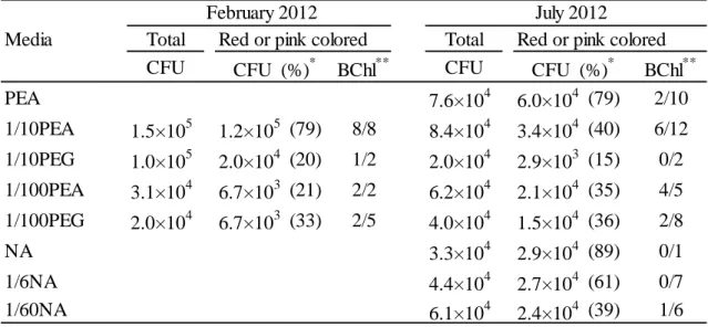
Table II-1 summarizes the results of isolation. 2.0 x 10⁴ to 1.5 x 10⁵ colonies per cm2 of the biofilm were totally detected from the samples used in this study. The CFUs did not vary widely among media. Many red or pink colored colonies were obtained from both the February and July samples.
Among the colored colonies obtained from the sample in February, 17 colonies were picked up to determine bacteriochlorophyll. Thirteen colonies within 17 colonies had bacteriochlorophyll. For the sample collected in July, bacteriochlorophyll was detected from 15 colonies among 51 colonies tested in total. Using 1/10 PEA and 1/100 PEA media, bacteriochlorophyll-containing colonies were frequently obtained.
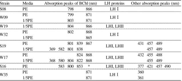
In vivo absorption spectra of whole cells of the 28 isolates showed absorption peaks within 800 nm to 875 nm. These peaks show the presence of a photosynthetic apparatus in which the bacteriochlorophyll is integrated. Fig. II-1 shows two characteristic in vivo absorption spectra of sonicated supernatant. The absorption spectrum of W09 showed absorption peaks at 803 nm and 871 nm, which indicate the presence of photochemical reaction center light-harvesting pigment-protein complex I (RC-LH I). W17 showed absorption peaks at 804 nm, 822 nm and 868 nm indicating the presence of RC-LH I and light-harvesting pigment-protein complex II (LH II) (4).
Absorption peaks of 455 nm and 489 nm of W17 shows absorbance of carotenoid.
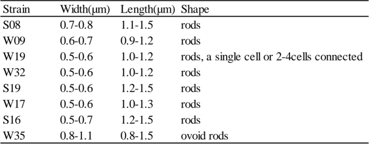
16S rRNA gene sequences of 28 bacteriochlorophyll-containing isolates were determined using the sequencing primer 27F2, and compared and classified the cut off
value of over 99.6% identity. 1409 positions of 16S rRNA gene sequences of representative 14 isolates (S08, W09, W32, W19, S19, W17, S16, S10, W45, S12, S20, W22, W14 and W35) were determined and finally these isolates were divided into 8 OTUs sharing 96% sequence identity. These OTUs were found in alpha-1, alpha-3, and alpha-4 subclasses of Alphaproteobacteria and Betaproteobacteria (details were shown in Fig. II-2).
Growth under anaerobic light conditions and aerobic dark conditions
Anaerobic growth was tested for representatives from phylogenetically distinct 8 OTUs and compared with that under aerobic conditions. Figure II-3 shows growth curves of three isolates, W35 (OTU8), S12 (OTU7) and W32 (OTU2) (Fig. II-3(A), (B) and (C)). All of these strains showed distinct growth under aerobic conditions in the dark (solid lines), and no growth under anaerobic conditions in the light (dashed lines).
Other 5 isolates in OTUs 1, 3-6, also showed growth under aerobic conditions and no growth under anaerobic conditions in the light (Fig. II-S3A, II-S3B, II-S3C). All the representative isolates in this study were confirmed to be aerobic anoxygenic photosynthetic bacteria.
Phylogenetic analysis of the isolates
Fig. II-2 shows a neighbor-joining tree based on 16S rRNA gene sequences from 14 representative isolates obtained in this study together with those from the database. A phylogenetic tree using the maximum-likelihood method showed tree topology roughly consistent with that in Fig. II-2 (data not shown). 8 OTUs were distributed to alpha-1,
36
Isolate S08 in OTU1 belonging to alpha-1 subclass was related to Roseomonas lacus, which had been isolated from freshwater sediment (10) with 98.1% identity.
Five isolates, W09, W32, W07, W40 and W19, i.e., OTU2 were found in alpha-3 subclass. Tabrizicola aquatica in this group had been isolated from a freshwater lake as a non-photosynthetic bacterium (25).
The alpha-4 subclass of Proteobacteria, OTUs 3-7, was the dominant group of the isolates. In the clade of OTUs 3-5, known aerobic anoxygenic photosynthetic bacteria such as Sandarakinorhabdus limnophila (5) was found.
OTU6 have neighbor Sphingopyxis wooponensis, isolated from freshwater of wetland.
Isolates belonging to OTU7 have aerobic anoxygenic photosynthetic relatives such as Porphyrobacter donghaensis.
An aerobic anoxygenic photosynthetic bacteria belonging to Betaproteobacteria, W35 (OTU8), was isolated. The nearest neighbor was aerobic anoxygenic photosynthetic bacteria Aquincola tertiaricarbonis isolated from German groundwater (16).
Nearest neighbors of the isolates and their similarities are listed in Table II-S1.
Growth properties in nutrient-rich medium and -poor medium
Growth affected by organic nutrients concentration in the medium was characterized. Three types of growth property were detected in 10 isolates (representatives of 8 OTUs and additional 2 isolates) under aerobic dark conditions (Fig.
II-3). First type was approximately equal growth rate in PE medium and 1/5 PE medium as seen in W35 (Fig. II-3A), W09, W17, S16 and S10 (Fig. II-S3A). Second type
showed higher growth rate in PE medium compared to that in 1/5 PE medium as seen in S12 (Fig. II-3B), S08 and W45 (Fig. II-S3B). And third type indicated to be oligotrophic, i.e., grown in 1/5 PE medium but no growth in PE medium (isolates W32 and W19; Fig.
II-3C and Fig. II-S3C). Differences in affinity to substrates may cause the difference in first type and second type.
Bacteriochlorophyll contents
Fig. II-4 shows bacteriochlorophyll contents of the isolates grown in 1/5 PE medium. The contents varied by strains from 0.13 to 3.58 nmol per mg of dry cell weight (Table II-S2). Within these, the highest content was comparable to that of anaerobic anoxygenic photosynthetic bacteria, Rhodobacter sphaeroides 2.4.1T. Strains containing LHII (S19 and S16) (Table III-2, Fig. III-S1(g), (h) and (k)) had a tendency to have higher bacteriochlorophyll content.
38
Discussion
In this study, I isolated 28 strains of bacteriochlorophyll-containing bacteria from the river biofilms and characterized those strains as aerobic anoxygenic photosynthetic bacteria.
Phylogenetic analyses of 16S rRNA sequences of the isolates indicated that 8 OTUs were widely distributed in alpha-1, alpha-3 and alpha-4 subclasses of the class Alphaproteobacteria and the class Betaproteobacteria (Fig. II-1). These results suggest that a large diversity of aerobic anoxygenic photosynthetic bacteria is present in the river biofilm.
Among the 8 OTUs, four of them were supposed to be novel lineages (OTU1, OTU3, OTU6 and OTU8), as isolates belonging to these OTUs have 16S rRNA sequence similarities of 97.3 % to 98.1 % to known species.
Only a few lineages of aerobic anoxygenic photosynthetic bacteria have been known in Betaproteobacteria, and two genera, Roseateles (22) and Aquincola (19), have been described. The 16S rRNA sequence identity between the isolate W35 (OTU8) and the nearest neighbor Aquincola tertiaricarbonis (accession no. JX094174) was only 97.3%, and the isolate was obviously distinct from A. tertiaricarbonis in the phylogenetic tree (Fig. II-1). Therefore, the isolate W35 would represent a novel genus and species in Betaproteobacteria. Other than the isolate W35, several additional strains of anoxygenic photosynthetic bacteria in Betaproteobacteria have been isolated from the river biofilm taken from the sampling site 20 km upstream of that in this study (Chapter IV).
Five strains (W09, W32, W07, W40 and W19) belonging to the alpha-3 subclass
of Alphaproteobacteria (OTU2) were isolated from the biofilm collected in winter (Fig.
II-1). This OTU of the isolates form a different clade from any known aerobic anoxygenic photosynthetic bacteria in the alpha-3 subclass such as Roseobacter denitrificans and Roseisalinus antarctis. The isolates form a clade with Rhodobacter species able to grow photosynthetically under anaerobic conditions. Until now, all aerobic anoxygenic photosynthetic bacteria belonging to the alpha-3 subclass of Alphaproteobacteria are marine or halophilic organisms classified Roseobacter clade (28, 29). Our new isolates in the alpha-3 subclass were the first freshwater organisms and also the first aerobic anoxygenic photostrophic bacteria belonged to the clade other than the Roseobacter clade.
The two isolates W19 and W32, belonging to the OTU2, showed significant growth in the low nutrient medium but no growth in the high nutrient medium (Fig.
II-2C, Fig. II-S2C). These strains may have adapted to the oligotrophic environments and lost the ability to grow in high-nutrient conditions. On the other hand, the rest of the isolates grew well in the high nutrient PE medium. The bacteriochlorophyll-content of isolate S10 was only 0.13 nmol/mg, whereas that of S16 was 3.58 nmol/mg.
Bacteriochlorophyll contents varied greatly depending on strains (Fig. II-3, Table II-S3).
These physiological diversities in nutrient requirement and bacteriochlorophyll contents may indicate that there are various microniches for aerobic anoxygenic photosynthetic bacteria in the river biofilm, i.e., diverse microenvironments such as different light conditions and concentrations of organic and inorganic chemicals depending on the space and time.
In conclusion, the present work showed the presence of phylogenetically and
40
References
1. Baik, K.S., H.N. Choe, S.C. Park, Y.M.Hwang, E.M. Kim, C.Park and C.N. Seong.
2013. Sphingopyxis rigui sp. nov. and Sphingopyxis wooponensis sp. nov., isolated from wetland freshwater, and emended description of the genus Shingopyxis. Int. J.
Syst. Evol. Microbiol. 63:1297-1303.
2. Beatty, J.T. 2002. On the natural selection and evolution of the aerobic phototrophic bacteria. Photosynth. Res. 73: 109–114.
3. Clayton, R.K. 1963. Toward the isolation of a photochemical reaction center in Rhodopseudomonas sphaeroides. Biochim. Biophys. Acta. 75:312-323.
4. Cogdell, R.J., A. Gall, and J. Köhler. 2006. The architecture and function of the light-harvesting apparatus of purple bacteria: From single molecules to in vivo membranes. Quarterly Reviews of Biophysics. 39:227-324.
5. Gich, F., and J. Overmann. 2006. Sandarakinorhabdus limnophila gen. nov., sp. nov., a novel bacteriochlorophyll a-containing, obligately aerobic bacterium isolated from freshwater lakes. Int. J. Syst. Evol. Microbiol. 56:847-854.
6. Hanada, S., A. Hiraishi, K. Shimada, and K. Matsuura. 1995. Isolation of Chloroflexus aurantiacus and related thermophilic phototrophic bacteria from Japanese hot springs using an improved isolation procedure. J. Gen. Appl.
Microbiol. 41:119-130.
7. Harashima, K., T. Shiba, T. Totsuka, U Shimidu, and N. Taga. 1978. Occurrence of bacteriochlorophyll a in a strain of an aerobic heterotrophic bacterium. Agric. Biol.
Chem. 42: 1627-1628.
8. Hirose, S., K.V.P. Nagashima, K. Matsuura, and S. Haruta. 2012. Diversity of purple