Posted at the Institutional Resources for Unique Collection and Academic Archives at Tokyo Dental College, Available from http://ir.tdc.ac.jp/
Title
Alignment of Biological Apatite Crystallites in Posterior Cortical Bone of Human Edentulous Mandible
Author(s) 岩田, 優行 Journal , (): ‑
URL http://hdl.handle.net/10130/3642
Right
Original Article
Alignment of Biological Apatite Crystallites in Posterior Cortical Bone
of Human Edentulous Mandible
Masayuki Iwata
Department of Oral and Maxillofacial Implantology
1
Abstract
The mandible is a unique bone with a two-layered structure comprising the alveolar area that
holds the teeth and the base of the mandible. It is subjected to various types of mechanical stress.
The mandible has a specialized structure enabling it to sustain localized stress exerted by the teeth.
When teeth are lost, the alveolar area is quickly resorbed, and the internal structure of the mandible
changes greatly. It is therefore imperative to quantitatively assess changes in the bone
microarchitecture that occur with tooth loss. We therefore quantitatively assessed bone mineral
density (BMD) and biological apatite (BAp) crystalline orientation in human edentulous mandibles
and elucidated the structural characteristics of human edentulous mandibles.
Japanese edentulous mandibles were divided into samples with a high and well-rounded alveolar
area and thin cortical bone in the alveolar area (α-type), thick cortical bone (β-type), and those with a
low and flat alveolar area (γ-type). BMD and BAp crystalline orientation were measured in the
alveolar area and base of the mandible of the site corresponding to the first molar (the region of
interest).
Although BMD did not differ by site, comparisons of the different types revealed BMD to be high
in the α-type and low in the β- and γ-types. BAp crystalline orientation in the alveolar area was
observed in the vertical direction to the virtual occlusal plane (Y-axis) and Buccalingual direction (Z
axis) in the α-type, whereas weak preferential orientation in the mesiodistal direction (X-axis) was 2
observed in the β- and γ-types. BAp crystals in the base of the mandible showed uniaxial preferential
alignment along the X-axis in all three sample types (p < 0.05).
High BMD values and orientation along the Y- and Z-axes were observed in the alveolar area of
α-type mandibles with an ultra-thin cortical bone, whereas preferential orientation along the X-axis
was observed in the alveolar areas of the β- and γ-type mandibles that have thick, morphologically
stable cortical bone. Moreover, comparisons of BAp crystalline orientation in the X-axis directions
in the alveolar area and base of the mandible revealed significantly lower values in the alveolar area.
These findings demonstrate that a most part of the human edentulous mandible develops long bone-
like characteristics with resorption of the alveolar area, and that orientation in the alveolar area
varies with morphological changes in the alveolar bone.
(382 words)
Keywords: human mandible, biological apatite crystallite, bone quality, microbeam x-ray diffraction, transmission method
3
Introduction
The human mandible is an extremely specialized bone with a masticatory function. It is subjected
to functional pressure exerted by the teeth and is said to have unique structural properties. Because
of this, loss of teeth drastically changes the external morphology of the mandible1,2). Individual
differences are observed in mandibular bone resorption, and the extent of absorption also varies by
site, even in the mandible. Changes are particularly notable in the alveolar area, where reductions are
seen in alveolar height and width3). Studies analyzing the bone structure of dentulous and edentulous
mandibles showed significant differences between the two in bone mineral density (BMD)4), and
studies are being carried out to explain the role of the load incurred via the teeth in changes in the
internal structure of the mandible5,6).
Meanwhile, bone assessment parameters have been expanded recently to include bone quality in
addition to bone mass, and the former is garnering attention as an indicator for assessing bone
strength that cannot be explained only by BMD7). Biological apatite (BAp) crystallites that are
hexagonal and extremely anisotropic orient preferentially along the c-axis in the direction of
collagen fibers, but this orientation responds strongly to the surrounding dynamic environment.
Observation of that characteristic can clarify mechanical stress intensity and orientation in the
crystallites. BAp crystalline orientation is therefore being studied as an important bone quality 4
indicator8-12).
Using neutron radiation to study human edentulous mandibles, Bacon et al. previously examined
the relationship between attached muscles and crystallites and found that BAp crystallites in the
mandible are oriented according to mechanical stress from the muscles13). More recently, a
microbeam X-ray diffraction system developed by Nakano et al. has been used to measure localized
BAp crystalline orientation. This system uses a collimator to focus X-ray beams from 10-100 μm on
a converging point, enabling quantitative evaluation of BAp crystalline orientation in microscopic
regions of bone8,14). They then used this system in animal experiments to visualize a BAp crystalline
orientation map in the mandible near the teeth8,15,16).
Morioka et al. 17) and Furuya et al. 18) then uncovered differences in BAp crystalline orientation in
the alveolar area and the base of the mandible in dentulous human mandibles that have more
complex masticatory function than the animals previously studied. They reported that the base of the
mandible exhibits long bone-like characteristics with the mandibular condyle constituting the head
of the bone, whereas crystallites in the alveolar area are oriented in the direction of the masticatory
force from mechanical stress exerted by the teeth. All studies on BAp crystalline orientation in
cortical bone to date have only focused on dentulous mandibles.
In recent years, implant treatment has become an essential part of dental practice. Despite the
critical role bone quality plays in determining the success of such treatment, no studies have yet been
5
conducted to examine three-dimensional BAp crystalline orientation or the relationship between
bone morphology and bone quality in human edentulous mandibles. Although the changes in human
edentulous mandibles vary greatly among individuals, they are characterized by loss of the alveolar
areas, resulting in changes in external morphology that are not generally seen in other bones.
We therefore assessed BMD and BAp crystalline orientation and the relationship between the two
to elucidate differences in BMD and bone quality with the extent of alveolar bone resorption in the
area corresponding to the first molar regions in human edentulous mandibles that are the main sites
for implants.
Materials and Methods
1. Samples
Edentulous mandibles from the Tokyo Dental College Department of Anatomy collection were
extracted from nine Japanese adult cadavers (mean age 79.3 years; seven men and two women) with
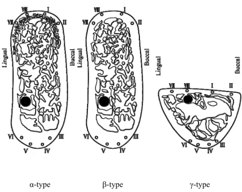
no history of metabolic bone disease. The morphology was classified for six samples with a high and
well-rounded alveolar area (α-type, β-type) and three samples with a low and flat alveolar area (γ
type). High and well-rounded samples were then further classified by cortical bone thickness (α
type: 0.80 mm or thinner , β-type: 1.60 mm or thicker). The posterior region from 9 mm from the
center of the mental foramen was cut along the coronal plane perpendicular to the mandibular plane
6
using a diamond cutter (FineCUT, Heiwa Technica, Japan). The mandibles of cadavers were fixed in
10% formalin before being dehydrated in ethanol for use in the study. This study was approved by
the Tokyo Dental College Ethics Committee (No. 326).
2. Micro-computed tomography (CT) scan
Micro-CT images of the samples (HMX-225 Actis4, Tesco Corporation, Japan) were acquired
with the following imaging conditions: tube voltage, 140 kV; tube current, 75 μA; magnification, ×3;
slice width, 50 μm; field of reconstruction, 60 mm; number of views, 1200; number of scans, 92;
number of slices, 20; matrix size, 512 × 512. Three-dimensional structure analysis software
(TRI/3D/BON, RATOC System Engineering, Japan) was used to create a 3D reconstruction, and the
internal structure was observed.
3. Measurement sites
The samples were placed with the measurement side facing downwards, and autopolymerizing
acrylic resin was injected and embedded from the top.
After embedding, the samples were sliced on the measurement side parallel to the coronal plane
using a saw microtome with a blade width of 300 μm (SP1600, Leica, Germany) to obtain one 200
μm slice. The remaining pieces of embedded samples were polished with waterproof sandpaper
7
(#400→#800→#1200) to eliminate roughness from the sectioned surfaces.
The area corresponding to the alveolar region (Al) and the base of the mandible (Ba) were
designated as the measurement areas, with four points set in each for measurement area, for a total of
eight measurement sites. The measurement sites on the samples cut along the coronal plane were two
sites on the buccal Al (I and II), two sites on the buccal Ba (III and IV), two sites on the lingual Ba
(V and VI), and two sites on the lingual Al (VII and VIII; Figure 1). Measurements of buccal Al site
I and lingual Al site VIII were taken in sites 4 mm from the alveolar crest. The region under the
cancellous bone in the Ba was bisected into the buccal side and the lingual side, which were used for
measurement of the buccal and lingual Ba, respectively.
4. BMD measurement
BMD was measured with a peripheral quantitative CT (pQCT) bone densitometer (XCT Research
SA+, Stratec Medizintech, GmbH, Pforzheim, Germany), with a voltage of 50.7 kV, current of 0.276
mA, and voxel size of 80 × 80 × 460 μm. Voxel data from each sample were scanned and read with
Microsoft Excel and exported as an ASCII CSV file.
BMD of the cortical bone was measured in areas with a threshold of at least 690 mg/cm3.
5. BAp crystalline orientation
8
Quantitative analysis of BAp crystalline orientation was performed with a microbeam X-ray
diffractor with a reflection-based optical system using Cu-Kα beams and a transmission-based
optical system using Mo-Kα beams (reflection system: RINT2500, Rigaku Corporation, Tokyo,
Japan; transmission system: Rigaku R-AXIS Bone Quality, Rigaku Corporation). Samples were
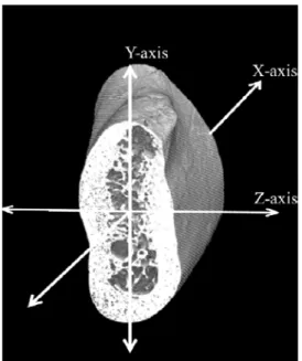
arranged with the mesiodistal direction along the X-axis, the vertical direction to the virtual occlusal
plane along the Y-axis, and the Buccalingual direction along the Z-axis (Figure 2). For the reflection
system, tube voltage was set at 40 kV and tube current at 200 mA. For the transmission system, tube
voltage was set at 50-kV holds and tube current at 90 mA. The incident beam was focused on a
minute 100-μm diameter spot using a collimator.
Samples were first measured along the X-axis using the diffractor of the reflection optical system.
The diffracted X-ray beams were detected using a curved position-sensitive proportional counter.
Samples were then measured along the Y- and Z-axes using the diffractometer of the transmission
optical system. Measurement conditions were the same as those used by Nakano et al.8,13).
The transmission diffractometer produced diffraction rings on the imaging plate with diffraction
lines. X-ray diffraction data out were recorded with Rigaku R-AXIS BQ software and evaluated by
calculating the intensity ratio of the (002) and (310) diffraction peaks. The means of three
measurements in each of the eight sites were calculated and used as measurement values.
9
6. Statistical analysis
For statistical analysis, measurements along the X-, Y-, and Z-axes were divided into Al data and
Ba data for the α-, β-, and γ-type samples, and the means of four points in Al and four points in Ba
were compared with Tukey’s multiple comparison tests. p < 0.05 was considered to be significant.
Results
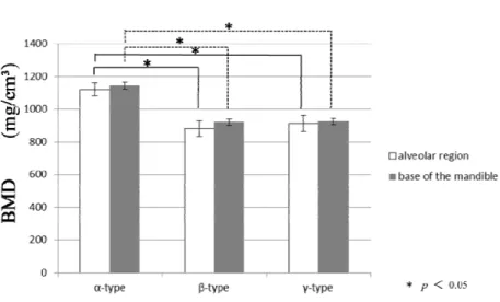
1. BMD
The results of quantitative BMD evaluation for the means of four points in Al and four points in Ba
with α-, β-, and γ-type measurements divided into Al data and Ba data are shown in Figure 3. No
site-based differences in BMD were observed between Al and Ba of the human edentulous mandible
in any of the three types. However, comparisons of the types revealed BMD to be higher in the α
type and lower in the β- and γ-types (p < 0.05).
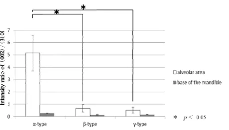
2. BAp orientation
Figure 4 shows the BAp crystalline orientation along the X-axis measured with the reflection
diffractometer for the means of four points in Al and four points in Ba with α-, β-, and γ-type
measurements divided into Al data and Ba data. The intensity ratio of HAp powder used as a control
was 1.53. BAp crystalline orientation along the X-axis in the α-, β-, and γ-type samples was stronger
in Ba and weaker in Al in all three samples, and both differed significantly among sample types (p <
10
0.05).
Figure 5 shows BAp crystalline orientation along the Y plane measured with the transmission x-
ray diffractometer for the means of four points in Al and four points in Ba with α-, β-, and γ-type
measurements divided into Al data and Ba data. Figure 6 shows the BAp crystalline orientation
along the Z-axis measured the same way. The intensity ratio of HAp powder used as a control was
0.60 for both the Y- and Z-axes. In Al of the α-type where orientation along the X-axis was
weakening, BAp crystalline orientation was high along the Y- and Z-axes. Moreover, comparisons of
BAp crystalline orientation in the Y- and Z-axes directions in Al and Ba revealed significantly higher
values in Al. In contrast, BAp crystalline orientation was weak along both the Y- and Z-axes in both
Al and Ba in β- and γ-type samples.
Discussion
1. BAp orientation
In the present study, we found a uniaxial preferential alignment along the X-axis in Ba and along
the Y- and Z-axes in Al in α-type samples. This observation suggests that the preferential orientation
was in the longitudinal direction of the bone, because Ba has a horseshoe-shape and long bone-like
structure with the mandibular condyle constituting the head of the bone. On the other hand, the
orientation from the dentulous period may remain in Al, leading to preferential orientation along the
11
Y- and Z-axes there. In addition, the extremely thin cortical bone in Al may have led to preferential
orientation along the Y- and Z-axes to supplement those structural characteristics.
In β- and γ-type samples, preferential orientation along the X-axis was observed in both Al and Ba.
Comparisons of BAp crystalline orientation in the X-axis directions in Al and Ba revealed
significantly lower values in Al. These findings demonstrate that the human edentulous mandible
becomes low response to mechanical stress with resorption of Al, resulting in a most part presenting
with long bone-like characteristics.
The present study also found marked differences in the crystalline orientation of α- and β-types
that were classified in the same group as mandibles with a high, well-rounded alveolar area. These
differences may be due to low response to mechanical stress with the thickening of cortical bone in
edentulous mandibles.
This observation suggests that Nano-scale-level bone evaluation using BAp is therefore extremely
useful. Further studies to determine the factors underlying differences between the α- and β-types
may help clarify details concerning the changes in bone morphology of edentulous mandibles.
2. Relationship between BAp orientation and BMD
Comparing BMD in α-, β-, and γ-type mandibles revealed significant differences between the α
type and the other two types, with α-type samples exhibiting a higher BMD. However, BMD did not
12
differ significantly between Al and Ba in any type of mandible. BMD is therefore useful for
distinguishing the α-type from the β- and γ-types, which differ dramatically in bone mass, but cannot
be used to make localized evaluations of each type of mandible. In contrast, BAp crystalline
orientation is effective for qualitative evaluation by site. This result is consistent with findings on
dentulous mandibles17,18). In the present study, we also found experimental evidence for the
usefulness of BAp crystalline orientation for evaluating edentulous mandibles.
3. Clinical implication
The mandible from which the teeth have been lost, BAp crystalline orientation becomes reset
regardless of the extent of bone resorption, resulting in the loss of preferential orientation in the
occlusal direction. When considering the clinical use of implants from the aspect of BAp crystalline
orientation, it is recommended that implants are placed while orientation along the Y- and Z-axes is
still maintained.
Acknowledgements
This research was supported by a Grant-in-Aid for Scientific Research (Challenging Exploratory
Research: 24390446 and 45463050) from the Japan Society for the Promotion of Science. The
13
authors would like to thank Mrs. Eiko Watanabe for her technical assistance.
References
14
1. Kingsmill V.J. Post-extraction remodeling of the adult mandible. Crit Rev Oral Biol Med
10:384-404,1999.
2. Merrot O, Vacher C, Merrot S, Godlewski G, Frigard B, Goudot P.Changes in the edentate mandible in the elderly. Surg Radiol Anat 27:265-270,2005
3. Hansson S and Halldin A. Alveolar ridge resorption after tooth extraction:A consequence of a fundamental principle of bone physiology. J Dent Biomech 3:1758736012456543,2012
4. Bassi F,Procchoio M,Fava C,Schierano G,Preti G. Bone density in human dentate and edentulous mandible using computed tomography. Clin Oral Impl Res 10:356-361,1999
5. Hart RT, Hennebel VV,Thongpreda N,Van Buskirk WC,Anderson RC. Modeling the biomechanics of the mandible:a three-dimensional finite element study.J Biomech 25: 261
286,1992
6. Ichim I,Kieser JA,Swain MV.Functional significance of strain distribution in the human mandible under masticatory load. Arch Oral Biol 52:465-473,2007
7. NIH Consensus Development Panel on Osteoporosis Prevention, Diagnosis, and Therapy. March 7-29, 2000:highlights of the conference. South Med J 94: 569-573, 2001
8. Nakano T, Kaibara K, Tabata Y, Nagata N, Enomoto S, Marukawa E, Umakoshi Y .Unique alignment and texture of biological apatite crystallites in typical calcified tissues analyzed by microbeam x-ray diffractometer system. Bone 31:479-487, 2002
9. Nakano T,Tabata Y, Umakoshi Y. Texture and Bone reinforcement.Encyclopedia of Materials,Science and Technology Updates,(Texture and Bone Reinforcement, Elsevier,O xford)MS2061:1-8,2005
10. Elliot JC. Structure and chemistry of the apatites and other calcium orthophosphates. Amsterdam Elsevier 1-389, 1994
11. Sasaki N, Matsushima N, Ikawa T, Yamamura H and Fukuda A. Orientation of bone mineral and
15
its role in the anisotropic mechanical properties of bone--transverse anisotropy. J Biomech 22:
157-164, 1989
12. Sasaki N and Sudoh Y. X-ray pole figure analysis of apatite crystals and collagen molecules in bone. Calcif Tissue Int 60: 361-367, 1997
13. Bacon GE, Bacon PJ and Griffiths RK.Orientation of apatite crystals in relation to muscle attachment in the mandible . J Appl Crystallogr 10: 124-126, 1977
14. Sasaki K, Nakano T, Ferrara JD, Lee JW, Sasaki T. New Technique for Evalution of Preferential Alignment of Bioligical Apatite(BAp) Crystallites in Bone Using Transmission X-ray Diffractometry. Materials Transactions 49 (9):2129-2135,2008
15. Ogai T, Morioka T, Matsunaga S, Nojima K, Nishii Y, Sueishi K and Yoshinari M. Relationship between Biological Apatite Alignment and Hemi-occlusion in Rabbit Mandibular Cortical bone.
J.Hard Tissue Biology 21: 165-172, 2012
16. Fujitani W, Nakano T, Change in Biological Apatite Orientation in Beagle Mandible. Materials Science Forum 654-656:2216-2219,2010
17. Morioka T, Matsunaga S, Yoshinari M, Ide Y, Nakano T, Sekine H and Yajima Y. Alignment of biological apatite crystallites at first molar in human mandible cortical bone. Cranio 30: 32-40, 2012
18. Furuya H, Matsunaga S, Tamatsu Y, Nakano T, Yoshinari M, Abe S and Ide Y. Analysis of biological apatite crystal orientation in the anterior cortical bone of the human mandible using microbeam X-ray diffractometry. Materials Transactions 53: 980-984, 2012
19. Ide Y, Agematsu H. Amorphological change of mandible and maxilla after loss of teeth. Jpn. J.
Oral Biol 39:79-90, 1997 [in Japanese]
20. Matsumoto T, Matsunaga S, Morioka T, Nakano T,Yoshinari M, Yajima Y. Relationship between Preferential Alignment of Biological Apatite and Young’s Modulus at First Molar in Human Mandible Cortical Bone. J.Hard Tissue Biology 22: 163-170, 2013
16
Figure legends
Figure 1. Structure of the coronal plane
α-type: High, well-rounded alveolar area (0.80 mm or thinner), β-type: High, well-rounded alveolar area (1.60 mm or thicker), γ-type: Low, flat alveolar area
I and II: sites on the buccal Al, III and IV: sites on the buccal Ba, V and VI: sites on the lingual Ba, VII and VIII: sites on the lingual Al
Figure 2. Setting of the coordinate axes
Measurement sites were designated for the samples. The mesiodistal direction was placed along the X-axis, the vertical direction to the virtual occlusal plane along the Y-axis, and the Buccalingual direction along the Z-axis.
Figure 3. BMD values (mean ± SD) for the Al and Ba.
Vertical axis: BMD value (mg/cm3)
No site-based differences in BMD were observed between the Al and Ba of the human edentulous mandible in any of the three types. However, comparisons of the types revealed BMD to be higher in the α-type and lower in the β- and γ-types (p < 0.05).
Figure 4. BAp crystalline orientation (mean ± SD) along the X-axis (mesiodistal direction)
Samples with a high and well-rounded alveolar area (α-type: about 0.80 mm or thinner; β-type:
about 1.60 mm or thicker) and samples with a low and flat alveolar area (γ-type) were compared.
Each sample was bisected into Al and Ba. The vertical axis shows the diffraction intensity ratio calculated from the(002)/(310) peaks.
BAp crystalline orientation along the X-axis in the α-, β-, and γ-type samples was stronger in Ba and weaker in Al in all three samples, and both differed significantly among sample types (p < 0.05).
17
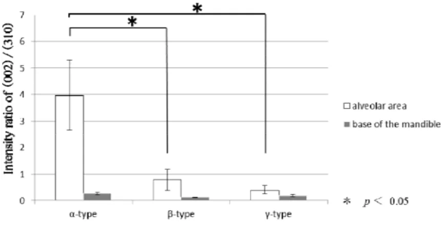
Figure 5. BAp crystalline orientation (mean ± SD) along the Y-axis (vertical direction to the virtual occlusal plane).
Samples with a high and well-rounded alveolar area (α-type: about 0.80 mm or thinner; β-type:
about 1.60 mm or thicker) and samples with a low and flat alveolar area (γ-type) were compared.
Each sample was bisected into Al and the Ba.
The vertical axis shows the diffraction intensity ratio calculated from the(002)/(310) peaks.
In Al of the α-type where orientation along the X-axis was weakening, BAp crystalline orientation was high along the Y-axes. Moreover, comparisons of BAp crystalline orientation in the Y-axis directions in Al and Ba revealed significantly higher values in Al. In contrast, BAp crystalline orientation was weak along both the Y- axes in both Al and the Ba in β- and γ-type samples.
Figure 6. BAp crystalline orientation (mean ± SD) along the Z-axis (Buccalingual direction) Samples with a high and well-rounded alveolar area (α-type: about 0.80 mm or thinner; β-type:
about 1.60 mm or thicker) and samples with a low and flat alveolar area (γ-type) were compared.
Each sample was bisected into Al and the Ba.
The vertical axis shows the diffraction intensity ratio calculated from the(002)/(310) peaks.
In Al of the α-type where orientation along the X-axis was weakening, BAp crystalline orientation was high along the Z-axes. Moreover, comparisons of BAp crystalline orientation in the Z-axis directions in Al and Ba revealed significantly higher values in Al. In contrast, BAp crystalline orientation was weak along both the Z- axes in both Al and Ba in β- and γ-type samples.
18
α-type β-type γ-type
Figure 1. Structure of the coronal plane
α-type: High, well-rounded alveolar area (0.80 mm or thinner) β-type: High, well-rounded alveolar area (1.60 mm or thicker) γ-type: Low, flat alveolar area
I and II: sites on the buccal Al III and IV: sites on the buccal Ba V and VI: sites on the lingual Ba VII and VIII: sites on the lingual Al
19
Figure 2. Setting of the coordinate axes
Measurement sites were designated for the samples. The mesiodistal direction was placed along the X-axis, the vertical direction to the virtual occlusal plane along the Y-axis, and the Buccalingual direction along the Z-axis.
20
Figure 3. BMD values (mean ± SD) for Al and Ba.
Vertical axis: BMD value (mg/cm3)
No site-based differences in BMD were observed between the Al and Ba of the human edentulous mandible in any of the three types. However, comparisons of the types revealed BMD to be higher in the α-type and lower in the β- and γ-types (p < 0.05).
21
Figure 4. BAp crystalline orientation (mean ± SD) along the X-axis (mesiodistal direction)
Samples with a high and well-rounded alveolar area (α-type: about 0.80 mm or thinner; β-type:
about 1.60 mm or thicker) and samples with a low and flat alveolar area (γ-type) were compared.
Each sample was bisected into Al and Ba. The vertical axis shows the diffraction intensity ratio calculated from the(002)/(310) peaks.
BAp crystalline orientation along the X-axis in the α-, β-, and γ-type samples was stronger in Ba and weaker in Al in all three samples, and both differed significantly among sample types (p < 0.05).
22
Figure 5. BAp crystalline orientation (mean ± SD) along the Y-axis (vertical direction to the virtual occlusal plane).
Samples with a high and well-rounded alveolar area (α-type: about 0.80 mm or thinner; β-type:
about 1.60 mm or thicker) and samples with a low and flat alveolar area (γ-type) were compared.
Each sample was bisected into Al and the Ba.
The vertical axis shows the diffraction intensity ratio calculated from the(002)/(310) peaks.
In Al of the α-type where orientation along the X-axis was weakening, BAp crystalline orientation was high along the Y-axes. Moreover, comparisons of BAp crystalline orientation in the Y-axis directions in Al and Ba revealed significantly higher values in Al. In contrast, BAp crystalline orientation was weak along both the Y- axes in both Al and the Ba in β- and γ-type samples.
23
Figure 6. BAp crystalline orientation (mean ± SD) along the Z-axis (Buccalingual direction) Samples with a high and well-rounded alveolar area (α-type: about 0.80 mm or thinner; β-type:
about 1.60 mm or thicker) and samples with a low and flat alveolar area (γ-type) were compared.
Each sample was bisected into Al and the Ba.
The vertical axis shows the diffraction intensity ratio calculated from the(002)/(310) peaks.
In Al of the α-type where orientation along the X-axis was weakening, BAp crystalline orientation was high along the Z-axes. Moreover, comparisons of BAp crystalline orientation in the Z-axis directions in Al and Ba revealed significantly higher values in Al. In contrast, BAp crystalline orientation was weak along both the Z- axes in both Al and Ba in β- and γ-type samples.
24