INTRODUCTION
Bone augmentation is often accomplished by using artificial materials in combination with oral implant treatment.1−4Moreover, placement of implants in the maxilla may require the use of artificial materials for elevation of the maxillary sinus.5−8 Beta-tricalcium phosphate (β-TCP) has a high level of osteo-con- ductivity and is absorbed relatively quickly, to be re- placed with autologous bone.9−14It is often used as an artificial material for bone augmentation in den- tistry.15−21For bone healing in the extraction socket and growth of tissue around implants, the initial develop- ment of microvascular formation is followed by forma- tion of new bone.22, 23 Microvascular formation ad- vances into the interspaces between theβ-TCP gran- ules used to fill the bone defects, resulting in the for- mation of new bone around the granules.24, 25Accord- ingly, the granule interspaces are considered impor- tant for microvascular and new bone formation. We speculated that microvascular formation in the granu- lar interspaces might vary depending on the size of the granules used. In the present study, we used β-
TCP granules of different sizes to fill bone defects in experimental animals, and investigated the relation- ship between granule interspace size and microvas- cular formation.
MATERIALS AND METHODS Materials
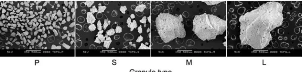
Four sizes ofβ-TCP granules were used ; Types P (50−150μm), S (200−500μm), M (500−1,000μm) and L (1,000−2,000μm) (Fig. 1). The Type P granules (Kyocera Medical, Kyoto, Japan) were the compact type, while the Type S, M and L granules (BIORE- SORBⓇMacro Pore ; ORALTRONICS, Bremen, Ger- many) each had a porosity of 30%, as they consisted of micropores of 0.5−10μm (mean 5μm) and macro- pores of 50−700μm (mean 500μm), both of which are referred to as ‘macropores’ for convenience in the present study.
Experimental animals
Five adult male crab-eating monkeys with an average body weight of 5 kg and healthy permanent dentition in both the upper and lower jaws were used. The bilat-
Microvascular formation in interspaces with different sizes ofβ-tricalcium phosphate granules used to fill bone defects
Hidehito Yasumitsu, Isumi Toda and Fumihiko Suwa
Department of Anatomy, Osaka Dental University, 8-1 Kuzuhahanazono-cho, Hirakata-shi, Osaka 573-1121, Japan
We investigated how granule interspaces affected vascular formation when a bone defect is filled with different sizes ofβ-TCP granules. Bone defects in the experimental animals were filled with four sizes ofβ-TCP granules. Bone and microvascular corrosion cast specimens were then prepared for SEM, and the volume of the newly-formed vascular network was cal- culated from the SEM images. Newly-formed vasculature was observed in the granule inter- spaces with all sizes of granules. In particular, the newly-formed vasculature in smaller gran- ules was uniformly distributed. The volume of the newly-formed vasculature was approxi- mately equal in both the experimental and control animals. Whenβ-TCP granules were used to fill bone defects, newly-formed vessels were more uniformly distributed in defects filled with small granules, which are considered advantageous for microvascular formation. (J Osaka Dent Univ 2015 ; 49 : 157−164)
Key words : Microvasculature ; Granule interspaces ; β-Tricalcium phosphate ; Bone de- fect
eral lower molars were used as experimental sites.
The animals were given a standard primate diet (PS- A, Oriental Yeast, Tokyo, Japan) and water. The pre- sent experiment was approved by the Animal Re- search Committee of Osaka Dental University (Ap- proval numbers 13-02001 and 14-02001) and per- formed under the provisions of the Regulations for Animal Experiments of Osaka Dental University.
Methods
Surgical procedures
The five monkeys were given general anesthesia us- ing 0.1 mL/kg injection of 2% xylazine hydrochloride (SelactalⓇ; Bayer Yakuhin, Tokyo, Japan) as a seda- tive and 0.2 mL/kg ketamine hydrochloride (Keta- larⓇ; Daiichi Sankyo Propharma, Tokyo, Japan) as an anesthetic. The bilateral lower molars (second pre- molar, first molar and second molar) were extracted to minimize surgical invasion of the surrounding tissue.
For 1 week after surgery, mouth rinsing was per- formed daily with a 0.01% benzethonium chloride so- lution (Neostelin Green Gargle 0.2%Ⓡ; Nippon Shika Yakuhin, Shimonoseki, Japan) and the animals were given a soft diet. As an antimicrobial agent for pre- venting infection, injection of 15 mg/kg lincomycin hy- drochloride hydrate (LincocinⓇ; Pfizer Japan, To- kyo, Japan) was administered intramuscularly.
At eight weeks after tooth extraction, the animals were again anesthetized using the same procedure as that for tooth extraction. The gingiva in the experi- mental region was incised and detached to expose the bone, and a total of 6 bone defects, 3 on each side, were formed by drilling to a depth of 6 mm in the buccal socket margin using a 3.5-mm implant drill (ITI implant system ; STRAUMANN, Basel, Switzerland) under irrigation with physiological saline (Otsuka nor-
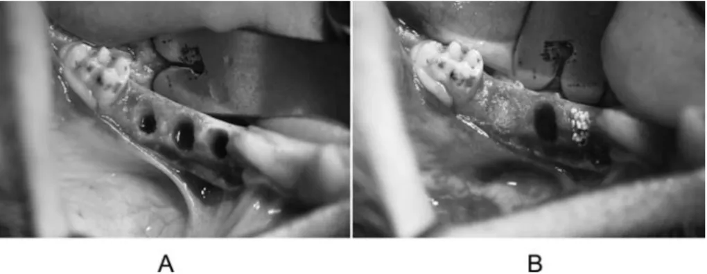
mal saline injection ; Otsuka Pharmaceutical, Toku- shima, Japan). Granules in physiological saline were used to fill the defects, with Type L and S granules separately placed in the two defects on one side, and Type P and M granules on the other side. The remain- ing two defects on each side were left unfilled to serve as controls. After filling with the granules, a gingival flap was sutured over the wound to complete the pro- cedure. Similar to the situation after tooth extraction, intraoral rinsing was performed and a soft diet was given for the first week after surgery, while an antibac- terial agent was administered intramuscularly to pre- vent infection. The suture was removed at 1 week af- ter surgery and the soft diet was replaced with a stan- dard primate diet. The animals were then housed for 1 more week (Fig. 2).
Preparation of samples
At two weeks after the second procedure, the five monkeys were euthanized by injection of an overdose of 50 mg/kg pentobarbital sodium salt (Somnopen- tylⓇ; Kyoritsu Seiyaku, Tokyo, Japan), and then acrylic resin was injected through the bilateral carotid artery using a microvascular injection method.26, 27Af- ter the resin hardened, the animals were soaked in 10% neutral buffer formalin solution and the experi- mental regions were extracted using a large diamond band saw (BS-310 CP ; EXAKT, Norderstedt, Ger- many). Blocks of each bone defect, both filled and un- filled, were prepared using a small diamond band saw (BS-3000 ; EXAKT).
Granule interspace measurement
Among the cut blocks, those filled with granules were radiographed using a micro focus X-ray CT system (micro-CT) (SMX-130 CT ; SHIMADZU, Kyoto, Ja-
Fig. 1 SEM images ofβ-TCP granule types P, S, M and L (Bars=500μm).
pan) with a tube voltage of 45 kV, a tube current of 120 μA, and a slice thickness of 27.5μm. Slice images of only the bone defect portions were separated and processed into 3D images using 3D image analysis software (VGStudio MAXⓇVer.1.2.1 ; Volume Graph- ics, Heidelberg, Germany). Furthermore, to highlight only the granule interspaces in the images, a colum- nar portion 3 mm in diameter and 5 mm in height was extracted by removing the surrounding area contact- ing the socket wall of the defect. The volume of the granules and that of the interspaces in the columnar portion was then determined. The dimension of the in- terspaces was calculated from the mean for the X, Y and Z axes.
Preparation of specimens for scanning electron microscopy
The bone defect portion of each block obtained from one of the experimental animals was cut at the center to obtain frontal sections. One was left untreated and the other was decalcified with 5% hydrochloric acid.
For specimens from the remaining four animals, each block was cut horizontally into four slices of thickness 1.5 mm, and the bottom and middle slices were se- lected as samples (Fig. 3). The sample slices were soaked in 5% sodium hypochlorite to dissolve and re- move soft tissue, then rinsed with water and dried.
These were used as bone and microvascular cast specimens. After evaporating with osmium tetroxide using a plasma multi coater (PMC-5000 ; Meiwa, Osaka, Japan), the specimens were observed under a scanning electron microscope (SEM) (JSM-5500 ;
JEOL, Tokyo, Japan) at an accelerating voltage of 5 kV and working distance of 48 mm. Digital images were obtained for observation from a cross-section of frontal sections and from the top surface of horizontal sections. Furthermore, horizontal section specimens were soaked in 5% hydrochloric acid to remove hard tissue. After rinsing in water and drying, they were used as microvascular cast specimens. Following gold coating (JFC-1500 ; JEOL, Tokyo, Japan), they were observed with an SEM and digital images were obtained.
Image analysis of newly-formed vessels in gran- ule interspaces
Based on SEM images of the microvascular casts as decalcified horizontal-section specimens, the bone defect portion was extracted using photo retouch soft- ware (PhotoshopⓇ; Adobe Systems, San Jose, CA, USA) on a personal computer (MacBook Pro, Apple
Fig. 2 Bone defects in an experimental animal (right side). (A) Immediately after creation of defects (three defects), (B) Defects filled with granules (Type M, Control, and Type P from medial to distal).
Fig. 3 Schematic illustration of the bone defect re- gions. The bone defect was separated horizontally into four regions (superficial, middle, deep and bottom) to make the specimens.
Computer, Cupertino, CA, USA). The ratio of the area of newly-formed vessels to the area of bone defect was calculated using binary images of the vessels and public domain image analysis software (Image J ; National Institutes of Health, Bethesda, MD, USA). The mean dimensions for the interspace be- tween each type of granule were calculated and used as the amount of newly-formed vessels.
RESULTS
Granule interspace measurements(Table 1) The mean dimensions of the interspace between granules used to fill the bone defects in the experi- mental animals were 96.4±4.3, 151.2±5.6, and 243.2±15.4μm for Types S, M and L, respectively. It was impossible to measure the interspace between the Type P granules using the 3D image analysis soft- ware employed in the present study.
SEM images(Figs. 4, 5 and 6) Type P granules
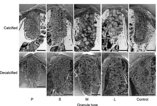
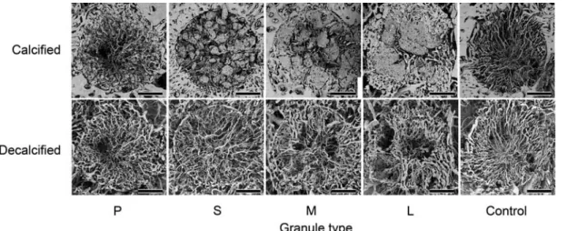
Although SEM images of the non-decalcified speci- mens showed newly-formed vessels in the granule in- terspaces, granules were missing in some areas at the time of specimen preparation. SEM images of the decalcified specimens showed newly-formed vessels oriented toward the center of the bone defect in a uni- form manner (Figs. 4 and 5).
Type S granules
SEM images of the non-decalcified specimens showed newly-formed vessels in the granule inter- spaces, some of which had invaded the macropores in the granules. SEM images of the decalcified speci- mens showed newly-formed vessels oriented toward the center of the bone defects in a uniform manner (Figs. 4, 5 and 6).
Table 1 Granule interspace dimensions
Granule type Average X-axis Y-axis Z-axis
S M L
96.4± 4.3 151.2± 5.6 243.2±15.4
98.2± 3.4 153.0± 8.1 240.0±16.0
99.8± 7.7 152.8± 6.1 240.0±16.2
92.0± 4.5 148.4±11.1 251.0±20.8 Mean±SD, n=5 (μm)
Fig. 4 SEM images of sagittal sections (Bar=2 mm).
Type M granules
SEM images of the non-decalcified specimens showed newly-formed vessels in the granule inter- spaces, some of which had invaded the macropores in the granules. SEM images of the decalcified speci- mens showed that areas of newly-formed vessels were absent, which corresponded to the granules.
New vessels formed in the interspaces appeared to encircle the granules (Figs. 4, 5 and 6).
Type L granules
SEM images of the non-decalcified specimens showed newly-formed vessels in the granule inter- spaces in the same manner as observed for the Type M granules. Some of the vessels had invaded the macropores in the granules. SEM images of the de- calcified specimens showed that areas of newly- formed vessels were absent, which corresponded to the granules, the same as seen with Type M gran-
ules. New vessels formed in the interspaces ap- peared to encircle the granules (Figs. 4, 5 and 6).
Controls
SEM images of the non-decalcified specimens show- ed formation of new vessels oriented nearly uniformly toward the center of the bone defect. Similarly, SEM images of the decalcified specimens showed newly- formed vessels that appeared to be nearly the same as those seen in the non-decalcified specimens (Figs. 4 and 5).
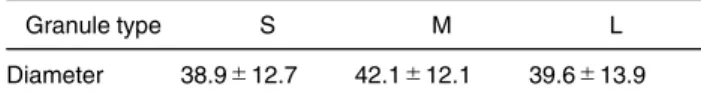
Vascular invasion of macropores
SEM images of the non-decalcified specimens for the Type S, M and L granules, in which macropores were present, showed newly-formed vessels invading the macropores (Fig. 6). Measurements of the invaded macropores using image analysis software (Image ProⓇ Plus, Ver. 5.1 ; Media Cybernetics, Rockville,
Fig. 5 SEM images of horizontal sections (Bar=1 mm).
Fig. 6 Close-up SEM images of horizontal sections of calcified specimens showing the granules filling the defect. Newly formed vessels (arrow heads) invaded the macropores in the granules (Bar=500μm).
MD, USA) showed them to be 38.9±12.7, 42.1±
12.1, and 39.6±13.9μm in diameter for the Type S, M and L granules, respectively. One-way ANOVA re- vealed no significant differences among the three sizes of granules (Table 2). In contrast, no macro- pores or vascular invasion was observed in speci- mens with the Type P granules.
Ratio of newly-formed vessels as determined by image analysis(Table 3)
The ratio of newly-formed vessels in the granule inter- spaces calculated on the basis of SEM images of hori- zontally sectioned decalcified specimens was 20.0±
5.0%, 20.0±4.5%, 17.5±4.6%, 17.9±6.7%, and 20.0±2.9% for Type P, S, M and L granules and the controls, respectively. One-way ANOVA revealed no significant difference among the specimens for differ- ent granule sizes.
DISCUSSION Animal experiment
The period from 1 to 2 weeks after surgery is consid- ered to be the stage of microvascular formation during bone healing around the extraction socket and im- plant, while the stage of new bone formation and bone remodeling begins at about 2 weeks, after which bone healing is completed.22, 23Accordingly, based on find- ings of animal experiments of bone defects filled with β-TCP granules, we think that microvascular forma- tion in the granule interspaces could be confirmed by preparing samples obtained at 2 weeks after sur- gery, which would be the stage of microvascular for-
mation just prior to new bone formation. Further- more, we think that newly-formed vessels in the bone defects could be clearly determined by use of speci- mens prepared following acrylic resin microvascular injection.26, 27
Granule interspace measurements
Granule interspace measurements based on micro- CT findings performed at 2 weeks showed that the in- terspaces in defects filled with Type S, M and L gran- ules became larger in accordance with granular size.
We also found that the mean interspace measure- ment for Type S granules was greater than 90μm, while it was previously reported that granules larger than 50μm are required for microvascular formation in the pores.28Thus, we concluded that microvascular formation could be sufficiently attained with use of the Type S, M and L granules employed in the present study.
On the other hand, determination of interspa- ces with the Type P granules was impossible, even though imaging was performed under the same condi- tions as for the other types. The cause for this failure seems to be related to the compact nature of the Type P granules, as radiolucency was low with the high granule density in the bone defects. As a result, inter- spaces smaller than 27μm were unrecognizable, since the slice thickness of the specimens subjected to micro-CT was 27.5μm. Granule interspaces smal- ler than 27μm could not be clearly visualized.
Relationship between granule interspaces and microvascular formation
SEM images of the non-decalcified specimens con- firmed newly-formed vessels in the granule inter- spaces of all defects filled with Type P, S, M and L granules. Even with Type P granules, for which inter- space measurements based on 3D image analysis was impossible, newly-formed vessels were ob- served in the interspaces. Accordingly, we speculate that even a granule interspace size of 27μm or less may be adequate for microvascular formation.
SEM images of the decalcified specimens showed areas free from newly-formed vessels corresponding to the granules in those with Type M and L granules,
Table 2 Diameter of macropores invaded by newly formed ves- sels
Granule type S M L
Diameter 38.9±12.7 42.1±12.1 39.6±13.9 Mean±SD, n=8 (μm)
Table 3 Ratio of newly formed vessels in the bone defect Granule
type P S M L Control
Area (%) 20.0±5.0 20.0±4.5 17.5±4.6 17.9±6.7 20.0±2.9 Mean±SD, n=8
in which newly-formed vessels in the interspaces were observed encircling the granules. Meanwhile with Type S and P granules there were no newly- formed vessels encircling the granules, as the vessels were spread throughout the bone defects. This was thought to be the result of microvascular formation in the finely distributed interspaces of the defects filled with Type S and P granules, which were relatively small in size.
Relationship between granular interspaces and the amount of microvascular formation
There was no significant difference in the amount of newly-formed vessels among the granule types. The ratio was about 20% for Types P and S, which was the same as that of the controls, while the ratios were a bit smaller for Types M and L, at 17% and 18%, respec- tively. These findings suggest that the size of the inter- spaces is not always proportional to the amount of newly-formed vessels.
In the controls, new vessels were formed nearly uniformly in the bone defects, as there was no factor to inhibit microvascular formation. As shown in the SEM images of decalcified specimens, microvascular formation occurred nearly uniformly in specimens with Types P and S. It seems that the amount of newly- formed vessels with this size of granules is approxi- mate, as microvascular formation was uniform in the uniformly distributed interspaces formed by small- sized granules, as was also seen in the controls.
Meanwhile, in specimens with Types M and L, it seems that the amount of newly-formed vessels was less, because microvascular formation occurred in the disproportionately larger interspaces formed by the larger granules.
Vascular invasion of macropores in granules The Type S, M and L granules used in the present study had a porosity of 30%, and contained intra- granular macropores of two size ranges, 0.5−10μm and 50−700μm. SEM images of the non-decalcified specimens confirmed vascular invasion of intra- granular macropores. The average size of macro- pores that showed invasion was 40μm, with no sig- nificant differences among the types. It has been re-
ported that macropores larger than 50μm are re- quired for microvascular formation.28However, in the present specimens, vessel invasion was noted in mi- cropores 40μm in diameter. Even at 2 weeks after surgery, when granular resorption had already been initiated and the macropores in the granules were as small as 0.5−10μm, the macropores were thought to grow larger to make invasion by new vessels possi- ble.
When using β-TCP granules to fill bone defects, larger granules with higher porosity and larger macro- pores are considered better for microvascular forma- tion than small granules like Type P and S. This is be- cause the newly-formed vessels are more uniformly distributed in defects filled with the larger Type M and L granules, making a greater amount of newly-formed vessels. We intend to perform another study in the fu- ture on the relationship between microvascular forma- tion and new bone formation during the new bone for- mation stage using the present methods to determine the optimum conditions for β-TCP granule filling of bone defects, such as granule size, porosity, and in- terspace size.
We are grateful to the members of the Department of Anat- omy for their kind advice and assistance.
REFERENCES
1. Hempton TJ, Fugazzotto PA. Ridge augmentation utilizing guided tissue regeneration, titanium screws, freeze-dried bone, and tricalcium phosphate : clinical report.Implant Dent 1994 ; 3: 35−37.
2. Hardwick R, Hayes BK, Flynn C. Devices for dentoalveolar re- generation : an up-to-date literature review. J Periodontol 1995 ; 66: 495−505.
3. Mercier P, Bellavance F, Cholewa J, Djokovic S. Long-term stability of atrophic ridges reconstructed with hydroxylapatite : a prospective study.J Oral Maxillofac Surg1996 ; 54: 960−
968.
4. Scarano A, Carinci F, Assenza B, Piattelli M, Murmura G, Piat- telli A. Vertical ridge augmentation of atrophic posterior mandi- ble using an inlay technique with a xenograft without minis- crews and miniplates : case series.Clin Oral Implants Res 2011 ; 22: 1125−1130.
5. Yildirim M, Spiekermann H, Biesterfeld S, Edelhoff D. Maxillary sinus augmentation using xenogenic bone substitute material Bio-OssⓇin combination with venous blood. A histologic and histomorphometric study in humans.Clin Oral Implants Res 2000 ; 11: 217−229.
6. Tadjoedin ES, de Lange GL, Holzmann PJ, Kuiper L, Burger EH. Histological observations on biopsies harvested following sinus floor elevation using a bioactive glass material of narrow
size range.Clin Oral Implants Res2000 ; 11: 334−344.
7. Hallman M, Cederlund A, Lindskog S, Lundgren S, Sennerby L. A clinical and histologic study of bovine hydroxyapatite in combination with autogenous bone and fibrin glue for maxillary sinus floor augmentation. Results after 6 to 8 months of heal- ing.Clin Oral Implants Res2001 ; 12: 135−143.
8. Szabó G, Suba Z, Hrabák K, Barabás J, Németh Z. Autoge- nous bone versusβ-tricalcium phosphate graft alone for bilat- eral sinus elevations (2- and 3-dimensional computed to- mographic, histologic, and histomorphometric evaluations) : Preliminary results.Int J Oral Maxillofac Implants2001 ; 16: 681−692.
9. Nagahara K. Osteogenesis in response to tricalcium phos- phate (TCP) and hydroxyapatite ceramics (HAP) implanted into bone tissues.Shika Kiso Igakkai Zasshi (Jpn J Oral Biol) 1987 ; 29: 131−155. (Japanese)
10. Oyake Y, Beppu M, Ishii S, Takagi M, Takashi M. Intramedul- lary anchoring strength of titanium rod with mixedβ-tricalcium phosphate and fibrin adhesive.J Orthop Sci2002 ; 7: 123−
130.
11. Ozawa M. Experimental study on bone conductivity and ab- sorbability of the pureβ-TCP.Seitai Zairyo (J Jpn Soc Bioma- terial)1995 ; 13: 167−175. (Japanese)
12. Saito M, Shimizu H, Beppu M, Takagi M. The role of β- tricalcium phosphate in vascularized periosteum.J Orthop Sci 2000 ; 5: 275−282.
13. Dong J, Uemura T, Shirasaki Y, Tateishi T. Promotion of bone formation using highly pure porousβ-TCP combined with bone marrow-derived osteoprogenitor cells. Biomaterials 2002 ; 23: 4493−4502.
14. Morikawa S. Comparative study on β-tricalcium phosphate and hydroxyapatite as bioactive artificial bone fillers.Tokyo Jikei-kai Ika Daigaku Zasshi (Tokyo Jikeikai Med J) 2000 ; 115: 193−207. (Japanese)
15. Ormianer Z, Palti A, Shifman A. Survival of immediately loaded dental implants in deficient alveolar bone sites augmented with β-tricalcium phosphate.Implant Dent2006 ; 15: 395−403.
16. Brkovic BMB, Prasad HS, Konandreas G, Milan R, Antunovic D, Sándor GKB, Rohrer MD. Simple preservation of a maxillary extraction socket using beta-tricalcium phosphate with type I collagen : preliminary clinical and histomorphometric observa- tions.J Can Dent Assoc2008 ; 74: 523−528.
17. Horowitz RA, Mazor Z, Miller RJ, Krauser J, Prasad HS, Ro- hrer MD. Clinical evaluation of alveolar ridge preservation with aβ-tricalcium phosphate socket graft.Compend Contin Educ Dent2009 ; 30: 588−603.
18. Honda K. Clinical study of a novel bone augmentation withβ- tricalcium phosphate.Okayama Shigakkai Zasshi (J Okayama Dent Soc)2009 ; 28: 1−9. (Japanese)
19. Frenken JWFH, Bouwman WF, Bravenboer N, Zijderveld SA, Schulten EAJM, ten Bruggenkate CM. The use of StraumannⓇ Bone Ceramic in a maxillary sinus floor elevation procedure : a clinical, radiological, histological and histomorphometric evaluation with a 6-month healing period.Clin Oral Implants Res2010 ; 21: 201−208.
20. Wakimoto M, Ueno T, Hirata A, Iida S, Aghaloo T, Moy PK. His- tologic evaluation of human alveolar sockets treated with an artificial bone substitute material. J Craniofac Surg 2011 ; 22: 490−493.
21. Hirota M, Matsui Y, Mizuki N, Kishi T, Watanuki K, Ozawa T, Fukui T, Shoji S, Adachi M, Monden Y, Iwai T, Tohnai I. Combi- nation with allogenic bone reduces early absorption of β- tricalcium phosphate (β-TCP) and enhances the role as a bone regeneration scaffold. Experimental animal study in rat mandibular bone defects.Dent Mater J2009 ;28: 153−161.
22. Suwa F. Bone healing around dental implant −microvasculari- zation and bone formation in functional or non functional condi- tions−.Nihon Shika Igakkai Shi (J Jpn Assoc Dent Sci)1998 ; 17: 124−129. (Japanese)
23. Suwa F. What is discovered from microvascular corrosion cast-bone specimens. Denshi Kenbikyo (Electron Micros- copy)1999 ; 34: 168−172. (Japanese)
24. Kuroki K, Toda I, Suwa F. Experimental study of bone defect repair process with different sizes ofβ-TCP granules.Nihon Koku Implant Gakkai Shi (J Jpn Soc Oral Implant)2008 ; 21: 21−31. (Japanese)
25. Toda I, Yasuda K, Ehara D, Kuroki K, Suwa F. The effects on extraction sockets filled with small β-tricalcium phosphate granules : An experimental histomorphometric study. Nihon Koku Implant Gakkai Shi (J Jpn Soc Oral Implant)2013 ; 26: 228−235. (Japanese)
26. Ohta Y, Okuda H, Suwa F, Okada S, Toda I. Plastic injection method for preparing microvascular corrosion casts for SEM and its practical application.Okajimas Folia Anat Jpn1990 ; 66: 301−312.
27. Suwa F, Uemura M, Takemura A, Toda I, Fang YR, Xu YJ, Zhang ZY. Acrylic resin injection method for blood vessel in- vestigation.Okajimas Folia Anat Jpn2013 ; 90: 23−29.
28. Ehara Y, Suwa F. Experimental studies on the osseous resto- ration and microvascular formation after dental implantation of the pored almina ceramics.Shika Kiso Igakkai Zasshi (Jpn J Oral Biol)1994 ;36: 471−485. (Japanese)