胎児心不全:不整脈の重症度をどう評価する
前野 泰樹
雪の聖母会 聖マリア病院新生児科
Assessment of the Severity of Heart Failure in Fetuses with Fetal Arrhythmias Yasuki Maeno
Department of Neonatology, St. Mary
ʼs Hospital, Fukuoka, Japan
Fetal arrhythmia is a known cause of fetal heart failure that can lead to fetal death. Although assessment of the severity of heart failure is important for managing affected fetuses, it is often difficult in fetuses with arrhyth- mias. Usually, Doppler waveform, which is accepted in the cardiovascular profiling (CVP) score, is the most use- ful finding for assessing cardiac function. However, arrhythmia changes the Doppler waveform regardless of the cardiac function, so many of these waveforms may not reveal heart failure. Hence, other fetal echocardiographic findings, such as cardiomegaly assessed by the cardiothoracic area ratio, presence of atrioventricular valve regur- gitation, and assessment of cardiac output calculated from the velocity-time integral, are used to detect early signs of cardiac failure. The Biophysical Profiling Score is another method useful for assessing the well-being of affected fetuses. Regarding the fetal heart rate, fetuses often develop heart failure with a fetal heart rate
>220 bpm in fetal supraventricular tachycardia,
>200 bpm in fetal ventricular tachycardia, and
<55 bpm in fetal bradycardia. However, most fetuses with arrhythmia also have cardiac dysfunction, such as tachycardia-induced cardiomyopathy in fetal tachycardia and myocarditis caused by anti-SS-A antibody in fetal bradycardia. Hence, fetal heart rate may not reflect the severity of heart failure in many fetuses. Assessment by combining multiple assessment strategies and sequential follow-up of these findings are essential for assessing heart failure in fetuses with arrhythmias.
Keywords: fetal arrhythmias, fetal heart failure, fetal echocardiography, fetal tachycardia, fetal brady- cardia
胎児不整脈では,心不全が進行すると胎児水腫となり胎内死亡をきたすため,心不全の重症度評価は 重要な課題となるが,実際には正確な評価は難しい.通常,胎児心エコー検査で胎児心不全を評価す
る
CVP (cardiovascular profiling
)スコアなどで使用されるドプラ血流波形は,不整脈により変化するため評価に使用できない項目が多い.そこで,心拡大の程度や房室弁閉鎖不全の出現,特に僧帽弁閉 鎖不全に着目したり,血流波形の
VTI (velocity-time integral
)による心拍出量の算出で心不全の兆候 を検出する.胎児の一般的な全身状態の評価であるBPS (biophysical profiling score
)も参考にできる.心拍数との関連では,頻脈性不整脈では,上室頻拍では心拍
220
回/
分以上,心室頻拍では200
回/
分以 上,胎児徐脈性不整脈では心拍55
回/
分未満が心不全進行の目安となる.しかし,不整脈症例では,頻 脈時の頻拍源性心筋症の併発や,徐脈時には心奇形や抗SS-A
抗体による心筋炎/
心筋症の合併によっ て心機能が低下してくることが知られており,心拍数のみでは心不全進行の予測は不十分である.複 数の指標を合わせて,継時的な経過観察により症例ごとの計測値の変化を評価しながら,管理方法を 判断していくことが重要である.著者連絡先:〒
830
‒8558
福岡県久留米市津福本町422
雪の聖母会 聖マリア病院新生児科 前野泰樹doi: 10.9794/jspccs.35.221
222
はじめに
胎児不整脈では,心不全が進行すると胎児水腫とな り胎内死亡をきたす.したがって心不全の重症度評価 は重要な課題となるが,実際には正確な評価は難し い.胎児心エコーでは,通常はドプラ血流波形を使用 することが多いが,不整脈があると,波形が不整脈自 体により変化するため評価に使用できないことが多 い.そのなかで,どのような所見に注目しながら評 価して,それぞれの症例の周産期管理を行っていくの か,解説する.
胎児不整脈について 胎児不整脈の分類
胎児不整脈は,徐脈性不整脈,頻脈性不整脈,およ びその他の脈の不整の
3
つに大きく分類され1‒3),こ の中で徐脈と頻脈のとき胎児心不全の評価が必要とな る.徐脈性不整脈は,胎児心拍数が100
回/
分未満,頻脈性不整脈は
200
回/
分以上と定義されることが多 い.ただし,頻脈性不整脈では,急激な心拍数の変 化などで不整脈の存在が示唆されるときには180
回/
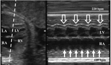
分以上であれば頻脈性不整脈と定義している文献も多 い.胎児期の不整脈診断は,主に心エコー検査によっ て行う.M
モード法(Fig. 1
),ドプラ法による心房Fig.
1
M-mode fetal echocardiogram in fetal atrial flutter
Simultaneous record of the left ventricle (LV) and the right atrium (RA) reveales 2 : 1 relation of atrial (white arrow) and ventricular contraction (hollow arrow). bpm, beats per minute; LA, left ventricle; RV, right ventricle.
Fig.
2 Fetal echocardiographic Doppler wave form of the simultaneous record of the superior vena cava (SVC) and the ascending aorta (aAo)
Right panel reveals the wave form of fetuses with normal sinus rhythm. Starting point of small reversal flow at SVC
represents the timing of atrial contraction (A), and the starting point of forward flow at aAo represents the timing of
ventricular contraction (V). The arrow reveals atrio-ventricular interval. IVC, inferior vena cava; LA, left atrium; RA, right
atrium; RV, right ventricle.
心室収縮を同時に評価する画像を描出し(
Fig. 2
),心房心室収縮の関連を見ることで不整脈の診断を行 う1‒3).
徐脈性不整脈では,まれに洞性徐脈もあるが,多く は房室伝導が乖離した完全房室ブロックであり,およ そ半数ほどは先天性心奇形(多脾症候群や修正大血管 転位など)に起因する.残りの半数ほどは心内構造が 正常であり,この場合は母体の抗
SS-A
抗体に起因し ていることが多い.抗SS-A
抗体陽性母体の1
〜7
%に 房室ブロックを発症するが,実際には母体はシェーグ レン症候群やSLE
は発症しておらず,胎児房室ブロッ クの発症後検査で抗体陽性に気づかれることが多い.頻脈性不整脈では,上室頻拍と心房粗動が多く,
そのほか心室頻拍もある.上室頻拍は心房心室が
1 : 1
伝導のものであり,胎児頻脈性不整脈のおよそ2/3
を占める.多くが房室回帰性頻拍(
WPW
症候群)で,その他,異所性心房頻拍や房室結節回帰性頻拍も認め る.心房心室が
2 : 1
で伝導する心房粗動が次に多く,この
2
つで胎児頻脈の大部分を占める.心室拍数のみ 頻拍となる心室頻拍は稀である.頻脈性不整脈では,持続性のものと,間歇的な頻脈があり(
Fig. 3
),頻脈 出現時間が全体の50
%以上のときに心不全をきたす 危険性が出てくるとされている.胎児不整脈と心不全評価
1.
CVP
(cardiovascular profiling
)スコアの限界 通常,胎児心エコー検査で胎児心不全を評価す る 時 に は,CVP
ス コ ア が 使 用 さ れ る こ と が 多 い(
Table 1
)4).しかし,胎児不整脈のときに問題となるFig.
3 Trace of fetal heart rate monitor in a fetus with intermittent supraventricular tachycardia reveals
sudden onset of fetal tachycardia with the heart rate of 190 bpm (hollow arrow) and sudden recov- ery to the sinus rhythm (black arrow)
Fetuses with the tachycardic episode more than 50
%are thought to be a risk for developing fetal hydrops.
Table
1
Cardiovascular Profile Scoring System
4)Parameter Cardiovascular Profile Score
0 points
−1 point −2 pointsHydrops None Ascites or pericardial
or pleural effusion
Skin edema
Ductus venosus and umbilical vein Doppler DV atrial flow reversal UV atrial pulsations Heart size (CTAR: heart area/chest area)
>0.20 to
<0.35 0.35 to 0.50
<0.20 or
>0.50
Cardiac function Normal TV and MV, RV
and LV SF
>0.28, biphasic diastolic filling
Holosystolic TR or MR, RV or LV SF
<0.28, ventricular hypertrophy
dP/dT at AV valve
<400 or monophasic filling
Umbilical artery Doppler Absent end-diastolic flow
with brain sparing
Reversed end-diastolic flow
AV, atrioventricular; CTAR, cardiothoracic area ratio; dP/dT, change in pressure/time; DV, ductus venosus; MR, mitral regurgita-
tion; MV, mitral valve; SF, shortening fraction;TR, tricuspid regurgitation; TV, tricuspid valve; UV, umbilical vein.
224
のは,
CVP
スコアでは6
項目中3
項目にドプラ血流 波形(臍帯静脈,静脈管,臍帯動脈)が使用されてい る点である.胎児不整脈では,血流波形は心不全の程 度にかかわらず不整脈自体により変化してしまう.こ のため,このCVP
スコアそのまま使用することがで きない.静脈の血流波形で心不全を評価する時には,心房収 縮時の逆流波が増大するのを指標として使用してい
る.しかし不整脈があると,心不全がなくても,この 大きな逆流波が出現する.不整脈では,心房と心室の 収縮時相にずれが生じるため,心室が収縮し房室弁が 閉鎖しているタイミングで心房が収縮することがあ る.このとき心房収縮による血流は心室に入ることが できず,静脈に向かって大きく逆流する(
Fig. 4, 5
).また,
CVP
スコアでは,房室弁流入波形が正常の2
峰性(E
波とA
波)から1
峰性になることを心不全 の指標として使用している.しかし頻脈により心拍数 が上昇していると,心不全がなくても1
峰性であり,心不全の程度の評価の参考にならない.
したがって,
CVP
スコアからは,血流波形以外の 項目,胎児水腫の程度,心拡大の程度(cardiothoracic area ratio: CTAR
)と,房室弁閉鎖不全が,胎児心不 全の指標として使用できる項目となる.ただし,臨床 的には,心不全による胎児水腫への進行を予見するこ とを求められることが多いため,それ以外のCTAR
と房室弁閉鎖不全が,評価に有用な所見であろう.2.
心不全評価に使用できる項目上記の通り,
CVP
スコアの中にあるCTAR
と房室 弁閉鎖不全があり,そのほか,心エコー検査関連の 項目では心拍出量も使用できる.一方,超音波検査 以外の所見も合わせた,胎児の全身状態を評価するBiophysical Profile Score
(BPS
)も心不全の重要な指 標となる.①
CTAR
心不全では心拡大をきたすことがよく知られている が,特に胎児不整脈のときにはこの心拡大が心不全の
Fig.
4 Schematic explanation of the relationship
between atrial contraction and reversal venous blood flow
A. In fetuses with normal sinus rhythm, the right atrium (RA) contract during diastolic phase of the right ventricle (RV). Most of the flood flow made by the RA contraction goes into the RV, and only small amount of the flood flow goes back to the vena cava. B. In fetuses with the arrhythmia, RA can contract during systolic phase. In this situation, blood in the RA can not go into the RV since the tricuspid valve is closed. Hence a large amount of the blood go back to the vena cava regardless the cardiac function.
Fig.
5 Simultaneous record of the superior vena cava (SVC) and the ascending aorta (aAo) in a fetus with short VA supraventricular tachycardia
SVC flow reveals large reversal flow (arrow) made by the atrial contraction started at a line A, since the atria contracts
during aAo flow which represents the ventricular systolic phase started at a line V.
指標として重要な所見となる.これは,胎児頻脈性不 整脈では頻脈誘発性心筋症が,胎児徐脈性不整脈では 心筋炎
/
心筋症の発症が急激な心不全の進行に重要な 要因となるためである.頻脈時の心不全の原因では,頻脈自体で
1
回拍出 量が次第に低下し時間当たりの心拍出量が低下するこ ともあるが,より重要なものに頻脈誘発性心筋症があ る.頻脈が持続すると拡張型心筋症のごとく心筋が菲 薄化し心拡大をきたす.これにより,頻拍の拍数は変 わらなくても急激に心不全が進行するため,胎児頻脈 症例では心拡大の程度を継時的に評価することが重要 となる.徐脈時は,
1
回拍出量の増加で心拍出量を維持する ために両心室が拡大しているため,もともと少なから ず心拡大を認める.しかし,それ以上に重要な要因と して,抗SS-A
抗体による胎児の心筋炎/
心筋症によ る心拡大がある.胎児房室ブロックのとき高頻度に心 筋炎/
心筋症が併発しており,心機能が低下し心不全 がより悪化する5).したがって,徐脈の症例を管理す るときにも,心拡大の程度を継時的に評価する.②房室弁閉鎖不全
心不全では,種々の要因により房室弁閉鎖不全が出 現し,さらにこの逆流により心不全も増悪するため,
この三尖弁や僧帽弁閉鎖不全は心不全の重要な指標と なる3).心不全が増悪してくると,心筋の機能が低下
して乳頭筋不全となり,房室弁の閉鎖不全をきたす.
また心拡大により弁輪拡大やテザリングのため房室弁 閉鎖不全をきたす.したがって房室弁の逆流の出現自 体が心不全の指標となる.そしていったん房室弁に逆 流が起きると,大血管方向への心拍出量が減少するこ と,および心房の容量負荷からやがて心房圧が上昇 することにより,さらに心不全を悪化させる.このた め,房室弁に逆流が認められれば,その後に心不全が 増悪してくることが予測される.
房室弁閉鎖不全では,三尖弁閉鎖不全より僧帽弁閉 鎖不全のほうが,心不全の指標としては重要となる.
正常心機能のときには,僧帽弁閉鎖不全は認められな いため,この所見が認められれば左室機能の低下が進 行していることが示唆される(
Fig. 6
).心拡大の項目 で示した通り,胎児頻脈性不整脈でも胎児徐脈性不整 脈でも心筋の機能低下が心不全増悪の重要な要因とな るため,僧帽弁閉鎖不全の出現は重要な指標となる.一方,三尖弁閉鎖不全では,胎児期には心機能が正 常のときにも高頻度に認められることは知られてい る.正常時の多くは収縮期早期のみの短い持続のもの であるため,全収縮期に認めることを心不全の兆候と して使用することもある.しかし正常心でもこの全収 縮期の逆流を認めることがあるため,この所見のみで 強い心不全があるとは判断できず,経時的な観察で増 悪傾向の有無を観察する.
Fig.
6 Clinical course of a fetus with supraventricular tachycardia with the relation to the mitral regurgita- tion (MR)
Maternal administration of digoxin was started at 32 weeks of gestation, but the tachycardia persisted. MR was found
at 33 weeks of gestation and fetus developed to fetal hydrops (FH) after that. Shortly after adding sotalol, the fetus
became to sinus rhythm. Of note, the MR persisted and FH worsened after for a few days of recovery of sinus rhythm,
suggested the presence of tachycardia induced cardiomyopathy. LA, left atrium; LV, left ventricle; RA, right atrium; RV,
right ventricle; TR, tricuspid regurgitation.
226
そのほか,房室弁閉鎖不全があるときは,
dP/dT
が計測できる.胎児期には収縮期圧が低いため,房 室弁逆流血流の流速が0.5 m/sec
から2.5 m/sec
となる
24 mmHg
の変化にかかった時間を計測して算出する.
dP/dT
が400 mmHg/sec
未満が心筋収縮能低下 の一つの指標となるが4),これも経時的な変化を評価 する.③心拍出量
心エコーでは,ドプラ波形により心拍出量が算出で きるため,特に徐脈のときに心拍出量が維持できてい るかの指標として使用される.ドプラ波形では,(血 管の断面積)
×
(ドプラ波形のVTI
)で血流量が算出でき(
Fig. 7
),これに心拍数を掛けると一分間の心拍出量が算出される.これを大動脈弁部と肺動脈弁部で算 出すると両心室からの総心拍出量が得られる.正常で は,体重
1 kg
あたり400 mL/min
の総心拍出量があ る.心拍出量も1
日の計測による絶対値の評価より,継時的な推移を見て判断する.
④
BPS
(Table 2
)BPS
は,産科ではよく使用されている胎児の全身状 態を評価するスコアである.スコアとしては超音波検 査所見のほか,胎児心拍モニタ所見を使用して算出さ れるが,胎児不整脈があるときには,胎児心拍モニタ の評価が使用できないため,超音波所見からの評価を 使用することとなる.とくにすでに胎児水腫へと進行 した症例などでは,BPS
による胎児の全身状態の評価 を行い,胎児死亡をきたす前に児の娩出による体外治 療への移行のタイミングを計るときなど,指標として 使用できる.胎児の心拍数と心不全へ進行するリスク 頻脈性不整脈のときも徐脈性不整脈のときも,上記 の通り合併する心機能の低下なども心不全をきたす大 きな要因であり,心不全へ進行する心拍数の限界のよ うな値は明確なものはない.しかし,ある程度の参考
Fig.
7
Calculation of the flow volume by Doppler echocardiography
Flow volume of a single beat is calculated from the velocity time integral (VTI) measured by the area of the Doppler signal, multiplied by the area of the vessel (R represents the diameter of the vessel). The flow volume in a minute is cal- culated by the flow volume in a single beat multiplied by the heart rate (HR). Total cardiac output during fetal period is obtained by adding the flow volume at the aortic valve and the pulmonary valve.
Table
2
Biophysical Profiling Score (BPS)
Parameter Normal (2 points) Abnormal (0 points)
NST/Reactive FHR At least two accelerations in 20 minutes Less than two accelerations to satisfy the test in 20 minutes US: Fetal breathing movements At least one episode of
>30 s in 30 minutes None or less than 30 s US: Fetal activity/gross body
movements
At least three movements of the torso or limbs Less than three movements
US: Fetal muscle tone At least one episode of active bending and straightening of the limb or trunk
No movements or movements slow and incomplete
US: Qualitative amniotic fluid At least one vertical pocket
>2 cm in the vertical axis Largest vertical pocket
</
=2 cm
FHR, fetal heart rate; NST, non-stress test; US, ultrasound
近くへ下がってくれば,胎児水腫が徐々に改善する,
ということはしばしば経験される.一方,心室頻拍で は
200
回/
分以上の,より低い心拍数で心不全へ進行 する.徐脈性不整脈では,
55
回/
分未満という値が心不全 へ進行するラインとしてよく使用される.ただし,実 際の臨床の場においては,徐脈のときは頻脈のとき以 上に,症例により差が大きい.心奇形に伴う徐脈では 心不全へ進行する症例が多く,60
回/
分程度でもリス クが高い.逆に,心奇形を伴わず抗SS-A
抗体も陰性 の症例では,37
回/
分まで低下しても胎児水腫へ進行 しなかったとの報告もある6).ま と め
胎児不整脈では,解説してきたように,どれか一つ の指標で正確に心不全を評価できるというものはな い.したがって,心拍数や心機能などから心不全へ進 行する危険性を判断しながら,複数の指標を使用して 心不全の程度を評価する必要がある.そのときには,
引用文献