九州大学学術情報リポジトリ
Kyushu University Institutional Repository
A Low Ankle Brachial Index is Associated with an Increased Risk of Cardiovascular Disease:The Hisayama Study
小嶋, 巌
https://doi.org/10.15017/2534521
出版情報:九州大学, 2019, 博士(医学), 論文博士 バージョン:
権利関係:
966 Journal of Atherosclerosis and Thrombosis Vol.21, No.9
Original Article
A Low Ankle Brachial Index is Associated with an Increased Risk of Cardiovascular Disease: The Hisayama Study
Iwao Kojima1, 2, Toshiharu Ninomiya1, 3, Jun Hata1, 3, Masayo Fukuhara1, 3, Yoichiro Hirakawa1, 3, Naoko Mukai1, 3, Daigo Yoshida1, Takanari Kitazono3 and Yutaka Kiyohara1
1Department of Environmental Medicine, Graduate School of Medical Sciences, Kyushu University, Fukuoka, Japan
2Research and Development Department, Omron Healthcare, Co., Ltd., Kyoto, Japan
3Department of Medicine and Clinical Science, Graduate School of Medical Sciences, Kyushu University, Fukuoka, Japan
Aim: Peripheral artery disease (PAD), defined as a decreased ankle brachial index (ABI), is a risk fac- tor for cardiovascular disease; however, few studies have assessed the relationship between a low ABI and cardiovascular risks in Asian populations. We herein examined the relationship between the ABI and the development of cardiovascular disease in a Japanese community.
Methods: A total of 2,954 community-dwelling Japanese individuals without prior cardiovascular disease ≥ 40 years of age were followed up for an average of 7.1 years. The subjects’ ABIs were cate- gorized into the three groups: low (≤ 0.90), borderline (0.91-0.99) and normal (1.00-1.40). We esti- mated the relationship between the ABI and cardiovascular risk using a Cox proportional hazards model.
Results: During the follow-up period, 134 subjects experienced cardiovascular events. The incidence of cardiovascular disease across the ABI values was significantly different (p<0.001). After adjusting for confounding factors, namely age, sex, systolic blood pressure, use of anti-hypertensive drugs, dia- betes, total cholesterol, high-density lipoprotein cholesterol, obesity, smoking, alcohol intake and regular exercise, individuals with a low ABI were at 2.40-fold (95% confidence interval [CI] 1.14- 5.06) greater risk of cardiovascular disease and 4.13-fold (95% CI 1.62-10.55) greater risk of coro- nary heart disease.
Conclusions: Our findings suggest that individuals with an ABI of ≤ 0.90 have an increased risk of cardiovascular events, independent from traditional risk factors, in the general Japanese population.
J Atheroscler Thromb, 2014; 21:966-973.
Key words: Peripheral artery disease, Ankle brachial index, Cardiovascular disease, Prospective study, Epidemiology
Introduction
Peripheral artery disease (PAD) of the lower extremities is an atherosclerotic disease that can cause intermittent claudication, limb ischemia, gangrene,
Address for correspondence: Toshiharu Ninomiya, Department of Medicine and Clinical Science, Graduate School of Medical Sciences, Kyushu University, 3-1-1 Maidashi, Higashiku, Fukuoka 812-8582, Japan
E-mail: [email protected] Received: November 19, 2013
Accepted for publication: March 10, 2014
amputations and subsequent decrements in the patient’s functional capacity and quality of life1, 2). In addition, the presence of PAD is an indicator of sys- temic atherosclerosis in other vascular territories, such as the coronary, carotid and cerebrovascular arteries3). Some epidemiological evidence suggests that individu- als with PAD are at an increased risk of cardiovascular mortality and morbidity, such as that involving coro- nary heart disease and brain infarction4-9). The mani- festations of PAD are thus attended by significant per- sonal, social and economic burdens, and PAD is increasingly being recognized as a health problem
Ankle Brachial Index and CV Disease 967
a medical history of CVD and 152 subjects with no data for the ABI. Two subjects with an ABI of >1.40 who were considered to have incompressible calcified arteries in the legs19) were also excluded because the number of subjects was too small to perform a reliable risk estimation. Finally, the remaining 2,954 partici- pants (1,262 men and 1,692 women) were enrolled.
Follow-Up
The subjects were followed up prospectively from the date of undergoing a comprehensive assessment until November 2009 using annual health examina- tions. The patient’s health status was checked yearly by mail or telephone for any subjects who did not undergo the annual examination in a given year or who moved out of the town. A daily monitoring sys- tem was also established among the study team, local physicians and the staff of the health and welfare office in the town. When a subject died, an autopsy was performed at the Department of Pathology of Kyushu University.
ABI Measurement
The ABI was measured with the subject in the supine position after at least five minutes of rest using an automatic oscillometric apparatus (BP-203PRE Ⅱ
form PWV/ABI; Omron Healthcare, Kyoto, Japan).
Four oscillometric cuffs were wrapped on both brachia and ankles. The cuffs were connected to a central unit that contained four pressure control pumps and four pressure sensors to automatically determine the blood pressure on the four limbs. The ABI was defined as the ankle SBP/brachial SBP ratio, for which the higher value of the brachial SBP between the right and left arms was used. Two readings of the ABI were mea- sured as the same time on the right side and left side, and the lower value was used in the present study. We categorized the ABIs into three groups: low (≤ 0.90), borderline (0.91-0.99) and normal (1.00-1.40), according to the guidelines of the ACC Foundation and AHA16).
Risk Factor Measurement
Self-administered questionnaires concerning the subject’s current use of anti-hypertensive agents, insu- lin and oral glucose-lowering agents, as well as smok- ing habits and alcohol intake were checked by trained interviewers at the time of screening. These variables were classified as being either habitual or not. The subjects engaging in sports or other forms of exertion
≥ 3 times a week during their leisure time made up a regular exercise group. Blood pressure was measured three times using an automated sphygmomanometer worldwide.
Screening to identify individuals with asymp- tomatic PAD is important, not only for preventing complications directly related to PAD, but also inhib- iting the development of cardiovascular disease (CVD). However, the findings of clinical examina- tions (e.g., skin color and temperature, peripheral pulse and bruits) have poor sensitivity for the detec- tion of asymptomatic PAD10). The ankle brachial index (ABI), which is calculated as the ratio of the ankle systolic blood pressure (SBP) to the brachial SBP, is a simple, noninvasive and relatively cost-effec- tive measurement for assessing individuals with asymptomatic PAD11).
Several epidemiological studies have demon- strated that a lower ABI is associated with a higher cardiovascular risk4-9). Meta-analyses of population- based cohort studies conducted in Western countries have also suggested that individuals with an ABI below 0.90 have a significantly greater risk of coronary heart disease, stroke and cardiovascular death12, 13). Recent guidelines from the European Society of Car- diology, European Society of Hypertension, American College of Cardiology Foundation (ACC) and Ameri- can Heart Association (AHA) recommend estimating the ABI to detect and manage asymptomatic PAD14-16). However, the relationship between the ABI and the incidence of CVD has not been fully addressed in general Asian populations. It is important to deter- mine whether the cut-off values for the ABI used in epidemiological studies conducted in Western popula- tions are applicable to Asian populations. We herein present the findings of a prospective cohort study that investigated the relationship between the ABI and the incidence of CVD in a general Japanese population.
Methods Study Population
The Hisayama Study is an ongoing prospective cohort study for CVD and its risk factors in the town of Hisayama, a suburb of the Fukuoka metropolitan area on Kyushu Island, Japan. The population of the town is approximately 8,000, and full community sur- veys of the residents have been repeated since 196117). In 2002 and 2003, a screening survey for the present study was performed. The detailed description of this survey has been published previously18). Briefly, a total of 3,328 residents 40 years of age or older (77.6% of the total population of this age group) participated in the examination and underwent a comprehensive assessment. We excluded 30 subjects who did not con- sent to participate in the study, 190 subjects who had
968 Kojima et al.
Statistical Analysis
We analyzed the linear trends in the mean values and frequencies of the risk factors across the ABI groups using a linear regression analysis and logistic regression analysis, respectively. The event-free survival rates for CVD based on the ABI group were calculated according to the Kaplan-Meier method and compared using a log-rank test. We calculated the incidence of CVD using the person-year method. The hazard ratios (HRs) with 95% confidential intervals (CIs) of cardio- vascular events according to the ABI were estimated using a Cox proportional hazards model. The SAS software package (SAS Institute, Cary, NC) was used to perform all statistical analyses. Two-sided p values of <0.05 were considered to be significant in all anal- yses.
Ethical Considerations
This study was conducted with the approval of the Kyushu University Institutional Review Board for Clinical Research. Written informed consent was obtained from all participants.
Results
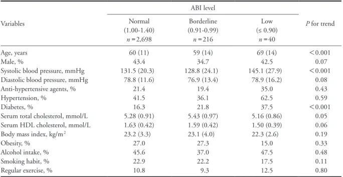
The baseline characteristics of the study popula- tion according to the ABI are shown in Table 1. The prevalence of low, borderline and normal ABI values were 1.4% (n=40), 7.3% (n=216) and 91.3%
(n=2,698), respectively. The subjects with a low ABI were significantly older than those with borderline or normal ABI values. The mean SBP and frequency of diabetes were significantly increased among the sub- jects with a low ABI.
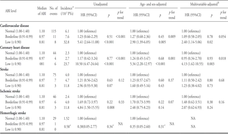
During the average 7.1-year follow-up period, 134 subjects experienced cardiovascular events, includ- ing 85 stroke events and 54 coronary heart disease events. The event-free survival rates for CVD accord- ing to the ABI are shown in Fig. 1A. The cardiovascu- lar event-free survival rates across the ABI values were significantly different (log-rank p<0.001). The sub- jects with a low ABI had a 5.41-fold (95% CI: 2.64- 11.08) higher risk of cardiovascular events compared to those with a normal ABI, whereas there was no evi- dence of any differences between the subjects with borderline and normal ABI values (p=0.51) (Table 2).
This relationship remained substantially unchanged after adjusting for age and sex (HR 2.90 [95% CI:
1.39-6.05]) as well as potential confounding factors, namely, age, sex, systolic blood pressure, anti-hyper- tensive agents, diabetes, serum total cholesterol, serum HDL cholesterol, obesity, smoking habits, alcohol intake and regular exercise (HR 2.40 [95% CI: 1.14- with the subject in the sitting position after at least
five minutes of rest. The mean of three readings was used in this study. Hypertension was defined as a blood pressure of ≥ 140/90 mmHg and/or the current use of anti-hypertensive agents. The plasma glucose levels were measured according to the glucose oxidase method. Diabetes was defined as a fasting plasma glu- cose level of ≥ 7.0 mmol/L (126 mg/dL), two-hour post-loaded or casual glucose level of ≥ 11.1 mmol/L (200 mg/dL) and/or the current use of insulin or oral glucose-lowering agents. The serum total and high- density lipoprotein (HDL) cholesterol concentrations were determined enzymatically. Body height and weight were measured with the subject in light cloth- ing without shoes, and the body mass index (BMI) was calculated as body weight/body height2 (kg/m2).
Obesity was defined as a BMI of ≥ 25.0 kg/m2. All clinical examinations and blood tests were conducted on the same day as the ABI measurements.
Definition of Cardiovascular Disease
The main outcomes of this study were first-ever events of CVD and its subtypes (coronary heart dis- ease, ischemic stroke and hemorrhagic stroke). Coro- nary heart disease was defined as acute myocardial infarction, silent myocardial infarction, sudden cardiac death within one hour after the onset of acute illness or the use of coronary interventions (coronary artery bypass surgery and angioplasty). Acute myocardial infarction was diagnosed when a subject met at least two of the following criteria: (1) typical symptoms, including prolonged severe anterior chest pain, (2) abnormally high levels of cardiac enzymes confirmed more than twice, (3) evolving diagnostic electrocardio- graphic changes and (4) morphological changes, including local asynergy of the cardiac wall motion on electrocardiography, persistent perfusion defects on cardiac scintigraphy or myocardial necrosis and/or the presence of scars measuring 1 cm or longer accompa- nied by coronary atherosclerosis at autopsy.
Silent myocardial infarction was defined as myo- cardial scarring without any historical indication of clinical symptoms or abnormal changes in cardiac enzymes. Stroke was defined as the sudden onset of nonconvulsive and focal neurological deficits that con- tinued for >24 hours and subclassified as either isch- emic or hemorrhagic. All cardiovascular events were assessed based on the findings of a physical examina- tion, review of all available clinical data, including medical records and brain imaging, and an autopsy by a panel of study members who remained blind to the information of the ABI values of the subjects.
Ankle Brachial Index and CV Disease 969
Table 1. Baseline characteristics of the study population according to the ABI (ankle brachial index)
Variables
ABI level
P for trend Normal
(1.00-1.40) n=2,698
Borderline (0.91-0.99)
n=216
Low (≤ 0.90)
n=40 Age, years
Male, %
Systolic blood pressure, mmHg Diastolic blood pressure, mmHg Anti-hypertensive agents, % Hypertension, %
Diabetes, %
Serum total cholesterol, mmol/L Serum HDL cholesterol, mmol/L Body mass index, kg/m2 Obesity, %
Alcohol intake, % Smoking habit, % Regular exercise, %
60 (11) 43.4 131.5 (20.3)
78.8 (11.6) 21.4 41.5 16.3 5.28 (0.91) 1.63 (0.42) 23.2 (3.3)
27.0 45.6 22.9 10.8
59 (14) 34.7 128.8 (24.1)
76.9 (13.4) 19.4 36.1 21.8 5.43 (0.97) 1.59 (0.42) 23.1 (4.0)
27.3 37.0 22.2 9.3
69 (14) 42.5 145.1 (27.9)
78.9 (16.2) 35.0 62.5 37.5 5.16 (0.86) 1.50 (0.39) 22.3 (2.6)
15.0 47.5 17.5 12.5
<0.001 0.07
<0.001 0.08 0.43 0.59
<0.001 0.05 0.06 0.19 0.33 0.48 0.11 0.80 ABI, ankle brachial index; HDL, high-density lipoprotein.
The values are presented as the mean (standard deviation) or percentage.
60 70 80 90 100
0 1 2 3 4 5 6 7
Follow up (years)
Event-free survival rate, free from CVD (%)
1.00-1.40 0.91-0.99
≤ 0.90 Log-rank: p<0.001
60 70 80 90 100
0 1 2 3 4 5 6 7
Follow up (years)
Event-free survival rate, free from CVD (%)
1.00-1.40 0.91-0.99
≤ 0.90 Log-rank: p<0.001
60 70 80 90 100
0 1 2 3 4 5 6 7
Follow up (years)
Event-free survival rate, free from Stroke (%)
1.00-1.40 0.91-0.99
≤ 0.90 Log-rank: p=0.15
60 70 80 90 100
0 1 2 3 4 5 6 7
Follow up (years)
Event-free survival rate, free from Stroke (%)
1.00-1.40 0.91-0.99
≤ 0.90 Log-rank: p=0.15
60 70 80 90 100
0 1 2 3 4 5 6 7
Follow up (years)
Event-free survival rate, free from CHD(%)
1.00-1.40 0.91-0.99
≤ 0.90 Log-rank: p<0.001
60 70 80 90 100
0 1 2 3 4 5 6 7
Follow up (years)
Event-free survival rate, free from CHD(%)
1.00-1.40 0.91-0.99
≤ 0.90 Log-rank: p<0.001
60 70 80 90 100
0 1 2 3 4 5 6 7
Follow up (years)
Event-free survival rate, free from CVD (%)
1.00-1.40 0.91-0.99
≤ 0.90 Log-rank: p<0.001
60 70 80 90 100
0 1 2 3 4 5 6 7
Follow up (years)
Event-free survival rate, free from CVD (%)
1.00-1.40 0.91-0.99
≤ 0.90 Log-rank: p<0.001
60 70 80 90 100
0 1 2 3 4 5 6 7
Follow up (years)
Event-free survival rate, free from Stroke (%)
1.00-1.40 0.91-0.99
≤ 0.90 Log-rank: p=0.15
60 70 80 90 100
0 1 2 3 4 5 6 7
Follow up (years)
Event-free survival rate, free from Stroke (%)
1.00-1.40 0.91-0.99
≤ 0.90 Log-rank: p=0.15
60 70 80 90 100
0 1 2 3 4 5 6 7
Follow up (years)
Event-free survival rate, free from CHD(%)
1.00-1.40 0.91-0.99
≤ 0.90 Log-rank: p<0.001
60 70 80 90 100
0 1 2 3 4 5 6 7
Follow up (years)
Event-free survival rate, free from CHD(%)
1.00-1.40 0.91-0.99
≤ 0.90 Log-rank: p<0.001
B. Coronary heart disease
C. Stroke A. Cardiovascular disease
Fig. 1. Event-free survival rates from cardiovascular disease and its subtypes according to the ankle-brachial index (ABI) during an average follow-up of 7.1 years
970 Kojima et al.
number of events was zero in the low ABI group.
Discussion
The present results clearly demonstrated that an ABI of 0.90 or lower is significantly associated with an increased risk of cardiovascular events. This relation- ship remained significant after adjusting for potential confounding factors. To the best of our knowledge, this is the first study to prospectively investigate the relationship between the ABI and cardiovascular events in a community-based cohort study of a general Asian population.
The ABI has been investigated as a risk factor in several population-based cohort studies, primarily in North America and Europe4-8). Almost all of these studies found that an ABI of ≤ 0.90 is significantly associated with an increased risk of total mortality4-6), cardiovascular mortality4-6) and incident coronary heart disease4, 7). The ABI Collaboration, an individ- 5.06]).
With regard to the subtype of CVD, the event- free survival rate for coronary heart disease was signifi- cantly different across the ABI groups (log-rank p< 0.001), whereas that for stroke was not (log-rank p=0.15) (Fig. 1B, C). The multivariable-adjusted risk of coronary heart disease was significantly increased (by 4.13-fold; 95% CI: 1.62-10.55) in the subjects with a low ABI compared to that observed in the sub- jects with a normal ABI, while there was no evidence of a significant relationship between the ABI values and the risk of stroke. Among the stroke subtypes, however, the univariate analysis revealed that those with a low ABI were at an increased risk of ischemic stroke (HR 4.84; 95% CI: 1.50-15.55), although the significance of the relationship disappeared after adjusting for the above-mentioned confounding fac- tors. No clear relationships between the ABI values and hemorrhagic stroke were observed when the bor- derline and low ABI groups were combined, as the
Table 2. Relationships between ankle brachial index (ABI) and the development of cardiovascular disease, stroke and coronary heart disease during the 7.1-year average follow-up period
ABI level Median
of ABI No. of events
Incidencea (/103 PYs)
Unadjusted Age- and sex-adjusted Multivariable-adjustedb
HR (95%CI) p p for
trend HR (95%CI) p p for
trend HR (95%CI) p p for
trend Cardiovascular disease
Normal (1.00-1.40) Borderline (0.91-0.99) Low (≤ 0.90) Coronary heart disease
Normal (1.00-1.40) Borderline (0.91-0.99) Low (≤ 0.90) Stroke
Normal (1.00-1.40) Borderline (0.91-0.99) Low (≤ 0.90) Ischemic stroke
Normal (1.00-1.40) Borderline (0.91-0.99) Low (≤ 0.90) Hemorrhagic stroke
Normal (1.00-1.40) Borderline (0.91-0.99) Low (≤ 0.90)
1.10 0.97 0.81 1.10 0.97 081 1.10 0.97 0.81 1.10 0.97 0.81 1.10 0.97 0.81
115 11 8 44 4 6 75 7 3 46
6 3 29 1 0
6.1 7.6 32.8 2.3 2.7 23.7 4.0 4.7 11.8 2.4 4.0 11.8 1.52 0.58c
1.00 (reference) 1.23 (0.66-2.29) 5.41 (2.64-11.08) 1.00 (reference) 1.17 (0.42-3.24) 10.50 (4.47-24.64) 1.00 (reference) 1.21 (0.56-2.62) 2.96 (0.93-9.38) 1.00 (reference) 1.69 (0.72-3.97) 4.84 (1.50-15.55) 1.00 (reference) 0.38(0.05-2.77)
0.51
<0.001
0.77
<0.001
0.63 0.07
0.22 0.008
0.34c
<0.001
<0.001
0.12
0.33
NA
1.00 (reference) 1.27 (0.68-2.36) 2.90 (1.39-6.05) 1.00 (reference) 1.24 (0.45-3.47) 5.34 (2.20-12.97) 1.00 (reference) 1.23 (0.57-2.67) 1.60 (0.49-5.16) 1.00 (reference) 1.70 (0.73-3.99) 2.48 (0.75-8.23) 1.00 (reference) 0.35 (0.05-2.60)
0.45 0.005
0.68
<0.001
0.60 0.43
0.22 0.14
0.31c 0.009
0.001
0.37
0.07
NA
1.00 (reference) 1.09 (0.58-2.05) 2.40 (1.14-5.06) 1.00 (reference) 0.95 (0.34-2.70) 4.13 (1.62-10.55) 1.00 (reference) 1.11 (0.50-2.42) 1.23 (0.38-4.02) 1.00 (reference) 1.48 (0.62-3.51) 2.07 (0.62-6.93)
NA NA
0.78 0.02
0.93 0.003
0.80 0.73
0.38 0.24
0.054
0.018
0.68
0.16
PYs, person-years; HR, hazard ratio; CI, confidence interval; NA, not analyzed.
aValues were unadjusted.
bThe risk estimates were adjusted for age, sex, systolic blood pressure, anti-hypertensive agents, diabetes, serum total cholesterol, serum high-den- sity lipoprotein cholesterol, obesity, smoking habits, alcohol intake and regular exercise.
cThe borderline and low groups were combined.
Ankle Brachial Index and CV Disease 971
low ABI to be a significant risk factor for stroke events5, 19, 23). The Atherosclerosis Risk in Communi- ties Study also found that a low ABI is associated with a higher incidence of ischemic stroke after adjusting for age, race, sex and center; however, this significant relationship disappeared after adjusting for other con- founding factors8). These results are similar to the present findings, although the relationship in the pres- ent study did not reach a level of statistical significance and was largely attenuated compared to that observed in previous studies, possibly because lacunar stroke is the dominant type of ischemic stroke in Japan, whereas large-artery atherothrombotic brain infarction is more prevalent in Western countries24). Given the etiology of each stroke subtype, the risk of athero- thrombotic brain infarction, as well as coronary heart disease, may be expected to increase in subjects with a low ABI because PAD is an atherosclerotic disease of the large arteries. Atherosclerotic changes in cerebral arteries, as well as coronary arteries, may already be advanced among subjects with PAD defined as an ABI of ≤ 0.9, as PAD is attributable to atherosclerotic remodeling of lower extremity peripheral arteries. The risk of atherothrombotic brain infraction may thus be expected to increase in subjects with a low ABI. Large- scale studies are therefore required to elucidate the relationship between a low ABI and the risk of stroke subtypes.
Several limitations of the present study should be noted. First, the generalizability of our findings may be limited, as 152 subjects without available ABI data were excluded from the study. Compared to the sub- jects who were included, the excluded subjects were older (mean age: 81 years for the excluded subjects vs.
60 years for the included subjects) and more likely to have hypertension (73.7% vs. 43.0%) or a history of cardiovascular events (21.5% vs. 4.5%). This bias would lead to underestimation of the relationship between the ABI and cardiovascular risks. Second, only a single ABI measurement was obtained at the baseline examination. This may have caused the mis- classification of subjects into the wrong ABI group.
Such misclassification, if present, would weaken the relationship found in this study, biasing the results toward the null hypothesis. Third, we were unable to obtain information regarding the subjects’ medical treatment during the follow-up period. The lack of this information may have reduced the accuracy of our findings to some extent. In addition, we used an automatic device to measure blood pressure in the four limbs and the oscillometric method instead of the standard doppler method. The standard doppler method is recommended in the ACC/AHA guidelines ual-based meta-analysis of 16 community-dwelling
cohorts conducted in Western countries, clearly dem- onstrated that a lower ABI (≤ 0.90) approximately doubles the risk of total mortality, cardiovascular mor- tality and major coronary events compared to an ABI of 1.11-1.20, independent of the Framingham risk score13).
With regard to Asian populations, a significant relationship between the ABI and cardiovascular risks has been found in several hospital-based prospective studies conducted among patients with risk factors such as metabolic syndrome, ischemic heart disease and chronic kidney disease20-22). For example, the China ABI Cohort Study, a hospital-based study of 3,210 Chinese patients with two or more cardiovascu- lar risk factors, showed that the multivariable-adjusted risk of cardiovascular mortality increased significantly (by 2.0 times) in the subjects with an ABI below 0.90 compared to that observed in the subjects with an ABI above 0.9020). However, limited studies have addressed this issue in the general Asian population. Our find- ings suggest that an ABI of ≤ 0.90 is an independent risk factor for the incidence of CVD in a community- based Asian population.
In the ABI Collaboration13), subjects with an ABI from 0.91 to 1.10 and those with a high ABI (> 1.40), which may be related to poor arterial compress- ibility resulting from stiffness and calcification, were at slightly increased absolute risks of total mortality, cardiovascular mortality and major coronary events.
These results suggest that subjects with an ABI of 0.91 to 1.10 or greater than 1.40 may have slightly higher risks of these outcomes than those with a normal ABI, although the widely accepted high-risk cut-off value of 0.90 is reasonable11). Based on these findings, the ACC/AHA practice guidelines define ABI categories as low ABI (severe and mild to moderate), borderline ABI, normal ABI and high ABI16). In the present study, however, we did not find that borderline ABI was related to the risk of cardiovascular events, likely due to the limited statistical power. Likewise, we were unable to address the relationship between an ABI of
>1.40 and cardiovascular risk, because only two sub- jects had an ABI of >1.40. The relationship between a borderline or high ABI and the risk of cardiovascular events should be addressed in large samples of Asian populations.
When we investigated the subtypes of cardiovas- cular events, we observed that, after adjusting for con- founding factors, a low ABI was significantly related to an increased risk of coronary heart disease events, but not the risk of stroke. Several epidemiological studies conducted in Western countries have shown a
972 Kojima et al.
Nichisho-Ippan-004).
References
1) Dumville JC, Lee AJ, Smith FB, Fowkes FG: The health- related quality of life of people with peripheral arterial disease in the community: the Edinburgh Artery Study.
Gen Pract, 2004; 54: 826-831
2) Yan BP, Moran D, Hynes BG, Kiernan TJ, Yu CM:
Advances in endovascular treatment of critical limb isch- emia. Circ J, 2011; 75: 756-765
3) Ikeda N, Kogame N, Iijima R, Nakamura M, Sugi K:
Impact of carotid artery ultrasound and ankle-brachial index on prediction of severity of SYNTAX score. Circ J, 2013; 77: 712-716
4) Ogren M, Hedblad B, Isacsson SO, Janzon L, Jungquist G, Lindell SE: Non-invasively detected carotid stenosis and ischaemic heart disease in men with leg arteriosclero- sis. Lancet, 1993; 342: 1138-1141
5) Leng GC, Fowkes FG, Lee AJ, Dunbar J, Housley E, Ruckley CV: Use of ankle brachial pressure index to pre- dict cardiovascular events and death: a cohort study. BMJ, 1996; 313: 1440-1444
6) Newman AB, Shemanski L, Manolio TA, Cushman M, Mittelmark M, Polak JF, Powe NR, Siscovick D: Ankle- arm index as a predictor of cardiovascular disease and mortality in the Cardiovascular Health Study. The Car- diovascular Health Study Group. Arterioscler Thromb Vasc Biol, 1999; 19: 538-545
7) Weatherley BD, Nelson JJ, Heiss G, et al.: The associa- tion of the ankle-brachial index with incident coronary heart disease: the Atherosclerosis Risk in Communities (ARIC) Study, 1987-2001. BMC Cardiovasc Disord, 2007; 7: 3
8) Tsai AW, Folsom AR, Rosamond WD, Jones DW: Ankle- brachial index and 7-year ischemic stroke incidence: the ARIC Study. Stroke, 2001; 32: 1721-1724
9) Abbott RD, Rodriguez BL, Petrovitch H, Yano K, Schatz IJ, Popper JS, Masaki KH, Ross GW, Curb JD: Ankle- brachial blood pressure in elderly men and the risk of stroke: the Honolulu Heart Program. J Clin Epidemiol, 2001; 54: 973-978
10) Khan NA, Rahim SA, Anand SS, Simel DL, Panju A:
Does the clinical examination predict lower extremity peripheral arterial disease? JAMA, 2006; 295: 536-546 11) Hirsh AT, Haskal ZJ, Hertzer NR, Bakal CW, Creager
MA, Halperin JL, et al.: ACC/AHA 2005 Practice Guide- lines for the management of patients with peripheral arte- rial disease (lower extremity, renal, mesenteric, and abdominal aortic): a collaborative report from the Ameri- can Association for Vascular Surgery/Society for Vascular Surgery, Society for Cardiovascular Angiography and Interventions, Society for Vascular Medicine and Biology, Society of Interventional Radiology, and the ACC/AHA Task Force on Practice Guidelines (Writing Committee to Develop Guidelines for the Management of Patients With Peripheral Arterial Disease): Circulation, 2006; 113:
e463-e654
12) Doobay AV, Anand SS: Sensitivity and specificity of the
because some devices using oscillometric blood pres- sure have been found to overestimate the actual ankle blood pressure, especially in subjects with a very low ankle blood pressure25). However, the precision of our device has been validated in both Japanese and Euro- pean populations26, 27), and thus, although this limita- tion may have reduced the precision of the ABI values to some extent, it is unlikely to have altered the find- ings substantially.
In conclusion, the present findings indicate that, in a general Japanese population, the subjects with an ABI of ≤ 0.90 had an increased risk of cardiovascular events, independent of traditional cardiovascular risk factors. Because the ABI is a noninvasive and user- friendly method for estimating the extent of systemic atherosclerotic disease, it may be applied more broadly in general practice to identify individuals at a high risk of developing CVD.
Acknowledgments
We thank the staff of the Division of Health and Welfare of Hisayama for their cooperation in this study.
Disclosures
I. Kojima is a former employee of Omron Healthcare Co., Ltd. There are no other conflicts of interest to declare.
Conflicts of Interest
The first author, Iwao Kojima is a former employee for Omron Healthcare, Co., Ltd., which produces the device used to measure the ABI. All remaining authors report no conflicts of interest.
Grant Support
This study was supported in part by Grants-in- Aid for Scientific Research on Innovative Areas (22116010) and Scientific Research (A) (25253048) from the Ministry of Education, Culture, Sports, Sci- ence and Technology of Japan and Health and Labour Sciences Research Grants from the Ministry of Health, Labour and Welfare of Japan (Comprehensive Research on Life-Style Related Diseases including Car- diovascular Diseases and Diabetes Mellitus: H22- Junkankitou [Seishuu]-Ippan-005, H23- Junkankitou [Seishuu]-Ippan-005, H25- Junkankitou [Seishuu]- Ippan-005 and H25- Junkankitou [Seishuu]-Sitei-022;
and Comprehensive Research on Dementia: H25-
Ankle Brachial Index and CV Disease 973
dreau R, Harris T, Thompson, et al.: Relationship of ankle blood pressures to cardiovascular events in older adults. Stroke, 2008; 39: 863-869
20) Li X, Luo Y, Xu Y, Li J, Hu D: Relationship of ankle-bra- chial index with all-cause mortality and cardiovascular mortality after a 3-year follow-up: the China ankle-bra- chial index cohort study. J Hum Hypertens, 2010; 24:
111-116
21) Cang Y, Li J, Li YM, Zhou Y, Wu YZ, Li XK, et al.: Rela- tionship of a low ankle-brachial index with all-cause mor- tality and cardiovascular mortality in Chinese patients with metabolic syndrome after a 6-year follow-up: a Chi- nese prospective cohort study. Intern Med, 2012; 51:
2847-2856
22) Zheng L, Li J, Hu D, Luo Y, Li X, Xu Y, Sun Z, Sun Y:
Association of low ankle-brachial index with mortality in patients with ischemic heart disease. J Atheroscler Thromb, 2010; 17: 759-767
23) Meves SH, Diehm C, Berger K, Pittrow D, Trampisch HJ, Burghaus I, et al.: Peripheral arterial disease as an independent predictor for excess stroke morbidity and mortality in primary-care patients: 5-year results of the getABI study. Cerebrovasc Dis, 2010; 29: 546-554 24) Ueshima H, Sekikawa A, Miura K, Turin TC, Takashima
N, Kita Y, et al.: Cardiovascular disease and risk factors in Asia: a selected review. Circulation, 2008; 118: 2702- 25) Aboyans V, Criqui MH, Abraham P, Allison MA, Creager 2709
MA, Diehm C, et al.: Measurement and interpretation of the ankle-brachial index: a scientific statement from the American Heart Association. Circulation, 2012; 126:
2890-2909
26) Koji Y, Tomiyama H, Ichihashi H, Nagae T, Tanaka N, Takazawa K, et al.: Comparison of ankle-brachial pressure index and pulse wave velocity as markers of the presence of coronary artery disease in subjects with a high risk of atherosclerotic cardiovascular disease. Cardiology, 2004;
94: 868-872
27) Richart T, Kuznetsova T, Wizner B, Struijker-Boudier HA, Staessen JA: Validation of automated oscillometric versus manual measurement of the ankle-brachial index.
Hypertens Res, 2009; 32: 884-888 ankle-brachial index to predict future cardiovascular out-
comes: a systematic review. Arterioscler Thromb Vasc Biol, 2005; 25: 1463-1469
13) Fowkes FG, Murray GD, Butcher I, Heald CL, Lee RJ, Chambless LE, et al.: Ankle brachial index combined with Framingham Risk Score to predict cardiovascular events and mortality: a meta-analysis. JAMA, 2008; 300: 197- 14) Tendera M, Aboyans V, Bartelink ML, Baumgartner I, 208
Clément D, Collet JP, et al.: ESC Guidelines on the diag- nosis and treatment of peripheral artery diseases: Docu- ment covering atherosclerotic disease of extracranial carotid and vertebral, mesenteric, renal, upper and lower extremity arteries: the Task Force on the Diagnosis and Treatment of Peripheral Artery Diseases of the European Society of Cardiology (ESC). Eur Heart J, 2011; 32:
2851-2906
15) Mancia G, Fagard R, Narkiewicz K, Redon J, Zanchetti A, Bohm M et al.: 2013 ESH/ESC Guidelines for the man- agement of arterial hypertension: The Task Force for the management of arterial hypertension of the European Society of Hypertension (ESH) and of the European Soci- ety of Cardiology (ESC). J Hypertens, 2013; 31: 1281- 16) Rooke TW, Hirsch AT, Misra S, Sidawy AN, Beckman 1357
JA, Findeiss LK, et al.: 2011 ACCF/AHA Focused Update of the Guideline for the Management of Patients With Peripheral Artery Disease (updating the 2005 guideline):
a report of the American College of Cardiology Founda- tion/American Heart Association Task Force on Practice Guidelines. J Am Coll Cardiol, 2011; 58: 2020-2045 17) Ohmura T, Ueda K, Kiyohara Y, Kato I, Iwamoto H,
Nakayama K, et al.: Prevalence of type 2 (non-insulin- dependent) diabetes mellitus and impaired glucose toler- ance in the Japanese general population: the Hisayama Study. Diabetologia, 1993; 36: 1198-1203
18) Ninomiya T, Kojima I, Doi Y, Fukuhara M, Hirakawa Y, Hata J, et al.: Brachial-ankle pulse wave velocity predicts the development of cardiovascular disease in a general Japanese population: the Hisayama Study. J Hypertens, 2013; 31: 477-483
19) Sutton-Tyrrell K, Venkitachalam L, Kanaya AM, Bou-