57:378
はじめに
2010年に Pittock らは橋を中心とした脳幹・小脳病変が亜
急性に進行し,ステロイドに良好な反応を示す炎症性疾患 を chronic lymphocytic inflammation with pontine perivascular enhancement responsive to steroids(CLIPPERS)として提唱 した.その後後脳領域のみならず大脳基底核・大脳白質にも 病変を生じた症例が報告されているが,一般的に後脳から離 れるに従って病変は縮小すると報告されている1).我々は既 報告と異なり,側頭葉に最大病変を示し,亜急性の経過で脳 幹・小脳へ病変が拡大した CLIPPERS の 1 例を経験したので 報告する. 症 例 症例:49 歳,男性 主訴:発熱,左眼の視野異常 既往歴:2 型糖尿病,脂質異常症,不眠症. 家族歴:特記すべきことなし. 現病歴:来院 22 日前頃から 37°C 台の発熱,悪寒,頭痛が 出現した.来院 6 日前の仕事中に,突然左眼に黒い部分が見 えるようになったが,自然に改善した.来院 1 日前に前医を 受診し,頭部 MRI で右側頭葉に造影効果を伴う病変を認めた ため,精査目的に当院へ入院した. 一般身体所見:身長 165 cm,体重 60 kg,血圧 109/79 mmHg, 脈拍 106/ 分,体温 37.3°C,SpO2 99%.表在リンパ節は触知 せず.その他一般身体所見に異常を認めなかった. 神経学的所見:意識清明.左眼視野の中心部に暗点を認め た.瞳孔径 4/4 mm.対光反射は両側迅速であったが,左 Marcus Gunn瞳孔を認めた.眼球運動障害,複視を認めなかった.顔 面筋力,顔面知覚に異常を認めなかった.運動系,感覚系, 協調運動に異常を認めなかった. 検査所見:一般血液検査ではフェリチンが 401 ng/ml と軽 度高値を示す以外に異常を認めなかった.抗核抗体,抗 SSA 抗体,抗 SSB 抗体,抗アクアポリン 4 抗体,抗 GAD 抗体, MPOANCA,PR3ANCA は陰性であった.ACE,可溶性 IL2 受容体の上昇を認めなかった.また各腫瘍マーカー(AFP, CEA,CA199,PSA)は正常範囲内であった.単純ヘルペス ウイルス,水痘帯状疱疹ウイルス,サイトメガロウイルスの 抗 体 価 は 既 感 染 パ タ ー ン で あ っ た. 髄 液 検 査 は 初 圧 170 mmH2Oと正常であったが,細胞数 47/μl(単核球 46/μl), 蛋白 57 mg/dl と上昇を認めた.ミエリン塩基性蛋白の上昇を
症例報告
側頭葉に最大病変を認めた chronic lymphocytic inflammation with pontine
perivascular enhancement responsive to steroids(CLIPPERS)の 1 例
松浦 啓
1)蒔田 直輝
1)石井亮太郎
1)*
藤田 泰子
2)古野 優一
3)水野 敏樹
1)要旨: Chronic lymphocytic inflammation with pontine perivascular enhancement responsive to steroids (CLIPPERS)の病変は一般的に後脳から離れるに従って縮小することが多いが,側頭葉に最大病変を示す症例を 経験した.症例は 49 歳男性.来院 22 日前から発熱が,その後左眼視野障害が出現し入院となった.神経学的に 左眼視野の中心部に暗点を認め,MRI で右側頭葉に粒状のガドリニウム造影効果を伴う FLAIR 高信号病変を認め た.その後,橋及び小脳に病変が拡大し,複視・滑動性眼球運動障害・輻輳麻痺が出現した.右側頭葉病変から開 頭脳生検し,CLIPPERS と診断した.ステロイド投与により病変・症状ともに軽快した.病初期に後脳から離れ た病変が大きい症例であっても,CLIPPERS の可能性を考慮する必要がある. (臨床神経 2017;57:378-382)
Key words: CLIPPERS,側頭葉病変,ステロイド
*Corresponding author: 京都府立医科大学大学院医学研究科神経内科学〔〒 6028566 京都市上京区河原町広小路上ル梶井町 465〕
1)京都府立医科大学大学院医学研究科神経内科学
2)京都府立医科大学附属病院細胞分子機能病理学
3)京都府立医科大学大学院医学研究科脳神経機能再生外科学
(Received February 6, 2017; Accepted April 11, 2017; Published online in JSTAGE on June 21, 2017) doi: 10.5692/clinicalneurol.cn001007
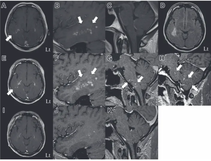
認めず,オリゴクローナルバンドは検出されなかった.眼科 的検査では,左眼底に乳頭浮腫を認め,Goldmann 視野検査で Mariotte盲点の拡大を認めた.フリッカー値,光干渉断層計 では異常を認めなかった.来院 1 日前の頭部 MRI で,右側頭 葉深部白質に FLAIR 高信号病変があり(Fig. 1D),同病変は ガドリニウム造影で粒状の造影効果を認めた(Fig. 1A~C). 全脊椎 MRI,下肢 MRI,ガリウムシンチグラフィー,FDGPET では異常を認めなかった. 入院後経過:入院 4 日目の頭部 MRI で,右側頭葉の病変が 拡大し,新規に粒状の造影病変が脳幹中心に橋,延髄,小脳 に出現した(Fig. 1E~H).入院 16 日目より右外転障害によ る右注視時の複視が出現し,また滑動性眼球運動障害,輻輳 麻痺を認めた.経過と検査結果から脱髄疾患,腫瘍性疾患を 鑑別するため,入院 23 日目に右側頭葉病変より開頭脳生検 を実施した.生検組織からは血管周囲腔への炎症細胞浸潤 を認め,炎症細胞は主に CD3,CD4 陽性リンパ球であった (Fig. 2A~H).脱髄斑2),悪性リンパ腫,腫瘍細胞像などを 認めなかったため,CLIPPERS と診断した.入院 26 日目より ステロイド大量療法(メチルプレドニゾロン 1,000 mg/ 日)を 3日間行った.治療開始後より症状は速やかに改善し,その 後プレドニゾロン 60 mg/ 日による後療法を行った.入院 38 日目の頭部 MRI では,右側頭葉,脳幹,小脳病変の縮小を認 めた(Fig. 1I~K).神経症状の増悪を認めないことを確認し ながら,プレドニゾロン 35 mg/ 日まで漸減し,入院 40 日目 に退院した.退院時の神経学的所見としては軽度の輻輳麻痺 のみであった.退院後は 2 か月プレドニゾロン 35 mg/ 日を継 続し漸減した.内服プレドニゾロン 20 mg/ 日となった入院 3か月目に画像上,左側脳室三角部近傍に新規病変を認め,無 症候性再発と判断しステロイド大量療法(メチルプレドニゾ ロン 500 mg/ 日)を 3 日間行った.内服プレドニゾロンを Fig. 1 Brain MRI findings.
(A–C) On the day before admission, gadolinium enhanced T1weighted images showed an enhanced punctate area in the lesion in the right
temporal lobe (A: Axial, B, C: Sagittal, 1.5 T, TR: 500 msec, TE: 15 msec). (D) On the day before admission, fluid attenuated inversion recovery (FLAIR) image showed a hyperintense lesion in the right temporal lobe (Axial, 1.5 T, TR: 7,000 msec, TE: 120 msec). (E–H) At 4 days after admission, MRI showed enlargement of the lesion in the temporal lobe and new lesions in the pons, medulla oblongata, and cerebellar hemispheres (E: Axial, F–H: Sagittal, 1.5 T, TR: 10 msec, TE: 5 msec). (I–K) At 12 days after steroid pulse therapy (38 days after admission), MRI showed resolution of the enhancement (I: Axial, J, K: Sagittal, 1.5 T, TR: 9 msec, TE: 5 msec).
臨床神経学 57 巻 7 号(2017:7) 57:380 45 mg/日まで増量した.徐々に内服プレドニゾロンの減量を 行い入院 9 か月目に 20 mg/ 日となった.入院 10 か月目の頭 部造影 MRI にて右半卵円中心及び右被殻外側に造影効果を 伴う新規病変を認めた.ステロイド大量療法(メチルプレド ニゾロン 1,000 mg/ 日)を 3 日間行った後に内服プレドニゾ ロンを 25 mg/ 日まで増量した.入院 12 か月後の頭部造影 MRI にて左被殻外側に再発を認め,入院 14 か月目には右尾状核頭 部に再発を認めた.内服プレドニゾロンを 20 mg/ 日で継続 し,6 週間おきにステロイド大量療法(メチルプレドニゾロ ン 1,000 mg/ 日)3 日間を行っている.退院後神経学的には増 悪を認めず,また入院 14 か月目以降は画像上も再発を認めて いない. 考 察 2010年に Pittock らは炎症性中枢神経疾患の中から亜急性 に増悪する歩行失調,複視,構音障害などを認め,橋および その隣接部を主座としステロイドに良好な反応を示す一群を まとめ,CLIPPERS という疾患概念を提唱した3).現在も CLIPPERSの診断基準は定まっていないが,2012 年に Simon らが提唱した CLIPPERS の主要症状(Table 1)を踏まえて 診断されている.画像所見として,橋・小脳・小脳脚を中心 に,peppering と表現される粒状の造影効果がみられる1)3). CLIPPERSは一般的に大脳基底核および大脳白質まで病変が 拡大することはあるものの,後脳から離れるに従って病変は 縮小すると報告されている1).病理学的には免疫染色で CD3 または CD4 陽性リンパ球の浸潤を血管周囲腔に認めること が多いが,CLIPPERS に特異的な所見はなく,組織診断はそ の他の鑑別診断を除外するために有効である1).鑑別診断は 多岐にわたり,自己免疫性脳炎,血管炎,多発性硬化症,急 性散在性脳脊髄炎,視神経脊髄炎,神経ベーチェット病,神 経サルコイドーシス,シェーグレン症候群,全身性エリテマ トーデスなどの炎症性疾患や,ランゲルハンス細胞組織球症, ErdheimChester病,悪性リンパ腫,神経膠腫,リンパ腫様 肉芽腫症といった腫瘍性疾患,中枢神経感染症,傍腫瘍症候 群などが挙げられる1)3)4).高用量ステロイド投与により著明 に臨床所見,画像所見が改善することも重要な特徴である. CLIPPERSは 2010 年に Pittock らの提唱以降,2011 年に Gabilondoらが大脳白質内に peppering 様の造影効果を伴う病 変が散在した症例を報告した5).その後,2012 年に Simon ら は仮性球麻痺,認知機能障害,大脳萎縮をきたし,かつ Pittock らの報告した特徴を満たす 5 症例を報告した.その中では病 理学的に軸索障害を認め,大脳皮質の萎縮と関連している可 能性が高いと結論し,CLIPPERS の病変部位は橋,小脳だけ でなく大脳へも浸潤しうる疾患であるとその定義を拡大し た4).CLIPPERS の臨床症状についても,その中核となる構 音障害6)7),小脳失調4)8)~11)以外に眼球運動障害4)8)12)~14),回 転性めまい6)12),錐体路徴候3)4)15),顔面の感覚異常9)12)14)16), 頭痛4),全身倦怠感3)4)17)を示すことが報告された.すなわち 2010年以降 CLIPPERS として報告された症例の病変部位や 臨床症状は徐々に拡大されており,その多様性が認識される ようになってきている. 本症例は,左のうっ血乳頭によるものと思われる左眼の視 野異常を主訴に来院した.うっ血乳頭とは脳脊髄圧の上昇に よる視神経乳頭の腫大と定義されている18).しかし,脳脊髄 液圧が正常の場合にも,うっ血乳頭と同様の視神経乳頭の腫 大が観察されることが報告されており19)~22).くも膜や線維 柱帯の炎症などが視神経周囲くも膜下腔の局所内圧を上昇さ せる機序が想定されている23).本症例においても画像で描出 されていない軽微な炎症によって左視神経周囲くも膜下腔 Fig. 2 Neuropathological findings.
Biopsy of the right temporal lobe was performed. Hematoxylin and eosin stainings (A) demonstrate intense perivascular lymphocytic infiltrates. Immunostaining CD3 staining (B), CD4 staining (C), CD8 staining (D), CD68 staining (E), CD20 staining (F), and CD1a staining (G), demon strates that inflammatory cells are predominantly composed of CD3positive and CD4positive T cells. (H) KlüverBarrera staining demonstrates no demyelinated plaque. Bar = 100 μm.
の内圧上昇が起こった可能性が考えられた.次に本症例の髄 液細胞数は 47/μl であった.CLIPPERS における髄液細胞 増多は軽度であることが多く一般的に 5~50/μl と言われてお り1),本症例はそれに合致する.因みに CLIPPERS における 髄液細胞数は 67/μl24)や 108/μl25)と比較的高値を示す報告も存 在する. 本症例では小脳症状・脳神経障害が出現し,粒状造影病変 があり,ステロイドに対して良好な治療反応性を示し,ステ ロイドの減量に伴って再発を繰り返した点は CLIPPERS の特 徴と合致していた.一方で粒状造影効果を伴うものの,発症 早期は病変が右側頭葉にのみ認められ,亜急性の経過で脳幹, 小脳病変が出現した点や,後脳から離れるに従って病変が縮 小するという既報告と異なり,右側頭葉に最大病変を認めた 点が特徴的であった.本症例のように病変の分布や大きさが 側頭葉を主体とする場合であっても CLIPPERS を鑑別に挙げ 精査する必要があると考えられた. ※本論文に関連し,開示すべき COI 状態にある企業,組織,団体 はいずれも有りません. 文 献
1) Dudesek A, Rimmele F, Tesar S, et al. CLIPPERS: chronic lymphocytic inflammation with pontine perivascular enhance ment responsive to steroids. Review of an increasingly
recognized entity within the spectrum of inflammatory central nervous system disorders. Clin Exp Immunol 2014;175:385396. 2) Popescu BF, Pirko I, Lucchinetti CF. Pathology of multiple
sclerosis: where do we stand? Continuum (Minneap Minn) 2013;19:901921.
3) Pittock SJ, Debruyne J, Krecke KN, et al. Chronic lymphocytic inflammation with pontine perivascular enhancement responsive to steroids (CLIPPERS). Brain 2010;133:26262634.
4) Simon NG, Parratt JD, Barnett MH, et al. Expanding the clinical, radiological and neuropathological phenotype of chronic lymphocytic inflammation with pontine perivascular enhance ment responsive to steroids (CLIPPERS). J Neurol Neurosurg Psychiatry 2012;83:1522.
5) Gabilondo I, Saiz A, Graus F, et al. Response to immunotherapy in CLIPPERS syndrome. J Neurol 2011;258:20902092.
6) Ha AD, Parratt JD, Babu S, et al. Movement disorders associated with CLIPPERS. Mov Disord 2014;29:148150. 7) Marinho PB, Montanaro VV, Barroso Freitas MC, et al.
CLIPPERS syndrome: Case report in a Brazilian patient with a long term disease evolution. Mult Scler Relat Disord 2015; 4:311314.
8) Biotti D, Deschamps R, Shotar E, et al. CLIPPERS: chronic lymphocytic inflammation with pontine perivascular enhance ment responsive to steroids. Pract Neurol 2011;11:349351. 9) Kastrup O, van de Nes J, Gasser T, et al. Three cases of
CLIPPERS: a serial clinical, laboratory and MRI followup study. J Neurol 2011;258:21402146.
Table 1 Core features of chronic lymphocytic inflammation with pontine perivascular enhancement responsive to steroids (CLIPPERS)4).
Core features of CLIPPERS* Clinical
Subacute progressive ataxia and dysarthria.†
Radiological
(a) Numerous punctate or nodular enhancing lesions bilaterally in at least two of the three following anatomical locations: pons, brachium pontis, cerebellum.
(b) Individual radiological lesions are small but may coalesce to form larger lesions (mass effect has not been seen and to date suggests an al ternative diagnosis).
(c) Lesions may occur in the spinal cord, basal ganglia of cerebral white matter but should be of decreasing density with increasing distance from the hindbrain.
(d) Absence of the following radiological features (i) Restricted diffusion on diffusion weighted imaging (ii) Marked hyperintensity on T2 weighted images
(iii) Abnormal cerebral angiography Corticosteroid responsiveness
Prompt and significant clinical and radiological response to corticosteroids. Histopathological
(a) White matter perivascular lymphohistiocytic infiltrate with or without parenchymal extension. (b) Infiltrate contains predominantly CD3+ and CD4+ lymphocytes.
(c) Absence of the following histopathological characteristics: (i) Monoclonal of atypical lymphocyte population
(ii) Necrotising granulomas of giant cells
(iii) Histological features of vasculitis (destruction of the vessel wall, fibrinoid necrosis, leukocytoclasia, fibrin thrombi).
*Differential diagnoses, including neurosarcoidosis, Sjögrenʼs syndrome, neuroBehcetʼs disease, vasculitis and lymphoma should be excluded.
臨床神経学 57 巻 7 号(2017:7) 57:382
10) Fujisawa N, Oya S, Mori H, et al. Chronic lymphocytic inflammation with pontine perivascular enhancement responsive to steroids with a significant elevation of beta2 microglobulin levels. J Korean Neurosurg Soc 2015;58:487490.
11) 谷 裕基,中嶋秀人,山根一志ら.高度の脳幹浮腫を呈した chronic lymphocytic inflammation with pontine perivascular enhancement responsive to steroids(CLIPPERS)の 1 例.臨 床神経 2014;54:581584.
12) Jones JL, Dean AF, Antoun N, et al. ‘Radiologically compatible CLIPPERS’ may conceal a number of pathologies. Brain 2011; 134:e187.
13) Suer D, Yusifova L, Arsava EM, et al. A Case Report of CLIPPERS (Chronic Lymphocytic Inflammation with Ponto cerebellar Perivascular Enhancement Responsive to Steroids) Syndrome. Clin Neuroradiol 2015;25:6163.
14) Wijntjes J, Wouda EJ, Siegert CE, et al. Need for prolonged immunosupressive therapy in CLIPPERS—a case report. BMC Neurol 2013;13:49.
15) Limousin N, Praline J, Motica O, et al. Brain biopsy is required in steroidresistant patients with chronic lymphocytic inflamma tion with pontine perivascular enhancement responsive to steroids (CLIPPERS). J Neurooncol 2012;107:223224.
16) Tan BL, Agzarian M, Schultz DW. CLIPPERS: Induction and maintenance of remission using hydroxychloroquine. Neurol Neuroimmunol Neuroinflamm 2015;2:e56.
17) Blaabjerg M, Ruprecht K, Sinnecker T, et al. Widespread inflammation in CLIPPERS syndrome indicated by autopsy and ultrahighfield 7T MRI. Neurol Neuroimmunol Neuroinflamm
2016;3:e226.
18) Hayreh SS. Pathogenesis of oedema of the optic disc. Doc Ophthalmol 1968;24:289411.
19) Gass A, Barker GJ, RiordanEva P, et al. MRI of the optic nerve in benign intracranial hypertension. Neuroradiology 1996;38: 769773.
20) Killer HE, Flammer J. Unilateral papilledema caused by a frontotemporoparietal arachnoid cyst. Am J Ophthalmol 2001; 132:589591.
21) Killer HE, Mironov A, Flammer J. Optic neuritis with marked distension of the optic nerve sheath due to local fluid conges tion. Br J Ophthalmol 2003;87:249.
22) Hickman SJ, Miszkiel KA, Plant GT, et al. The optic nerve sheath on MRI in acute optic neuritis. Neuroradiology 2005;47: 5155.
23) Killer HE, Jaggi GP, Flammer J, et al. Cerebrospinal fluid dynamics between the intracranial and the subarachnoid space of the optic nerve. Is it always bidirectional? Brain 2007;130: 514520.
24) Tohge R, Nagao M, Yagishita A, et al. A case of chronic lymphocytic inflammation with pontine perivascular enhance ment responsive to steroids (CLIPPERS) in East Asia. Intern Med 2012;51:11151119.
25) Ma Y, Sun X, Li W, et al. Chronic lymphocytic inflammation with pontine perivascular enhancement responsive to steroids (CLIPPERS) with intracranial EpsteinBarr virus infection: A Case Report. Medicine (Baltimore) 2016;95:e5377.
Abstract
A case of chronic lymphocytic inflammation with pontine perivascular enhancement
responsive to steroids (CLIPPERS) with the largest lesion in the temporal lobe
Hiraku Matsuura, M.D.
1), Naoki Makita, M.D.
1), Ryotaro Ishii, M.D., Ph.D.
1),
Yasuko Fujita, M.D.
2), Yuichi Furuno, M.D., Ph.D.
3)and Toshiki Mizuno, M.D., Ph.D.
1)1)Department of Neurology, Kyoto Prefectural University of Medicine
2)Department of Pathology and Cell Regulation, Kyoto Prefectural University of Medicine 3)Department of Neurosurgery, Kyoto Prefectural University of Medicine