Title
Three-Dimensional Culture of Keratinocytes and the Formation of
Basement Membrane for Canine Footpad Substitute(Surgery)( 本
文(Fulltext) )
Author(s)
YAMAZOE, Kazuaki; MIYAMOTO, Shuji; HIKOSAKA, Yoko;
KITAGAWA, Koji; WATANABE, Kazuhiro; SAKAI, Hiroki;
KUDO, Tadaaki
Citation
[The journal of veterinary medical science] vol.[69] no.[6]
p.[611]-[617]
Issue Date
2007-06-25
Rights
The Japanese Society of Veterinary Science (社団法人日本獣医
学会)
Version
出版社版 (publisher version) postprint
URL
http://hdl.handle.net/20.500.12099/28737
Three-Dimensional Culture of Keratinocytes and the Formation of Basement
Membrane for Canine Footpad Substitute
Kazuaki YAMAZOE1), Shuji MIYAMOTO1), Yoko HIKOSAKA1), Koji KITAGAWA1), Kazuhiro WATANABE1),
Hiroki SAKAI2) and Tadaaki KUDO1)
1)Laboratories of Veterinary Surgery and 2)Veterinary Pathology, Department of Veterinary Medicine, Faculty of Applied Biological
Sciences, Gifu University, 1–1 Yanagido, Gifu 501–1193, Japan (Received 20 July 2006/Accepted 22 February 2007)
ABSTRACT. A pad equivalent for a dog was prepared as a substitute for the loss of footpad. In addition to the time course of formation
on epidermal morphogenesis, we investigated expressions of α6 integrin subunit as adhesive molecule, and laminin and type IV and VII
collagens as extracellular matrices of basement membrane components. Epithelium of the pad equivalent was thick enough to be easily confirmed at 5 days at the air-liquid interface, but many creases appeared on it at 7 days, and it shrank at 10 and 14 days. Keratinocytes were increased in 4 to 5 cell layers at 1 day at the air-liquid interface, differentiating into basal cell layer. Granular and corneal cell layers were confirmed until 5 days, and maintained their shape at least until 14 days. Alpha 6 integrin was expressed at almost the same fluorescent intensity as native pad tissue at 1 day at the dermal-epidermal junction. Laminin and type IV collagen were intermittently expressed at 5 and 10 days, respectively, at the dermal-epidermal junction, and at 14 days the fluorescence showed almost the same inten-sity as native pad tissue. The expression of type VII collagen was discontinuous at 2 days at the dermal-epidermal junction, but remained as it was at 14 days. The present findings suggested that although the formation of anchoring fibrils in basement membrane was incom-plete, the pad equivalent in the dog was reconstructed similar to a native pad by epidermal morphogenesis.
KEYWORDS: air-liquid interface, canine, epidermis, footpad, keratinocyte.
J. Vet. Med. Sci. 69(6): 611–617, 2007
The footpad of dogs is indispensable for a weight-bearing surface just as the heel is in humans. Footpad tissue forms a thick corneal cell layer, functioning to absorb the normal force received from the ground and to resist the frictional force of ambulation [5, 16]. It is often injured by trauma such as laceration and abrasion that require surgical treat-ment [33]. It is known that the optimal tissue reconstructing the injured pad is only the footpad tissue from other intact sites since haired skin can not bear such stress generated during ambulation [16]. When each of the metacarpal or metatarsal pads is severely injured, segmental pad grafting or pedicle pad replacement using the second or the fifth dig-ital pad can be performed [9, 24, 32]. In addition, if more extensive damage involving complete loss of all the foot-pads of a limb occurs, segmental pad grafting or delayed reimplantation using bipedicle direct flap has been reported to succeed [8, 25]. However, these surgical methods of grafting require a large amount of footpad tissue with lim-ited availability from donor sites. To resolve this problem, it is desirable to develop a footpad substitute grown in vitro using autologous pad tissue collected.
Recent advances in regenerative medicine have made cul-tured epithelial sheet grafting available for patients suffer-ing from large skin defects such as thermal burns, resultsuffer-ing in a low take rate less than 50% [28]. On the other hand, a skin equivalent Bell et al. [6] devised in 1983 has been clin-ically utilized as a skin substitute for acute and chronic wound closure [13, 22, 28]. It is comparatively easy to han-dle and has the advantage of a higher take rate than cultured epithelial sheet. It consists of a dermal equivalent made up of fibroblasts in a collagen matrix and an epidermis that is
differentiated from keratinocytes seeded on the dermal equivalent [3, 4, 7, 15, 26]. To prepare skin equivalents, dermal fibroblasts are needed not only in the proliferation and the differentiation of keratinocytes [10, 35], but also in the formation of basement membrane components [2, 11, 14, 18, 20]. Basement membrane at the dermal-epidermal junction is mainly constituted with laminin, and type IV and VII collagens, adhering to basal cell via hemidesmosome composed of α6β4 integrin, and to dermis via anchoring fibrils composed of type VII collagen [20]. Epithelium of skin equivalent forms a multilayered structure which is sim-ilar to that of normal skin [11, 12, 15, 34]. In addition, skin equivalent also forms basement membrane including the expression of α6β4 integrin, laminin, and type IV and VII collagens [2, 4, 12, 20, 29].
The present study deals with the preparation of a pad equivalent in the dog as a pad substitute, while investigating the time course of the epidermal formation and the expres-sion of α6 integrin, laminin, and type IV and VII collagens
in basement membrane zone components. MATERIALS AND METHODS
Animals: Four clinically healthy female beagle dogs (2 to
3 years old, weighing 7 to 10 kg) were used in this study. These dogs were treated in accordance with the guideline approved by the Animal Use Committee of Gifu University.
Harvest and isolation of fibroblasts: Each dog was
sedated with medetomidine before maintaining anesthesia with oxygen under isoflurane inhalation. One of the carpal pads was cleansed and disinfected after clipping hair around
K. YAMAZOE ET AL.
612
it. A full-thickness pad section was obtained by use of Φ6 mm biopsy trephine (Biopsy punch, Kai Industries Co., Ltd., Gifu, Japan) and was transferred into a dish with Dul-becco’s Ca(–)Mg(–) phosphate-buffered saline (PBS(–)). The wound was closed by suture before covering with dress-ing materials. The pad section was cleansed in a dish con-taining a 2% povidone-iodine solution (Meiji Seika, Co., Tokyo, Japan) from which the dermal connective tissue was trimmed off. The pad sample was cut into 8 pieces so as to leave the dermal side face down in a dish for 30 min. After that, 5 ml of Dulbecco’s modified Eagle’s medium (DMEM, Sigma, St. Louis, MO, U.S.A.) supplemented with 10% fetal bovine serum (FBS, lot No. 2B0018, JRH Biosciences, Lenexa, KS, U.S.A.) was gently poured into the dish to be incubated at 37°C in a humid 5% CO2 atmosphere. The
medium was changed every 3 days. When colonies reached 70 to 80% confluence in 10 days after confirming migration of fibroblasts from the pad pieces, the fibroblasts were pas-saged. The fifth to tenth passage was used in this study.
Harvest and isolation of keratinocytes: Each dog
identi-cal with the one harvested for the fibroblasts was anesthe-tized under the same conditions described above. Both metacarpal and metatarsal pads were cleansed and disin-fected after clipping hair around those sites. Six full-thick-ness pad sections were obtained by use of Φ6 mm biopsy trephine and were transferred into a dish with PBS(–). The wounds were closed by suture before covering with dressing materials. After the removal of dermal connective tissue and 5 to 6 rinsings in PBS(–), all the pad sections were cut into 4 pieces, respectively. They were incubated at 4°C for 12 to 16 hr in a dish containing 25 mg/ml dispase (Godo Shuzo Co., Tokyo) prepared in PBS(–). The epidermis was then separated from the dermis and placed in a flask contain-ing 0.125% trypsin/0.5 mM EDTA solution (Gibco, Grand Island, NY, U.S.A.). After gently stirring for 20 min, 10 ml of 60% FBS/PBS (–) was added followed by centrifugation at 200 g for 5 min. The supernatant was discarded, and the pellet was washed one more time with PBS(–). After deter-mination of the cell viability and the number by trypan blue staining, the cells were resuspended in William’s medium E supplemented with 10% Nu-serum IV (Becton & Dickinson Lab., Franklin Lakes, NJ, U.S.A.), 10-10 M cholera toxin
(Sigma), and 2 mM L-glutamine (Gibco). Then, 1 to 2 × 10⁶
cells in 5 ml were plated on a 60 mm collagen dish and incu-bated at 37°C in a humid 5% CO2 atmosphere. The medium
was changed initially at 4 days and then every 2 days. Pas-sage two was used in this study.
Organotypic cultures: Organotypic cultures of
kerati-nocytes on a collagen gel involved with fibroblasts were established in accordance with the method of Jakic-Razumovic et al. [17]. Briefly, the dermal equivalent col-lagen gel was prepared as follows. Seven volumes of type I collagen solution (Cellmatrix Type I-A, Nitta Gelatin Inc., Osaka, Japan) were mixed with one volume of E tissue medium (DMEM : Ham’s F-12=3:1), one volume of a reconstructive buffer (2.2 g NaHCO3 and 4.77 g HEPES in
100 ml 0.05 N NaOH, Nitta Gelatin Inc.) and one volume of
1 to 2 × 10⁶ cells/ml fibroblasts (5th to 10th passage) in FBS
on ice. Dermal equivalent solution was poured into a cul-ture insert dish with a nitrocellulose membrane base (Cell culture insert 6 wells, Becton & Dickinson Lab.). It was placed at 37°C in a humid 5% CO2 atmosphere for 1 hr to
induce gel formation in the insert. Then the gelatinized der-mal equivalent was overlaid with 0.5 × 10⁶ keratinocytes in
1 ml DMEM supplemented with 10% FBS, 10-8 M retinoid
acid (Sigma), 0.4 µg/ml hydrocortisone, 0.1 nM cholera toxin, 5 µg/ml transferrin (Sigma), 2 nM 3,3’-5 tri-iodothy-ronine (Sigma), 10 ng/ml epidermal growth factor (EGF), 5
µg/ml insulin (WAKO, Osaka), incubating at 37°C in a humid 5% CO2 atmosphere for 5 to 6 days. After that, the
medium was changed into DMEM supplemented with 10% FBS and 0.4 µg/ml hydrocortisone every day. When the keratinocytes were 80 to 90% confluent on the surface of the collagen gel, the medium was removed to wash the organo-typic cultures with PBS(–). Then an insert dish was placed in an outer dish, in which filter paper was laid at the base, and the medium was poured into the outer dish so as not to submerge the collagen gel. The organotypic cultures were incubated at 37°C in a humid 5% CO2 atmosphere for 14
days.
Gross and histological examination: Epidermal
morpho-logical findings were observed at 0, 1, 2, 3, 4, 5, 6, 7, 10, and 14 days after culturing at the air-liquid interface. Each orga-notypic culture was fixed in 10% formalin and embedded in paraffin. Three-micrometer-thick sections were stained with hematoxylin and eosin.
Immunofluorescence analysis: The expressions of cell
adhesion molecule (α6 integrin subunit) and basement
mem-brane components (laminin, type IV, and VII collagens) were observed at 0, 1, 2, 3, 4, 5, 6, 7, 10, and 14 days after culturing at the air-liquid interface by indirect immunofluo-rescence (IIF). Each culture was embedded in a fixative (OCT compound, TISSUE-TEK; Elkhart, IN, U.S.A.). Five-micrometer-thick frozen sections, which were obtained at –20°C, were air-dried and then fixed in acetone at 4°C for 10 min. The primary antibodies used in this study were: rat anti-human CD49f [integrin α6] monoclonal
anti-body (Chemicon, Temecula, CA, U.S.A.) at a dilution of 1:100; rabbit anti-laminin polyclonal antibody (Chemicon) at a dilution of 1:100; polyclonal antibody to collagen type IV (Quartett Immunodiagnostika und Biotechnologie GmbH, Berlin, Germany) at a dilution of 1:10; and mono-clonal anti-collagen type VII (Sigma) at a dilution of 1:100. After incubation with primary antibodies at 4°C overnight, sections were washed with PBS(–) and then incubated with FITC-conjugated goat anti-rat IgG (Zymed Laboratories Inc., San Francisco, CA, U.S.A.), goat anti-rabbit IgG (Zymed) and goat anti-mouse IgG (Zymed) for 1 hr at 4°C. The sections were again washed in PBS(–) and mounted with 10% glycerin before examining under the fluorescence microscope (Carl Zeiss, Oberkochen, Germany). As nega-tive control, PBS(–) was used instead of each primary anti-body.
RESULTS
Gross and histological appearances of pad equivalent:
Figure 1 shows the time course of gross appearance of the pad equivalent after culturing at the air-liquid interface. The epithelium of the pad equivalent presents a smooth surface, but is difficult to confirm at 0 day (Fig. 1-A). The epidermal surface of the pad equivalent is dried at 1 day and becomes
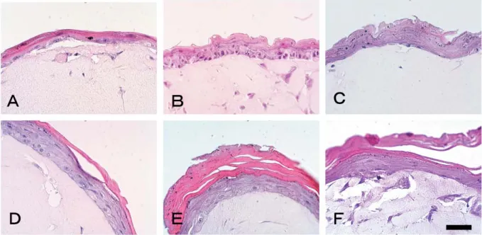
thick at 5 days (Fig. 1-B, C). After creasing at 7 days (Fig. 1-D), the epidermal equivalent on collagen gel shrank so as to be readily confirmed at 10 and 14 days (Fig. 1-E, F). Fig-ure 2 shows the time course of histological appearance of the pad equivalent after culturing at the air-liquid interface. Keratinocytes are piled up into only 2 to 3 cell layers on the dermal equivalent at 0 day (Fig. 2-A). At 1 day, epithelium becomes stratified into 4 to 5 cell layers, in which the basal
Fig. 1. Photographs of pad equivalents culturing at air-liquid interface for (A) 0, (B) 1, (C) 5, (D) 7, (E) 10, and (F) 14 days, respectively. Bar=5 mm.
Fig. 2. Photomicrographs of pad equivalents culturing at the air-liquid interface for (A) 0, (B) 1, (C) 3, (D) 4, (E) 5, and (F) 14 days, respectively. Hematoxylin and eosin stain. Bar=50 µm.
K. YAMAZOE ET AL.
614
cell layer is confirmed (Fig. 2-B). At 3 days, epithelium became stratified into 5 to 6 cell layers, in which basal and granular cell layers were confirmed (Fig. 2-C). The corneal cell layer appeared at 4 days, and became thick at 5 days (Fig. 2-D, E). After that, the corneal cell layer was main-tained until 14 days without exfoliation (Fig. 2-F).
Immunofluorescence analysis on basement membrane formation of pad equivalent: Time courses of the
expres-sions on α6 integrin subunit, laminin, type IV and VII
col-lagens after culturing at the air-liquid interface are shown in Figs. 3 to 6. The expression of α6 integrin subunit is
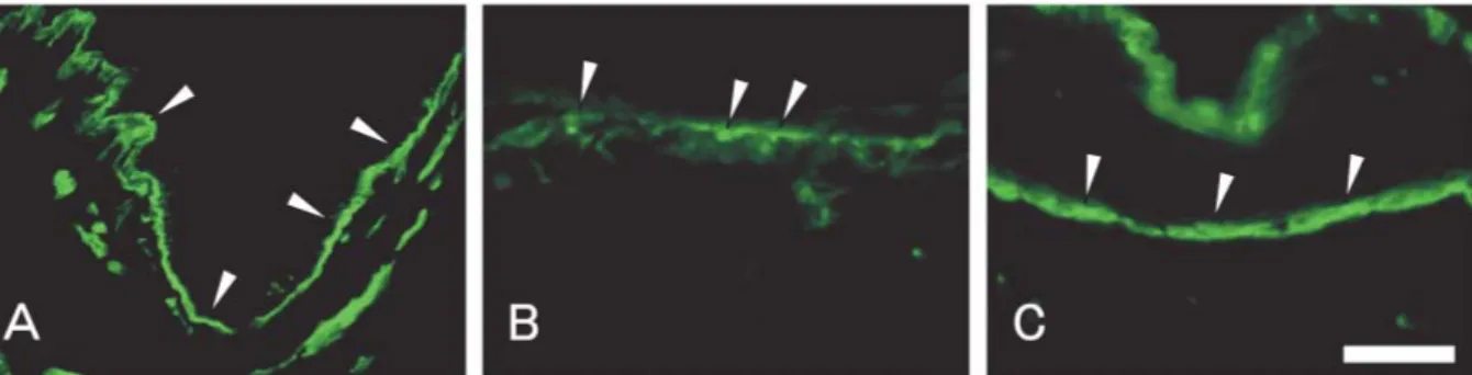
contin-uously observed at the dermal-epidermal junction in native pad tissue (Fig. 3-A). On the pad equivalent, α6 integrin
subunit is intermittently expressed even at 0 day at the der-mal-epidermal junction (Fig. 3-B). The fluorescence of α6
integrin subunit is continuously observed from 1 day through 14 days shows as much intensity as native pad tis-sue (Fig. 3-C). The expression of laminin is continuously observed at the dermal-epidermal junction in native pad tis-sue (Fig. 4-A). On the pad equivalent, laminin is discontin-uously expressed from 5 through 10 days at the dermal-epidermal junction (Fig. 4-B). After that, the fluorescence of laminin is continuously observed at 14 days with as much intensity as native pad tissue (Fig. 4-C). The expression of type IV collagen is continuously observed at the dermal-epi-dermal junction in native pad tissue (Fig. 5-A). On the pad equivalent, the fluorescence of type IV collagen started to be discontinuously observed at 10 days at the dermal-epider-mal junction (Fig. 5-B). At 14 days, the fluorescence of type IV collagen continuously shows as much intensity as native pad tissue (Fig. 5-C). The expression of type VII collagen is continuously observed at the dermal-epidermal junction in native pad tissue (Fig. 6-A). On the pad equivalent, type VII collagen fluorescence is intermittently observed at 2 days at the dermal-epidermal junction (Fig. 6-B). The intensity of the fluorescence is increased from 6 through 10 days (not shown). The fluorescence is still discontinuously observed at the dermal-epidermal junction at 14 days, but the intensity is inferior to that of native pad tissue (Fig. 6-C).
DISCUSSION
The epithelium of the pad equivalent, which was spread over collagen gel, was dried on the surface at 1 day at the air-liquid interface, and at 5 days it became thick enough to be readily confirmed. The skin equivalent based on this study has almost the same thickness as native skin, and is easier to handle than the epidermal sheet in the grafting [28, 30]. Moreover, the thick corneal cell layer of the skin equiv-alent functioned as a barrier against the external stresses on the graft, and that gives rise to a high take rate [28]. As for reconstruction of a pad whose structure is similar to skin, a thick corneal cell layer is supposed to be favorable for the subsequent grafting. The pad equivalent prepared in this study was thick enough to be clearly observed at 5 days, considering that it was applicable to the grafting. But there were many creases on the epithelium, which shrank at 10
and 14 days. Moreover, the epidermal part of the pad equiv-alent was mobile at 14 days, indicating that the adhesion at the dermal-epidermal junction was insufficient.
It is known that keratinocytes seeded on a collagen gel containing fibroblasts only proliferate to be piled up in sub-merged culture, while they are lifted up to the air-liquid interface to be multilayered and differentiated [3, 21]. In the present study, keratinocytes on a collagen gel proliferated and became confluent in submerged culture before differen-tiating at the air-liquid interface. Consequently, kerati-nocytes only piled up in 2 to 3 layers at 0 day at the air-liquid interface, and at 1 day they increased 4 to 5 layers so as to differentiate into the basal cell layer. The epithelium then became 5 to 6 layers including the granular cell layer at 3 days. The corneal cell layer, which appeared at 4 days, became thick at 5 days, maintaining the morphology until at least 14 days. Shirakata et al. [30] prepared a human skin equivalent, in which the number of layers of keratinocytes were 1 to 2 prior to culturing at the air-liquid interface, then differentiating into 5 to 8 layers until 7 days, and maintain-ing the shape at least until 14 days. In addition, since the structure of the epithelium of canine skin equivalent Jakic-Razumovic et al. [17] prepared was similar to that of normal skin in 8 days, it was considered that the epithelium of the pad equivalent was generally developed at the air-liquid interface in about one week. But they indicate at the same time that the skin equivalent does not form rete ridges as well as the pad equivalent prepared in this study [17]. More-over, Limat et al. [19] also reported that the epithelium of the human palmar skin equivalent developed a thick corneal cell layer, although not forming rete ridges without the grafting onto nude mouse. Therefore, in the present study, it was suggested that the structure of epithelium of the pad equivalent in the dog was generally developed, but it was difficult to prepare the pad equivalent in vitro comparable to a native pad.
Since basement membrane, which is at the dermal-epi-dermal junction, not only mediates the adhesion to each of them, but also influences the proliferation and the differen-tiation of keratinocytes [1], the time course of the expres-sions on α6 integrin subunit as adhesive molecule, and
laminin, and type IV and VII collagens as extracellular matrices were investigated during the formation of pad equivalent. Alpha 6 integrin subunit and laminin intermit-tently expressed at 0 and 5 days at the dermal-epidermal junction, respectively. Since α6 integrin subunit mediates
signal transduction from dermal extracellular matrix to keratinocytes to promote the reepithelialization such as the proliferation of keratinocytes and epidermal differentiation [23], the early expression of α6 integrin subunit in this study
could be supposed to be related to subsequent epidermal for-mation. In addition, laminin 5 bound to α6 integrin subunit
expressed at 2 days at the dermal-epidermal junction on human skin equivalent as reported by Amano et al. [2]. The expression of laminin in this study was observed at 5 days when the epidermal morphogenesis had almost been com-pleted, but the reason was not known. It followed that type
Fig. 3. Photomicrographs of immunofluorescence of α6 integrin subunit in native pad tissue for (A) control, and in pad equivalents for (B) 0, and (C) 14 days, respectively. Arrowheads indicate α6 integrin subunit. Bar=100 µm.
Fig. 4. Photomicrographs of immunofluorescence of laminin in native pad tissue for (A) control, and in pad equivalents for (B) 5, and (C) 14 days, respectively. Arrowheads indicate laminin. Bar=100 µm.
Fig. 5. Photomicrographs of immunofluorescence of type IV collagen in native pad tissue for (A) control, and in pad equivalents for (B) 10, and (C) 14 days, respectively. Arrowheads indicate type IV collagen. Bar=100 µm.
Fig. 6. Photomicrographs of immunofluorescence of type VII collagen in native pad tissue for (A) control, and in pad equivalents for (B) 2, and (C) 14 days, respectively. Arrowheads indicate type VII collagen. Bar=100 µm.
K. YAMAZOE ET AL.
616
IV collagen, the main component of basement membrane, discontinuously expressed at 10 days at the dermal-epider-mal junction, at which the time of expression almost coin-cided with the one reported by Amano et al. [2]. The expression of type VII collagen was intermittently observed at 2 days at the dermal-epidermal junction, and remained as it was until 14 days.
Amano et al. [2] also reported that type VII collagen was intermittently expressed at 5 days at the dermal-epidermal junction, but it was not continuously confirmed at 14 days. However, in the human skin equivalent, Smola et al. [31] reported that in spite of type VII collagen being expressed at 7 days and still being discontinuous at 14 days, it became continuous at 21 days. Moreover, ultrastructual observation revealed that basement membrane including anchoring fibrils mainly composed of type VII collagen was com-pletely formed at 21 days. In this way, the formation of basement membrane including anchoring fibrils was required for at least 3 to 4 weeks [27, 31], suggesting that its formation, including anchoring fibrils, was incomplete at 14 days on the pad equivalent prepared in this study.
After the grafting, considering that the structural improvement of epithelium of skin equivalent is dependent on the formation of basement membrane [19], an additional 1- to 2-week culturing at the air-liquid interface might be expected to form a more secure basement membrane. But it is not practical for clinical use because of the long prepara-tion time involved. On the other hand, as reported by Limat
et al. [19], a human palmar skin equivalent was transplanted
on nude mouse after culturing for 7 days at the air-liquid interface, so that the structure of the epithelium of the graft was improved with regard to the thickness of the corneal cell layer, and the basement membrane was formed 21 days after the grafting. With the pad equivalent prepared in this study, therefore, autologous grafting after 5 days when the corneal cell layer formed was supposed to promote the formation of basement membrane as well as the take of the graft.
In conclusion, it is suggested that the pad equivalent in the dog prepared in this study was reconstructed similar to a native pad by epidermal morphogenesis except for the incompleteness of basement membrane formation.
REFERENCES
1. Adams, J.C. and Watt, F.M. 1993. Regulation of development and differentiation by the extracellular matrix. Development 117: 1183–1198.
2. Amano, S., Akutsu, N., Matsunaga, Y., Kadoya, K., Nish-iyama, T., Champliaud, M.F., Burgeson, R.E. and Adachi, E. 2001. Importance of balance between extracellular matrix syn-thesis and degradation in basement membrane formation. Exp. Cell Res. 271: 249–262.
3. Asselineau, D. and Pruniéras, M. 1984. Reconstruction of ‘simplified’ skin: control of fabrication. Br. J. Dermatol. 111 (supp. 127): 219–222.
4. Asselineau, D., Bernard, B.A., Bailly, C., Darmon, M. and Pruniéras, M. 1986. Human epidermis reconstructed by cul-ture: is it “normal”? J. Invest. Dermatol. 86: 181–186. 5. Basher, A.W., Fowler, J.D. and Bowen, C.V.A. 1991. Free
tis-sue transfer of digital foot pads for reconstruction of the distal limb in the dog. Microsurgery 12: 118–124.
6. Bell, E., Sher, S., Hull, B., Merrill, C., Rosen, S., Chamson, A., Asselineau, D., Dubertret, L., Coulomb, B., Lapiere, C., Nus-gens, B. and Neveux, Y. 1983. The Reconstruction of Living Skin. J. Invest. Dermatol. 81 (Suppl.): 2s–10s.
7. Bernstam, L.I., Vaughan, F.L. and Bernstein, I.A. 1986. Kerat-inocytes grown at the air-liquid interface. In Vitro Cell. Dev. Biol. 22: 695–705.
8. Bradley, D.M., Scardino, M.S. and Swaim, S.F. 1998. Con-struction of a weight-bearing surface on a dog’s distal pelvic limb. J. Am. Anim. Hosp. Assoc. 34: 387–394.
9. Bradley, D.M., Swaim, S.F., Alexander, C.N. and Jull, B.A. 1994. Autogenous pad grafts for reconstruction of a weight-bearing surface: A case report. J. Am. Anim. Hosp. Assoc. 30: 533–538.
10. Coulomb, B., Lebreton, C. and Dubertret, L. 1989. Influence of human dermal fibroblasts on epidermalization. J. Invest. Der-matol. 92: 122–125.
11. El Ghalbzouri, A. and Ponec, M. 2004. Diffusible factors released by fibroblasts support epidermal morphogenesis and deposition of basement membrane components. Wound Rep. Reg. 12: 359–367.
12. El Ghalbzouri, A., Gibbs, S., Lamme, E., Blitterswijk, C.A.V. and Ponec, M. 2002. Effect of fibroblasts on epidermal regen-eration. Br. J. Dermatol. 147: 230–243.
13. Falanga, V., Margolis, D., Alvarez, O., Auletta, M., Maggia-como, F., Altman, M., Jensen, J., Sabolinski, M., Hardin-Young, J. and the Human Skin Equivalent Investigators Group 1998. Rapid healing of venous ulcers and lack of clinical rejec-tion with an allogeneic cultured human skin equivalent. Arch. Dermatol. 134: 293–300.
14. Fleischmajer, R., Utani, A., MacDonald II, E.D., Perlish, J.S., Pan, T.C., Chu, M.L., Nomizu, M., Ninomiya, Y. and Yamada, Y. 1998. Initiation of skin basement membrane formation at the epidermo-dermal interface involves assembly of laminins through binding to cell membrane receptors. J. Cell Sci. 111: 1929–1940.
15. Fuchs, E. 1990. Epidermal differentiation: the bare essentials. J. Cell Biol. 111: 2807–2814.
16. Hutton, W.C., Freeman, M.A.R. and Swanson, S.A.V. 1969. The forces exerted by the pads of the walking dog. J. Small Anim. Pract. 10: 71–77.
17. Jakic-Razumovic, J., Storb, R., Sandmaier, B.M. and Sale, G.E. 1994. An organotypic skin culture model in dogs. Trans-plantation 57: 285–287.
18. Lee, D.Y. and Cho, K.H. 2005. The effects of epidermal kerat-inocytes and dermal fibroblasts on the formation of cutaneous basement membrane in three-dimensional culture systems. Arch. Dermatol. Res. 296: 296–302.
19. Limat, A., Stockhammer, E., Breitkreutz, D., Schaffner, T., Egelrud, T., Salomon, D., Fusenig, N.E., Braathen, L.R. and Hunziker, T. 1996. Endogeneously regulated site-specific dif-ferentiation of human palmar skin keratinocytes in organotypic cocultures and in nude mouse transplants. Eur. J. Cell Biol. 69: 245–258.
20. Marinkovich, M.P., Keene, D.R., Rimberg, C.S. and Burgeson, R.E. 1993. Cellular origin of the dermal-epidermal basement membrane. Dev. Dyn. 197: 255–267.
21. Maruguchi, T., Maruguchi, Y., Suzuki, S., Matsuda, K. and Isshiki, N. 1992. Culture of keratinocytes on artificial skin der-mis. Jpn. J. Plast. Reconstr. Surg. 12: 73–82.
F., Sabolinski, M., Hardin-Young, J. and Eaglstein, W.H. 1999. Behavior of tissue-engineered skin. Arch. Dermatol. 135: 913–918.
23. Ngyuen, B.P., Ryan, M.C., Gil, S.G. and Carter, W.G. 2000. Deposition of laminin 5 in epidermal wounds regulates integrin signaling and adhesion. Curr. Opin. Cell Biol. 12: 554–562. 24. Olsen, D., Straw, R.C., Withrow, S.J. and Basher, A.W.P.
1997. Digital pad transposition for replacement of the metacar-pal or metatarsal pad in dogs. J. Am. Anim. Hosp. Assoc. 33: 337–341.
25. Pavletic, M.M. 1994. Foot salvage by delayed reimplantation of severed metatarsal and digital pads using a bipedicle direct flap technique. J. Am. Anim. Hosp. Assoc. 30: 539–547. 26. Prunéras, M., Régnier, M. and Woodley, D. 1983. Methods for
cultivation of keratinocytes with an air-liquid interface. J. Invest. Dermatol. 81 (Suppl.): 28s–33s.
27. Raghunath, M., Höpfner, B., Aeschlimann, D., Lüthi, U., Meuli, M., Altermatt, S., Gobet, R., Bruckner-Tuderman, L. and Steinmann, B. 1996. Cross-linking of the dermo-epidermal junction of skin regenerating from keratinocyte autografts. J. Clin. Invest. 98: 1174–1184.
28. Rennekampff, H.O., Kiessig, V. and Hansbrough, J.F. 1996. Current concepts in the development of cultured skin replace-ments. J. Surg. Res. 62: 288–295.
29. Rosdy, M., Pisani, A. and Ortonne, J.P. 1993. Production of
basement membrane components by a reconstructed epidermis cultured in the absence of serum and dermal factors. Br. J. Der-matol. 129: 227–234.
30. Shirakata, Y., Tokumaru, S. and Hashimoto, K. 1999. Histo-logical examination of three dimensional reconstituted skin and its clinical applications. Jpn. J. Dermatol. 109: 1165–1171. 31. Smola, H., Stark, H.J., Thiekötter, G., Mirancea, N., Krieg, T.
and Fusenig, N.E. 1998. Dynamics of basement membrane for-mation by keratinocyte-fibroblast interactions in organotypic skin culture. Exp. Cell Res. 239: 399–410.
32. Swaim, S.F., Bradley, D.M., Steiss, J.E., Powers, R.D. and Buxton, D.F. 1993. Free segmental paw pad grafts in dogs. Am. J. Vet. Res. 54: 2161–2170.
33. Swaim, S.F., Riddell, K.P. and Powers, R.D. 1990. Healing of segmental grafts of digital pad skin in dogs. Am. J. Vet. Res. 53: 406–410.
34. Tsunenaga, M., Adachi, E., Amano, S., Burgeson, R.E. and Nishiyama, T. 1998. Laminin 5 can promote assembly of the lamina densa in the skin equivalent model. Matrix Biol. 17: 603–613.
35. Wilson, J.L., Dollard, S.C., Chow, L.T. and Broker, T.R. 1992. Epithelial-specific gene expression during differentiation of stratified primary human keratinocyte cultures. Cell Growth Differ. 3: 471–483.