Title Isolation and Characterization of Novel Biocontrol Agents forControlling Tomato Bacterial Wilt( 本文(Fulltext) )
Author(s) Malek Khaled Mahmoud Marian
Report No.(Doctoral Degree) 博士(農学) 甲第701号 Issue Date 2018-09-30 Type 博士論文 Version ETD URL http://hdl.handle.net/20.500.12099/77267 ※この資料の著作権は、各資料の著者・学協会・出版社等に帰属します。
Isolation and Characterization of Novel Biocontrol
Agents for Controlling Tomato Bacterial Wilt
(䝖
䝖䝬䝖㟷ᯤ䛻ᑐ䛩䜛᪂つ⏕≀㜵㝖䜶䞊䝆䜵䞁䝖䛾ศ㞳䛸≉ᛶゎᯒ
)
2018
The United Graduate School of Agricultural Science,
Gifu University
Science of Biological Resources
(Gifu University)
Isolation and Characterization of Novel Biocontrol
Agents for Controlling Tomato Bacterial Wilt
(䝖
䝖䝬䝖㟷ᯤ䛻ᑐ䛩䜛᪂つ⏕≀㜵㝖䜶䞊䝆䜵䞁䝖䛾ศ㞳䛸≉ᛶゎᯒ
)
Contents
General introduction
4
Chapter 1
22
Screening of biocontrol bacteria for controlling tomato bacterial wilt
Chapter 2
76
Establishment of an effective application method of Mitsuaria sp.
TWR114 and non-pathogenic Ralstonia sp. TCR112
Chapter 3
103
Biocontrol mechanisms of Mitsuaria sp. TWR114 and non-pathogenic
Ralstonia sp. TCR112
General discussion
143
References 150
Summary
185
Summary in Japanese
190
Acknowledgements 192
Publications 194
General introduction
1. Tomato plant
The commercial tomato belongs to a species most frequently referred to as Lycopersicon esculentum Miller. Other alternative names Solanum lycopersicum L., or Lycopersicon lycopersicum L. Karsten have appeared also in the literature. Tomato plant is a member of the Solanaceae (nightshade) family and belongs to the genus Solanum, which include also other economically important crops such as potato (S. tuberosum L.) and eggplant (S. melongena L.). Tomato is native to South America, especially Peru and Galapagos Islands, being first domesticated in Mexico. The Nahuatl (Aztec language) word tomatl gave rise to the Spanish word "tomate", from which the English word tomato derived. The fruit was thought to be poisonous, like its relative, the deadly nightshade. Its importance as a vegetable has occurred only in the 19th century (14).
Tomato plant is grown for their edible fruit which is often red in color. The plants typically grow to a 1–3 meters in height and have a weak stem that sprawls. It is a perennial in its native habitat, and cultivated as an annual crop (14). Five million hectares of tomatoes are estimated to be grown annually worldwide, producing >177 million tons, with China accounting for 32% followed by India (10.4%), United Stated of America (7.4%), and Turkey (7%). In terms of productivity, the Netherlands and Belgium were the most productive countries, with a nationwide average of 507 tons per hectare (56). In Japan, tomatoes are grown on a total area of 12 thousand hectares, with an annual production of 727 thousand tons. Usually it is cultivated in two periods, summer (July – November) and winter (December – June), with
Hokkaido prefecture accounting for 15.5% of the total production in the summer season, and Kumamoto prefecture accounting for 26.4% of the total production in the winter season (125).
2. Ralstonia solanacearum species complex
Following its discovery, Ralstonia solanacearum was first classified as a member of the genus “Bacillus” (188). The application of DNA-based methods eventually resulted in its transfer to the genus Burkholderia (227) and then to the genus Ralstonia (228). The species is classified into races and biovars (75) and recently into phylotypes (57), according to host range, biochemical, and molecular characteristics, respectively. More recently, R. solanacearum species complex was taxonomically organized into three genomic species: (i) R. solanacearum, including phylotype IIA and IIB; (ii) R. pseudosolanacearum, including phylotype I and III; and (iii) R. syzygii, including the former R. solanacearum phylotype IV and the clove pathogen R. syzygii (162, 178). The phylotypes are subdivided into sequevars based on sequence variation in the endoglucanase (egl) partial gene (57).
3. Bacterial wilt
Bacterial wilt was among the first diseases that Smith (188) proved to be caused by a bacterial pathogen. Bacterial wilt is caused by the gram negative β-proteobacteria soil-borne pathogens R. solanacearum (228), R. pseudosolanacearum, and R. syzygii subsp. indonesiensis (formerly classified as R. solanacearum) (178), and is the second most destructive bacterial disease of plants
worldwide (126). Together, these pathogens infect more than 200 plant species belonging to an over 50 different plant families, mostly Solanaceae and Musaceae. The disease affects the yield of many economically important solanaceous crops, such as tomato, potato, tobacco (Nicotiana tabacum L.), eggplant, and Capsicum species (75). Direct yield losses by bacterial wilt vary widely according to the host, cultivar, climate, soil type, cropping pattern, and strain. For instance, yield losses vary from 0 to 91% in tomato, 33 to 90% in potato, 10 to 30% in tobacco, 80 to 100% in banana, and up to 20% in groundnut (Arachis hypogaea L.) (53). To date, in Japan, more than 46 species belonging to an over 24 families have been reported to be hosts (212), and new hosts continue to be found (233).
Plant Pathogenic Ralstonia species are well adapted to grow and survive in the bulk soil for many years in the absence of susceptible host plants. When the pathogen encounters a susceptible host, it enters the root through wounded roots or natural openings such as lateral root emergence points, colonizes the root cortex and then invades the xylem vessels by degrading the cell wall and produces large amounts of exopolysaccharides that block water flow (42). The most frequent external symptoms of the infected plants are wilting, stunting and yellowing of the foliage. Other symptoms are leaves bent downward showing leaf epinasty, adventitious roots growing in the stems, and the observance of narrow dark stripes corresponding to the infected vascular bundles beneath the epidermis. The most frequent internal symptoms are progressive discoloration of the vascular tissue, mainly the xylem, at early stages of infection, and of portions of the pith and cortex, as disease develops, until complete necrosis. Slimy viscous ooze typically appears on transverse-sectioned stems at the points corresponding to the vascular bundles.
As a result, collapse and death of the plant take place because of the degradation of occluded xylem vessels and the destruction of surrounding tissues (10).
3. Management of bacterial wilt
The current management strategies used against bacterial wilt includes chemical controls, cultural practices, and biological control methods. These approaches have been investigated for decades, extensively reviewed by Elphinstone (53) and Yuliar et al. (238). In general, the control of bacterial wilt is very difficult owing to the viability, adaptability, and genetic diversity of the responsible pathogen (53). Many attempts are made to control this devastating disease. The following approaches have been taken.
3.1 Chemical control
The chemical control methods have not been always efficient in eradicating R. solanacearum, and due to the environmental concerns, chemical control is being discouraged (177). Chemicals such as soil fumigants (1,3-dichloropropene, chloropicrin, and dazomet), bactericides (streptomycin sulfate), and plant activators generating systemic resistance on different plants (acibenzolar-S-methyl, DL-3-aminobutyric acid, and validamycin A) have been used to control bacterial wilt (13, 74, 87, 115, 127, 128). In addition, biofumigation using volatile plant essential oils from thyme (Thymus spp.), palmarosa (Cymbopogon martini Roxb.), Indian mustard (Brassica juncea L. Czern), lemongrass (C. citratus L.), eucalyptus (Eucalyptus
globulus), bergamot (Citrus aurantium var. bergamia L.), and sweet orange (Citrus sinensis L.) oils have also been used to control bacterial wilt (7, 11, 91, 153).
3.2 Cultural practices
The growth of plant cultivars which are resistant to bacterial wilt has been accomplished in the past (59, 114). However, resistance to bacterial wilt in many crops has generally been negatively correlated with yield and quality. Moreover, public acceptance in Japan is needed prior to the commercial use of such genetically modified crops. Thus, the release of resistant cultivars is difficult (238).
Crop rotation and intercropping has also been used for controlling bacterial wilt. However, in addition to being a high labor intensive method, it has been indicated that crop rotation and intercropping, depending on the companion plant used might have little suppressive effective on bacterial wilt (134, 135). Crop rotation of susceptible tomato line with corn (Zea mays L.), okra (Abelmoschus esculentus L. Moench), cowpea (Vigna unguiculata L. Walp.), or partially resistant tomato line has been shown to delay onset and reduce disease severity of the bacterial wilt (2). Potato cultivation rotated with wheat (Triticum aestivum L.), sweet potato (Ipomoea batatas L. Lam), maize, millet (Pennisetum glaucum L. R.Br., carrots (Daucus carota subsp. sativus Hoffm. Schübl. & G. Martens), sorghum (Sorghum bicolor L. Moench), or phaseolus beans (Phaseolus vulgaris L.) reduced the incidence of wilt by 64 to 94% while the yield of potatoes was 1- to 3-fold higher than that of monocultured potatoes (95). Intercropping tomato with Allium plants, such as Chinese chive (Allium tuberosum Rottler ex Spreng) and garlic (Allium sativum L.), has also been reported to suppress bacterial wilt (106, 235).
Previous studies evaluated the potential of soil amendments with rock dust, urea, mineral ash, calcium oxide, and fertilizers such as calcium and silicon on the incidence of bacterial wilt (13, 17, 113, 230). However, some of these components have been shown to be not effective. In addition, their biocontrol effect can be site-specific, and the generation of several toxic substances may also affect their potential use.
Cultural practices through commercially grafted seedlings (grafting resistant rootstock with susceptible scion) restrict pathogen multiplication and movement in the rootstock, thereby suppressing the infection and wilting in the scion, and through an anaerobic reductive soil disinfestation (RSD) method reduces the pathogen population in the soil and is widely adopted in Japan (137). However, grafting is expensive, requires more labor, and result in the production of fruits of inferior quality (taste, color, and sugar contents) (110). Furthermore, new virulent races of the pathogen might overcome the resistance, resulting in colonization and migration of the pathogen into susceptible scions and causing wilt symptoms (141). Moreover, for the RSD method, achieving sufficient disinfection in the deep soil layers where the pathogen might localize is difficult (137).
3.3 Biological control
Great interest in the biological control method has increased over the past decade due to concerns from the excessive use of chemicals (218). The advantages of using biological control agents (BCAs) is that; they are effective, safe, potentially self-sustaining, spread on their own after initial establishment, reduced input of non-renewable resources, and long-term disease suppression in an environmentally
friendly manner (136, 219). Among the natural soil microorganisms, researchers mainly focused on identifying bacteria with an in vitro antibacterial activity against RSSC, as potential BCAs for controlling bacterial wilt. Despite the large body of literature describing their use as BCAs in the past decade (Table. 1), there are only few commercialized biocontrol products available in the world, mostly in China, such as a wettable powder of Bacillus subtilis (Cohn) Y1336, a water suspension of Pseudomonas fluorescens (Migula), a mixture of wettable powder and granule of Paenibacillus polymyxa (Ash, Priest and Collins) (195). In Japan, rhizospheric P. fluorescens isolates were previously commercialized as a biocontrol product against bacterial wilt (Cell Nae Genki, Taki Chemical, Kakogawa, Japan); however this product was abolished and no longer exists, therefore it is necessary to develop a new biocontrol product against bacterial wilt.
4. Mechanisms employed by bacteria in the biocontrol of bacterial wilt
4.1 Competition for nutrients and niche
The root surface and the surrounding rhizosphere are significant carbon sinks. Photosynthate allocation to this zone can be as high as 40%. Thus, along root surfaces there are various suitable nutrient rich niches attracting a great diversity of microorganisms, including phytopathogens. Competition for these nutrients and niches is a fundamental mechanism by which biocontrol bacteria protect plants from phytopathogens (39). Recently, Huang et al. (82) showed that the bacterium Chryseobacterium nankingense sp. WR21 effectively suppresses R. solanacearum via intensive root exudates competition, particularly four amino acids (i.e.,
Asparagine, Glutamine, Histidine, and Leucine), against R. solanacearum. This effect might aid in the colonization of this isolate, thereby effectively suppressing tomato bacterial wilt. Additionally, Wu et al. (221) showed that the competitive ability of biocontrol bacteria Bacillus amyloliquefaciens SQYUV162 to use the tomato root exudate citric acid directly affected not only the population density of R. solanacearum but also its pathogenicity, thus efficiently suppressing the incidence of bacterial wilt.
Competition for niches has been also suggested to be one of the mechanism by which biocontrol bacteria suppress bacterial wilt. For instance, McLaughlin and Sequeira (131) suggested that the avirulent R. solanacearum strain B82 confer protection against bacterial wilt disease by competitive exclusion of the pathogen, particularly, in the crown region of the vascular system. Moreover, Etchebar et al. (54) suggested that competition for space in the xylem vessels is one of the possible explanations for the protective ability of a HrcVÀ mutant strain of R. solanacearum against subsequent invasion by the wild R. solanacearum strain. Through tagging the biocontrol bacteria B. amyloliquefaciens T-5 and R. solanacearum with different fluorescent protein markers, Tan et al. (204) revealed that the root colonization of pathogen was almost completely suppressed in the presence of biocontrol strain T- 5-GFP when both soil and plant seedlings were treated with T-5-GFP, suggesting that the biocontrol bacteria may prevent the pathogen infection through competitive exclusion.
● General in trodu cti o n Rece n t ex am ples o f th e us e o f bac te ria as p o te ntial bioco n tr ol ag ents ag ainst ba cteri a l w ilt reported in t he pa st deca de. P ot ential BCAs Host plant P roposed mechanism(s) Re fe rences Bacillus amyloliquefaciens Bg -C 31 Capsicum Produc
tion of antimicrobial protein
78 B. amyloliquefaciens BZ 6-1 Peanut ( Arachis hypogaea L. ) Produc
tion of antimicrobial compounds
(surfactin and fe ng ycin ) 214 B. amyloliquefaciens QL -5 a nd QL -1 8 + or ganic fe rtilizer Tomato ( Solanum lyc opersicum L. ) Antibiosis 216 B. amyloliquefaciens HR 62 + or ga nic fe rtilizer (BI O 62) Tomato Produc tion of antibacteri al compounds (Ma crolactin A a nd 7 -O -m al on yl mac rolactin A ) 81 B. amyloliquefaciens CM-2 and T-5 Tomato Antibiosis. Induced s yste m ic resistanc e (I SR). Competition for ni ches 203, 204 B. amyloliquefaciens S13-3 Tomato Antibiosis . 229 B. amyloliquefaciens WF0 2 Tomato
Antibiosis. Siderophore production. Biofilm formation.
IS R 79 B. amyloliquefaciens SQRT3 Tomato Antibiosis. Produc tion of siderophore a nd protea se. B iofilm formati on. I S R 37 B. amyloliquefaciens JK 6 Tomato
Production of antimicrobial compounds (surfactin, fe
ng ycin, and Y ndJ protein). Produc tion of siderophor e, indole ac etic acid (I A A ) and p roteas e. B iofilm for mation 223 B. amyloliquefaciens SQ YUV 162 Tomato
Competition for nutrients
221 B. amyloliquefaciens S20 + or ga nic fe rtilizer Egg plant ( Solanum me longena L. ) Produc
tion of antimicrobial compound
(iturin A) 33 B. amyloliquefaciens + organic fertilizer ( B IO 23 ) and B. subtilis + organic fe rtilizer (B IO 36) Potato ( Solanum tuberosum L. ) Antibiosis 44 B. amyloliquefaciens SQR-7 and SQR-101 and Bacillus methylotrophicus SQR-29 Tobacco ( Nicotiana tabacum L. ) Antibiosis. Produc tion of siderophore a nd indol e aceti c a cid 236 Bacillus thuringiensis Tomato IS R 85 B. thuringiensis UF V-56 and Bacillus cereus Eucal yptus Produc tion of siderophor e and volatiles 180
● General in trodu cti o n UF V-62 compounds B. cereus B C 1AW and P seudomonas putida PP3WT Tomato Antibiosis. Produc tion of siderophore, protease,
and quorum sen
sing inhibiting molecules. I S R 105 Bacillus subtilis L u144 Mulberr y ( Morus alba L. ) Antibiosis. Competi tion 92 Bacillus sp. (RCh6) and Pseudomonas mallei (RB G4) Egg plant
Antibiosis. Siderophore production
167 Bacillus vallismortis Ex TN -1 Tomato IS R 209 Bacillus velezensis Y6 a nd F 7 Tomato Produc
tion of antimicrobial compounds
(surfactin, iturin, and fen
gy cin) 28 B. subtilis PF MRI , Ps eu domonas fuorescens PF 20, and Paenibacillus mac erans BS -D FS Potato Antibiosis 8 B. subtilis 1J N 2, Myroides odoratimimus 3YW8, B. amyloliquefaciens 5Y N8, and Stenotrophomonas maltophilia 2J W 6. Ging er ( Zingiber officinale Roscoe ) A ntibiosis. Produc tion of siderophore a nd protea se 232 B. subtilis and P. fluores cens Coleus forskohli Antibiosis 185 Ps eudomonas brassicac earum J1 2 Tomato Produc
tion of antimicrobial compound
(2,4diac et yl phloro glucin ol). Produc tion of siderophore a nd protea se 242
Pseudomonas monteilii + Glomus fasciculatum
C. forskohli Antibiosis 186 P. fluoresc ens EB 69 Egg plant
Antibiosis. Siderophore production
166 Pseudomonas putida A1+or ganic fe rtilizer Tomato Antibiosis . B iofilm formation 194 Pseudomonas aeruginos a NXHG29 Tobacco Antibiosis 123 Streptomyces virginiae
E36 and Y30
Tomato Siderophore produ ction 202 Streptomyces m ycarof ac iens SS-2-243 and Streptomyces philanthi RL -1 -178 Chili peppe r ( Capsicum annuum L. ) Antibiosis 26 Brevibacillus brev is L-25 + Streptomyc es roche L-9 + or ganic f ertilizer Tobacco Antibiosis 117 Acinetobacter sp. Xa 6 an d Enterobacter sp. Xy 3 Tomato Rhiz ocompetence and ro ot coloniz ation 225
● General in trodu cti o n Flavobacterium johnsoniae W R 4 and Chryse obacterium sp. WR2 1 Tomato
Antibiosis and competiti
on for nutrients 80, 82 Serratia sp. XY21 Tomato Rhiz ocompetence and an tag onistic acti vit y. 226 Ralst onia pi ckett ii QL -A 6 Tomato Competition 217 Paenibacillus sp. Pb28, P. putida Pp17, and P. fluoresc ens Pf11 Potato Antibiosis. Produc tion of siderophore a nd protea se 97
4.2 Siderophore-mediated competition for iron
In general, the availability of soluble iron in the soil is extremely low (10-17 M),
whereas a minimum concentration of 10-6 M is commonly needed for
microorganisms to grow (143). Most microorganisms produce siderophores, which are low-molecular-weight metabolites with a high affinity for Fe3+, under conditions of low iron availability (18). These siderophores chelate Fe3+ from the environment and transport it into the microbial cells (142). Earlier studies have indicated that siderophore production may contribute to disease suppression of bacterial wilt, possibly by limiting iron availability to R. solanacearum (138, 168). In addition, it was suggested that the siderophore pseudobactin, produced by some fluorescent Pseudomonas spp., is one of the determinants responsible of triggering induced systemic resistance in Eucalyptus urophylla against bacterial wilt (169).
4.3 Antibiosis
The basis of antibiosis as a mechanism employed by biocontrol bacteria has become increasingly better understood over the past two decades (70, 159, 146, 218). Antibiotics encompass a chemically heterogeneous group of organic, low-molecular weight compounds produced as secondary metabolites by microorganisms. At low concentrations, antibiotics are deleterious to the growth or metabolic activities of other microorganisms (60). A variety of antibiotics such as
2,4-diacetylphloroglucinol, surfactin, iturin A, and fengycin, produced by different biocontrol bacteria have been identified to be involve in the suppression of bacterial wilt (33, 214, 223, 242). Recently, Huang et al. (81) identified two types of
polyketides Macrolactin A and 7-O-malonyl macrolactin A produced by the B. amyloliquefaciens HR62 which could inhibit R. solanacearum. The production of these polyketides was pursued as another possible mechanism behind the biocontrol of bacterial wilt by HR62. Moreover, Hu et al. (78) indicated that the antagonistic action responsible for the inhibition of R. solanacearum was due to an antimicrobial peptide LCI produced by the endophytic bacterium B. amyloliquefaciens Bg-C31.
4.4 Lytic enzymes
Many bacteria produce and release enzymes that can hydrolyze a wide variety of polymeric compounds, including chitin, proteins, cellulose, hemicellulose, and DNA. Expression and secretion of enzymes such as protease and polygalacturonase by different biocontrol bacteria can sometimes result in the direct suppression of plant pathogens or aid in their colonization ability (150). For example, Elhalag et al. (52) reported that the biocontrol activity of Stenotrophomonas maltophilia against bacterial wilt was due of the direct antagonism against R. solanacearum, which depends on the production of the enzyme protease. Moreover, the strong competitive ability of rhizobacteria to utilize pectin by producing extracellular pectinases may play a significant role in their rhizoplane competence, possibly resulting in suppressing the multiplication of R. solanacearum (86, 183). Cell wall-degrading enzymes such as pectinases also play some role in triggering defense mechanisms in plants, probably by releasing cell wall fragments (e.g., oligogalacturonides), which can act as elicitors of host defense responses (141).
Systemic acquired resistance (SAR) and induced systemic resistance (ISR) are two forms of induced resistance; in both SAR and ISR, plant defenses are preconditioned by prior infection or treatment that results in resistance (or tolerance) against subsequent challenge by a pathogen (213). ISR as a mechanism by which non-pathogenic rhizobacteria suppress plant diseases has been widely investigated during the last two decades (19, 100, 130, 159). Many studies have reported the ability of biocontrol bacteria to induce ISR against R. solanacearum in tomato plant (72, 94, 200). ISR is generally associated with a physiological state in which plant can react more efficiently to a pathogen attack; that is the priming of the plant defense mechanism (40). Primed defense reactions include an earlier oxidative burst and stronger upregulation of defense-related genes (4). Earlier studies have discussed the importance of host defense priming in the suppression of bacterial wilt by the treatment with biocontrol bacteria (4, 148).
5. Methods of improving the efficacy of biocontrol agents
The biocontrol effect exhibited by single BCAs can sometimes be low and/or last for only a short period of time, thus requiring uneconomically high inoculum densities or frequent applications in the field. These points were considered the most important disadvantages of BCAs in controlling bacterial wilt (238). Several sophisticated methods such as the combination of biocontrol bacteria with chemical pesticide (154), organic amendments (117), and other bacteria in a mixture (88) have been proven to be an effective way to overcome some of the drawbacks from the application of single BCAs against bacterial wilt. For example, the combined
application (foliar and/or soil drenching) of P. fluorescens Pf2 and the plant activator acibenzolarǦSǦmethyl achieved higher biocontrol effect compared with their application individually against tomato bacterial wilt (1). The mechanism behind the improved effect might be due to the enhanced expression of several tomato defense-related enzymes. Moreover, a previous study reported that the combination of endophytic bacteria Bacillus sp. or Serratia marcescens with the susceptible tomato cultivar Santa Clara could reduce bacterial wilt for up to 35%, however the disease reduction was improved for up to 65% when they were applied with the resistant cultivar Yoshimatsu (22). Furthermore, Nion (145) have demonstrated that the suppressive effects against tomato bacterial wilt were enhanced by the combinations of the biocontrol bacteria Burkholderia nodosa G5.2.rif1 with lysine and sucrose. And the addition of these nutrients also improved the root colonization of this isolate. Recently, there has been increasing interest among researchers in using the combination of BCAs to exploit potential synergistic effects on plant health (reviewed by; 181, 224). Many previous studies reported that the combined application of multiple microbes may enhance the biocontrol efficacy and reliability against bacterial wilt on tomato, (88, 90), tobacco (117, 237), bell pepper (Capsicum annuum L.) (116), and Coleus (Coleus forskohlii Briq.) (186) plants. Additionally, the combination may also lead to a broad-spectrum protection against multiple pathogens(46, 89, 170) and may improve the growth, yield and quality of different crops such as tomato, rice (Oryza sativa), and potato (122, 179, 192).
The main goal of this thesis is to develop a practical biocontrol strategy for controlling tomato bacterial wilt. In an attempt to achieve the main objective, experiments described in the following chapters were conducted:
In chapter one, bacteria were isolated from the rhizosphere soil of tomato, Chinese
chive, and Welsh onion. Rhizobacterial isolates were then screened for their antibacterial activity against Ralstonia pseudosolanacearum using the agar well diffusion assay. All isolates exhibiting antibacterial activity were identified based on the partial sequencing of the 16S rRNA gene. Antibacterial isolates were evaluated for their biocontrol effect against bacterial wilt using a tomato seedling bioassay. Isolates affiliated to the bacterial genera Ralstonia and Mitsuaria were assessed for their biocontrol effect in a series of pot experiments. Among the isolates, TCR112 (identified as non-pathogenic Ralstonia sp.) and TWR114 (identified as Mitsuaria sp.) showed consistent disease suppression in pot experiments, thus were selected for further evaluation under field conditions. We monitored the population dynamics of R. pseudosolanacearum in the rhizosphere and aboveground stem of TCR112- and TWR114-treated tomato plants. Moreover, the colonization capacity of both isolates in the same regions was also investigated.
In chapter two, we evaluated the biocontrol effect of the combined application of
TWR114 and TCR112 against bacterial wilt. In the first pot experiment, the effect of several inoculum ratios (i.e., 1:1, 1:2, and 2:1) of the TWR114+TCR112 were tested for their biocontrol ability against bacterial wilt under glasshouse conditions. In the second pot experiment, the effect of inoculum concentrations (i.e., original concentration [ca. 9 × 10⁸ CFU/ml], 2-fold [ca. 4.5 × 10⁸ CFU/ml] and 10-fold [ca. 9 × 10⁷ CFU/ml] dilutions of the original concentration) of the TWR114+TCR112 (at a ratio of 2:1) were tested for their biocontrol effect against the wilt disease. The
population dynamics of R. pseudosolanacearum, TWR114, and TCR112 in several regions of TWR114+TCR112-treated tomato plants and the individually-treated plants was monitored.
In chapter three, we investigated the different biocontrol mechanisms of TWR114
and TCR112. The production of siderophore, indole-3-acetic acid, protease, polygalacturonase, and hydrogen cyanide by these isolates was examined using in vitro assay. The effect of TWR114 and TCR112 individual treatments and their combination on the expression of six defense-related marker genes (i.e., PR1-a, GluA, GluB, Osmotin-like Le4, and LoxD) in the roots of tomato plants was examined. The expression levels of these genes were determined by using quantitative real-time PCR at 5 and 7 days after treatment (2 and 4 days post-challenge inoculation, respectively) in pathogen-uninoculated and -inoculated plants. To identify the genetic traits possibly involved in the biocontrol activity and to assess their taxonomical relationships, the genomes of TWR114 and TCR112 were sequenced and analyzed. Genome relatedness was computed using the average nucleotide identity and genome-to-genome distance (in silico DNA-DNA hybridization) analysis. Pan- and core-genomic analysis of Mitsuaria and non-pathogenic Ralstonia isolates was performed to identify shared and unique genetic components in TWR114 and TCR112 isolates. Additionally, a core-genome based phylogenetic analysis was constructed to obtain a higher resolution classification of our isolates.
Chapter 1
Screening of biocontrol bacteria for controlling tomato
bacterial wilt
Screening of biocontrol bacteria for controlling tomato bacterial
wilt
Abstract
In this chapter, we aimed to identify potential biocontrol agents capable of suppressing tomato bacterial wilt caused by Ralstonia pseudosolanacearum. In total, 442 bacteria were isolated from the rhizosphere soil of tomato, Chinese chive, and Welsh onion. Based on the results of the in vitro antibacterial activity assay, 276 isolates were selected and further evaluated using a tomato seedling bioassay. Nineteen isolates that belonged to that the genera Ralstonia and Mitsuaria exhibited a relatively higher disease suppression (>50% reduction in disease severity) than the other isolates. The isolate TCR112 of Ralstonia and 11 isolates of Mitsuaria were assessed for their biocontrol effect in a series of pot experiments. Among the isolates, TCR112 (identified as non-pathogenic Ralstonia sp.) and TWR114 (identified as Mitsuaria sp.), which showed a consistent disease suppression in pot experiments, were selected as final candidates for further evaluation under field conditions. The results showed that soil drenching at weekly intervals with isolates TCR112 and TWR114 reduced the wilt incidence in the first year by 57.2% and 85.8%, and in the second year by 57.2% and 35.3%, respectively, indicating that these isolates were promising biocontrol agents of tomato bacterial wilt. The isolates effectively reduced the pathogen population in the rhizosphere and crown of pot grown tomatoes. Monitoring the population dynamics of biocontrol isolates revealed that both isolates have stable rhizosphere and endophytic colonization capacities. This is the first study reporting the potential of Mitsuaria as a biocontrol agent against tomato bacterial wilt.
1. Introduction
Bacterial wilt is caused by Ralstonia solanacearum (228), R. pseudosolanacearum, and R. syzygii subsp. indonesiensis (formerly classified as R. solanacearum) (178), and is the second most destructive bacterial disease of plants worldwide (126). Bacterial wilt affects the yield of many solanaceous plants, such as tomato (Solanum lycopersicum), potato (S. tuberosum), tobacco (Nicotiana tabacum), and eggplant (S. melongena) (75). Five million hectares of tomatoes are estimated to be grown annually worldwide, producing >170 million tons (56). In Japan, tomatoes are grown on a total area of 12 thousand hectares, with an annual production of 740 thousand tons (56), and thus are listed as the second most important crop after rice.
The management of tomato bacterial wilt is difficult owing to the viability, adaptability, and genetic diversity of the responsible pathogen (53). In Japan, the current countermeasures used against bacterial wilt include chemical controls and cultural practices. However, chemical controls using soil fumigants such as chloropicrin are potentially harmful to the environment and have not been efficient in eradicating R. solanacearum (177). Cultural practices through commercially grafted seedlings (grafting resistant rootstock with susceptible scion) restrict pathogen multiplication and movement in the rootstock, thereby suppressing the infection and wilting in the scion, and through an anaerobic/reductive soil disinfestation (RSD) method reduces the pathogen population in the soil and is widely adopted in Japan (137). However, grafting is expensive, requires more labor, and result in the production of fruits of inferior quality (taste, color, and sugar contents) (110). Furthermore, new virulent races of the pathogen might overcome the resistance, resulting in colonization and migration of the pathogen into susceptible scions and
causing wilt symptoms (140). Moreover, for the RSD method, achieving sufficient disinfection in the deep soil layers where the pathogen might localize is difficult (137). Thus, other alternative or supplementary methods for controlling bacterial wilt are required. The biological control method of using beneficial microorganisms has been proposed as an effective, safe, and sustainable approach.
R. solanacearum is well adapted to grow and survive in the bulk soil for many years in the absence of susceptible host plants (165). When the pathogen encounters a susceptible host, it enters the root via wounded parts or natural openings such as lateral root emergence points and colonizes the root cortex (42). Therefore, antagonistic rhizobacteria were thought to be the best choice of biocontrol agents (BCAs) for controlling tomato bacterial wilt. Indeed, several studies in the past have successfully obtained rhizobacteria such as Pseudomonas spp. (112), Bacillus spp. (105), and Flavobacterium johnsoniae and Chryseobacterium daecheongens (80) that have strong biocontrol ability against bacterial wilt under laboratory and/or greenhouse conditions. In Japan, rhizospheric Pseudomonas fluorescens isolates were previously commercialized as a biocontrol product against bacterial wilt (Cell Nae Genki, Taki Chemical, Kakogawa, Japan); however this product was abolished and no longer exists, therefore it is necessary to develop new biopesticides against bacterial wilt.
Many researchers have screened rhizobacteria from host plants susceptible to pathogen infection to identify promising candidates as BCAs to control soil-borne diseases, as these bacteria have high affinity for the roots of host plant. We assumed that bacteria inhabiting the rhizosphere of non-host plants, particularly companion plants, are also a good source of BCAs. Intercropping has long been used for controlling soil-borne diseases. Companion plants used for intercropping
enhance antagonist populations in soil and reduce pathogen attack on host plants (77). Intercropping with Allium plants, such as Welsh onion, Chinese chive, and garlic, has been reported to suppress soil-borne diseases including bacterial wilt of tomato (106, 235). Nishioka et al. (146) have shown that antagonistic bacteria inhabiting the rhizosphere of Allium spp. play an important role in the suppression of cucumber Fusarium wilt. Although the mechanisms of bacterial wilt suppression due to Allium intercropping are unknown, this suppression can be attributed to the accumulation of antagonistic bacteria. Therefore, Allium spp. were thought to be a reservoir of potential BCAs.
In this study, we isolated antagonistic rhizobacteria from tomato and Allium plants, and then screened their biocontrol potential against tomato bacterial wilt to develop a new biocontrol product.
2. Materials and methods
2.1. Isolation of rhizobacteria
Bacteria were isolated from the rhizosphere soil of tomato (S. lycopersicum cv. Ohgata-Fukuju), Chinese chive (Allium tuberosum Rottler ex Spreng., cv. Super green belt), and Welsh onion (Allium fistulosum L., cv. Kujo-hoso), grown in fields at Gifu University (Yanagido, Gifu city, Gifu Prefecture, Japan). For isolating the bacteria from the rhizosphere, 3-month-old plants (tomato, Chinese chive, and Welsh onion) were uprooted, and loosely adhering soil was gently removed. Then, roots of each plant were suspended in sterile distilled water (SDW) and shaken on a rotary shaker at 150 rpm for 15 min. Serial dilutions of the soil suspension were
spread on the surface of tryptic soy agar medium and incubated at 30°C for 24 h. The purified colonies were suspended in 10% (w/v) skim milk (Difco, Sparks, MD, USA) supplemented with L-glutamic acid monosodium salt (16.5 g/l) and kept at −80°C until use.
2.2. Bacterial isolates and culture conditions
R. pseudosolanacearum isolate VT0801 (isolated from an infested tomato field in Tsu city, Mie prefecture, Japan) was used as the challenging pathogen. R. pseudosolanacearum and rhizobacterial isolates were cultured in casamino acid-peptone-glucose broth medium (76) and nutrient broth (NB) medium (Nissui Pharmaceutical Co., Tokyo, Japan), respectively, at 30°C for 24 h with shaking at 200 rpm.
2.3 In vitro antibacterial activity
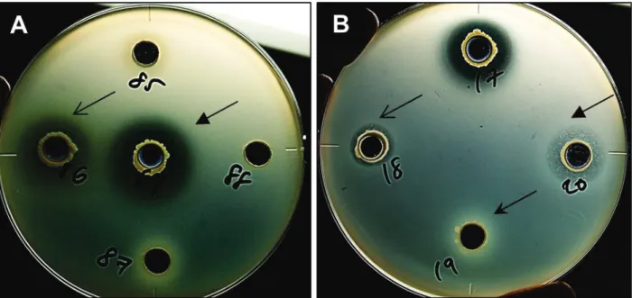
The antibacterial activity was assessed using the agar well diffusion assay (166). A 70-μl aliquot of 24-h-old culture broth (approximately 107–108 cells/ml) of each rhizobacterial isolate was applied to 7-mm-diameter well on solidified King’s B medium supplemented with washed cell suspension of isolate VT0801 and incubated at 30°C for 24 h. The inhibition of VT0801 growth was assessed based on the production of a clear halo zone surrounding the wells. Three replicates were used for each bacterial isolate.
Rhizobacterial isolates that exhibited antibacterial activity in the agar well diffusion assay were further screened for their disease suppressive activity against bacterial wilt using tomato seedling bioassay as described previously (6), with some modifications. Seeds of susceptible tomato (cv. Ponderosa) were surface sterilized with 70% (v/v) ethanol for 1 min, followed by 2% sodium hypochlorite for 5 min, and then thoroughly rinsed with SDW. After germination, 10 seeds were sown into a flat-bottom glass tube (25 mm × 100 mm; AGC Techno Glass Co. Ltd., Shizuoka, Japan) that contained 3.4 g of sterile vermiculite (autoclaved twice at 24-h intervals). The cells of rhizobacterial isolates harvested from 24 h were washed twice with SDW. A 2-ml aliquot of cell suspension of each isolate, adjusted to OD600 = 0.1 (ca. 108
CFU/ml) was added the above seeded tubes, followed by inoculation with 2 ml of pathogen suspension (ca. 8 × 10⁵ CFU/ml). The control treatment was prepared using 2 ml of SDW instead of the rhizobacterial cell suspension. All tubes were maintained in a controlled environmental chamber (Biotron, standard, Nippon Medical and Chemical Instruments Co., Ltd., Osaka, Japan) at 28°C under a 12-h light/12-h dark cycle for 7 days. In trial 1, three seedling tubes were used for each isolate. In trial 2, three tubes were used for each isolate, and the experiment was repeated thrice. The disease severity of the tomato seedlings was visually score on a scale of 0–2, where 0 represent no symptoms, 1 indicate small areas of the hypocotyl showing necrosis, 2 indicates wilted seedling or large areas of the seedling showing necrosis. The disease suppressive efficacy was calculated using the following formula: disease suppressive efficacy = [(mean disease scale of the control treatment) − (mean disease scale of bacterial treatment)/(mean disease scale of control treatment)] ×100%.
2.5. Evaluation of selected rhizobacterial isolates in pot experiments
2.5.1. Growth of plant and bacterial inoculation
The rhizobacterial isolates selected in the above seedling bioassay were evaluated for their biocontrol effect in a series of pot experiments (trial 1 to 3). As described later, we selected 1 isolate of Ralstonia and all of the Mitsuaria isolates, except for the TCR127 isolate, for pot experiments.
Tomato seeds (cv. Ponderosa) were surface sterilized and germinated as described above. The seeds were then sown in plastic trays (Bee pot Y-49; Canelon Kaka Co. Ltd., Japan) that contained a commercial potting soil mix “New star bed” (Zen-Noh, Tokyo, Japan) and grown in a glasshouse maintained at 30°C with a relative humidity of 70% until the seedlings reached fourth-leaf stage. Seedlings were transplanted into vinyl pots (9 cm in diameter) comprising three layers: top and bottom layers, each containing 150 g of commercial potting soil mix, and middle layer containing 20 g of river sand. Rhizobacterial cells were harvested from 24-h-old cultures, washed twice, and diluted with SDW to obtain a concentration of ca. 3 × 10⁸ CFU/ml. In trials 1 and 2, tomato plants were treated by bottom watering with the cell suspension of each rhizobacterial isolate (100 ml per pot) to obtain a final concentration of ca. 1 × 10⁸ CFU/g soil. Plants treated with an equal volume of SDW without the rhizobacteria were used as controls. One day after treatment, both control plants and those treated with rhizobacteria were challenged with 100 ml of VT0801 washed cell suspension (ca. 4 × 10⁷ CFU/ml) to obtain a final concentration of ca. 1 × 10⁷ CFU/g soil. The inoculated plants were maintained in the same glasshouse for 14 days. In trial 3, tomato plants were treated as above with the cell
suspension of rhizobacteria isolates (ca. 9 × 10⁸ CFU/ml) to obtain a final concentration of ca. 3 × 10⁸ CFU/g soil. Three days after the treatment, plants were challenged with 100 ml of VT0801 cell suspension (ca. 2 × 10⁷ CFU/ml) and grown for 14 days under the same glasshouse conditions. Five plants were used for each treatment in trial 1. By contrast, each treatment consisted of three replicates of nine plants per replicate and five replicates of ten plants per replicate in trial 2 and trial 3, respectively.
2.5.2. Development of disease symptoms
The symptoms of tomato bacterial wilt were monitored daily on the basis of a disease scale that ranged from 0 to 4, as described by Kempe and Sequeira (96), where 0 = no wilt symptoms (healthy), 1 = up to 25% of the leaves wilted, 2 = 25%– 50% of the leaves wilted, 3 = 50%–75% of the leaves wilted, and 4 = 75%–100% of the leaves wilted. The disease incidence, disease severity and the area under disease severity progress curve (AUDPC) were calculated using the following formulas:
Disease incidence = {[total number of diseased plants (scale 1–4) in the treatment/total number of plants investigated)]} × 100
Disease severity = [(the number of diseased plants in each scale × disease scale)/(total number of plants investigated × the highest disease scale)] × 100.
AUDPC was calculated on the basis of disease severity using the trapezoid integration of disease progress curve over time according to the following formula: AUDPC = ∑ [0.5 (xi + xi − 1)] (ti − ti − 1), where xi and xi − 1 are disease severity at time ti
and ti – 1, respectively, and ti and ti − 1 are consecutive evaluation dates, with ti and ti − 1 equal to 1.
2.6. Quantification of R. pseudosolanacearum
Tomato plants were treated with two final candidate isolates (TCR112 and TWR114) and challenged with R. pseudosolanacearum VT0801 as in trial 3 of pot experiments. The pathogen multiplication in the rhizosphere and crown (basal part of hypocotyl) of tomato plants was determined at 1, 3, 5, 7, and 14 days after challenge inoculation. Samples were obtained from a total of three plants at each time point. Rhizosphere soil samples were serially diluted with SDW. Crown samples of tomato plants (2 cm in length) were surface sterilized with 100% ethanol and flamed as described previously (Wei et al., 2013). The samples were then homogenized using mortar and pestle, and used to prepared serial dilutions in SDW. Dilutions of rhizosphere soil and crown homogenate were spread in triplicates onto the surface of modified semi-selective medium South Africa (M-SMSA) (61). Typical colonies of R. pseudosolanacearum that appeared elevated fluidal with a pink center were counted after incubation for 3 days at 30°C. The experiment was repeated thrice. The population was expressed as log colony-forming units per gram (wet weight) of soil (log CFU/g wet soil) or tissue (log CFU/g fresh tissue).
2.7. Colonization capacity of isolates TCR112 and TWR114
The populations of the isolates TCR112 and TWR114 in the rhizosphere and crown of tomato plants were simultaneously enumerated with the pathogen population. Dilutions of the rhizosphere soil and crown homogenate, used for pathogen enumeration, were spread onto the surface of isolation media that were
optimized for each isolate. A preliminary survey of the antibiotic resistance of the isolates revealed that TCR112 and TWR114 had resistance to six (kanamycin, ampicillin, hygromycin B, gentamicin, tobramycin and streptomycin) and three (kanamycin, ampicillin and hygromycin B) antibiotics, respectively. Accordingly, these antibiotics were added in respective combination to 1/10-strength TSA medium (5 mg/L each). Moreover, cycloheximide (50 mg/L) was also added to both media to prevent fungal contamination. These inoculated plates were incubated at 30°C for 48 h, and the number of representative colonies of each isolate (Fig. 1) was counted. The experiment was repeated thrice.
2.8. Field experiments
Field experiments were conducted in an experimental field at Gifu University, from August to October in 2016 and from September to October in 2017. Before transplanting, 30 tons/ha of organic fertilizer (60% of cow manure, 20% of pig manure, and 20% of horse manure) and 2.3 tons/ha of chemical fertilizer (N:P2O5:K2O in the ratio of 12:9:10 supplemented with Mg:B ratio of 2:0.1) (Nittofc
Co., Ltd., Japan) was added to the soil. Moreover, limestone was added at a rate of 2.3 tons/ha (55.4% CaO, pH 9.5) (Shinko Kogyo Co., Ltd., Japan). Furthermore, the population of the pathogen in the field was quantified for both years. Soil samples were obtained from 12 different locations distributed across the field. Ten grams of bulk soil was used to prepared serial dilutions in SDW. Dilution of bulk soil was spread onto the surface of M-SMSA medium and incubated as described earlier. The population was expressed as log colony-forming units per gram (dry weight) of soil (log CFU/g dry soil).
The field (11.5 m × 6.6 m) comprised eight rows, and each row (10.8 m length, 0.8 m width) was divided into three plots (3.6 m length). There were three and four replicate plots per treatment arranged in a randomized complete block design in the first and second year, respectively. Six tomato plants were transplanted in each plot with distances of 0.55 m between the plants. Standard agronomic practices were performed to grow tomato plants.
Field experiments comprised three treatments: (1) control, (2) TCR112, and (3) TWR114. Fourth-leaf-stage tomato seedlings (cv. TY Misora 86) grafted with the rootstock (cv. Magnet, moderately resistant to R. solanacearum and highly resistant to Fusarium oxysporum f. sp. radicis-lycopersici, F. oxysporum f. sp. lycopersici, Verticillium dahlia, and Pyrenochaeta lycopersici) and planted in vinyl pots containing commercial potting soil mix (300 g). These plants were then treated with TCR112 and TWR114 (final concentration ca. 3 × 10⁸ CFU/g soil) as described earlier. Tomato plants treated with SDW were served as control. All the plants were grown in a greenhouse at 28–30°C. After reaching the eight–ninth-leaf stage, the tomato plants were transplanted into the field, and then, 300 ml of the cell suspension (ca. 3 × 10⁸ CFU/ml) of each isolate or the same volume of distilled water was applied around the stem base of each plant. TCR112 and TWR114 were applied at weekly intervals until 42 and 28 days in the first and second year of the experiments, respectively. During the experiment, the number of wilted plants was recorded daily and disease incidence was calculated as described above. Moreover, at the end of the experiment in first year (8 days after the final application with the candidate isolates), three healthy plants from each treatment (one plant from each plot) were used to estimate the population of the TCR112 and TWR114 in the rhizosphere and crown tissues as described above.
2.9. Identification of selected rhizobacterial isolates
2.9.1 Partial and complete sequence of the 16S rRNA gene
Bacterial isolates showing in vitro antibacterial activity was tentatively identified based on the partial sequence of 16S rRNA gene, according to a protocol described previously (147). The isolates TCR112 and TWR114 were further identified by sequencing the full-length 16S rRNA gene. Primers 27f and 1492r (Lane, 1991), were used for sequencing the PCR products. PCR amplification and DNA sequencing were performed using the same conditions as described previously (147). The 16S rRNA gene sequences were compared with those of type strains in the EzBioCloud database (https://www.ezbiocloud.net/) (234). Additionally, the 16S rRNA gene sequences of the representative strains were downloaded from the GeneBank database and aligned with the sequences of TCR112 and TWR114 isolates. A phylogenetic tree was constructed with the neighbor-joining method using MEGA version 7.0.26 (201). The 16S rRNA gene sequences of the isolates were deposited in GeneBank database under the accession numbers MG877646– MG877664.
2.9.2 Multilocus sequence analysis
An additional phylogenetic analysis based on multiple protein coding genes was carried out to better characterize the final candidate isolate TCR112 at the species level.
Five protein-coding housekeeping genes gdhA (glutamate dehydrogenase, NADP-specific, oxidoreductase protein), mutS (methyl-directed DNA mismatch repair
protein), leuS (Leucyl-tRNA synthetase), rplB (50S ribosomal subunit protein L2), and gyrB (DNA gyrase, subunit B), were selected for the multilocus sequence analysis (MLSA) scheme of the isolate TCR112. Sets of primers used to amplify fragments of these five genes are listed in Table 1. PCR amplifications were carried out using One-Taq Hot Start 2X Master Mix with GC Buffer (New England BioLabs) to help the amplification of the GC-rich sequences. Primer concentrations (forward and reverse) were 0.2 μM and 10% One-Taq High GC enhancer was added to help the amplification of extremely difficult amplicons. The amplification conditions were at 94°C for 2 min (initial denaturation), followed by 30 cycles at 94°C for 1 min, the appropriate annealing temperature (Table 1), and an extension at 72°C for 1 min, with a final extension step at 72°C for 5 min. The amplification products were purified using GenElute PCR Clean-Up Kit (Sigma, MO, USA), as described in the manufacturer’s instructions. The cycle sequencing reaction was performed using BigDye Terminator v3.1 Cycle Sequencing Kit (Applied Biosystems, CA, USA) and the same primers used for the amplification process except for the gdhA gene (Table 1). The cycle sequencing conditions were 96°C for 1 min (initial denaturation), followed by 25 cycles at 96°C for 1 min, 96°C for 10 s, 50°C for 5 s, and 60°C for 4 min. Amplification and sequencing were performed in ABI PRISM 3100 Genetic Analyzers (Applied Biosystems, CA, USA). The raw sequences were assembled and then manually inspected and corrected using Chromas pro (version 2.1.6, Technelysium Pty. Ltd, Tewantin, Queensland, Australia). The partial sequences of the housekeeping genes of other representative strains were downloaded from the GeneBank database. The consensus sequence of each gene was aligned with the sequences of the other representative strains using CLUSTAL W within MEGA software (201). Gene alignments were concatenated with Geneious R11 software
platform (version 11.0.3; Biomatters Ltd., New Zealand). The phylogenetic tree was constructed using the maximum likelihood in PhyML (version 3.0, http://www.atgc-montpellier.fr/phyml/) (69). Bootstrap support with 1000 replicates was generated to estimate the reliability of the clusters. The protein-coding housekeeping genes sequences of TCR112 isolate were deposited in GeneBank database under the accession numbers MG878974–MG878978.
2.9.3 Physiological and biochemical characterization
The growth of TCR112 and TWR114 was tested by culturing the isolates on nutrient agar (NA) plates and incubating them under various temperature conditions (4°C, 15°C, 25°C, 30°C, 37°C, and 42°C). Tolerance to NaCl was tested on NA plates containing 0%, 1%, 3%, and 5% NaCl (w/v). Urease production was detected using urea agar as previously described by Christensen (35). Starch hydrolysis was examined using starch agar medium as previously described by Atlas et al., (15). Briefly, the bacterial isolates were grown on NA medium supplemented with 2% starch and incubated at 30°C for 4 days. After incubation, the zone of hydrolysis was detected by flooding the plates with iodine solution.
Additional biochemical tests were performed to assess the carbon source utilization pattern of TCR112 and TWR114 isolates. Briefly, overnight bacterial cultures were adjusted to an optical density of 0.1 (OD600) and were grown in 96-well
microtiter plates in 150-μl one-quarter strength of M63 minimal medium (VWR International, LLC, Solon, Ohio, USA) supplemented with each carbon source with a final concentration of 10mM. After 48-h growth at 30°C with agitation (200 rpm), the OD570 was measured using a Tecan Sunrise microplate reader (Tecan Austria
GmbH, Grödig, Austria). Wells with an OD570>0.05 were scored as positive for
growth on a given substrate.
2.10. Data analysis
The data of pot experiments were compared using Student’s t-test (P<0.05). The data of bacterial counts were transformed into logarithm numbers and compared by Student’s t-test (P<0.05). All analyses were performed using SigmaPlot 11.0 software (Systat Software Inc., USA).
3. Results
3.1. Isolation of antibacterial rhizobacteria
In total, 442 bacteria were successfully isolated from rhizosphere soil samples and used as a pool for antibacterial screening (Table 2). Of these isolates, 276 (62.4%) exhibited weak-to-very strong antibacterial activity against R. pseudosolanacearum in the agar well diffusion assay (Table 3 and Fig. 2A) and were selected for subsequent tomato seedling bioassay. By analyzing a partial sequence of 16S rRNA gene, these antibacterial isolates were assigned to 24 genera, including Burkholderia, Pseudomonas, Mitsuaria, Acinetobacter, Arthrobacter, Achromobacter, and Ralstonia (Table 3).
The suppressive effect of selected antibacterial isolates against tomato bacterial wilt was examined using the tomato seedling bioassay. In the first trial, 56 of 276 isolates reduced the disease severity in the treated plants compared with that in the untreated control (Table 3). In addition, rhizobacterial isolates from Chinese chive and Welsh onion plants exhibited stronger suppressive effect than those isolated from tomato plants (Fig. 3B). Among the 56 isolates, 19 showed 50–100% reduction in disease severity (Table 3). Therefore, the disease suppressive effect of these 19 isolates was further evaluated in the second trial of the seedling bioassay. All the tested isolates exhibited strong suppressive effects (ranging from 68.5% to 95.9% reduction in disease severity) (Table 3). These isolates comprised two genera, namely Ralstonia (isolates TCR111, TCR112, TCR113, TCR123, TCR124, TCR133, TCF143, and TCF148) and Mitsuaria (isolates TCR103, TCR127, TCR156, TCR158, TCR159, TCR167, TWR114, TWR120, TWR137, TWR165, and TWR167).
3.3. Evaluation of biocontrol efficacy of selected rhizobacteria in pot experiments
Because none of the isolates belonging to genus Mitsuaria has been reported as a biocontrol agent against R. pseudosolanacearum, we evaluated the biocontrol efficacy of all of our Mitsuaria isolates against tomato bacterial wilt in trials 1 and 2 of pot experiments. Furthermore, TCR112 was selected from eight Ralstonia isolates for trial 3, because this isolate showed the highest suppressive effect, both in the first and second trial of seedling bioassay (Table 3). In trial 1 performed in a glasshouse with 11 Mitsuaria isolates, ten isolates reduced disease severity, which was expressed as AUDPC (Table 4). In particular, four isolates, TCR103, TCR127,
TCR159, and TWR114, showed the lowest AUDPC. Therefore, the biocontrol effect of these four isolates was again evaluated in trial 2. In this trial, the highest reduction of AUDPC was achieved using the isolate TWR114 (45.7%)(Table 4); thus, this isolate was selected for the trial 3.
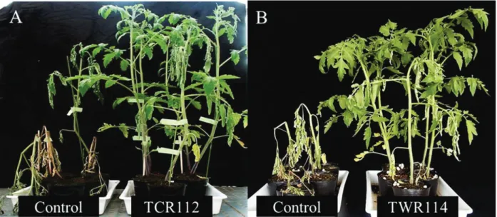
The evaluation of biocontrol efficacy of the final candidate isolates TCR112 and TWR114 in trial 3 proved that the isolates were highly effective in suppressing disease severity of tomato bacterial wilt, as shown by the significant reduction of AUDPC values of 66.4% and 55.3%, respectively (Table 5 and Fig. 4).
3.4. Quantification of R. pseudosolanacearum
The R. pseudosolanacearum population was effectively reduced following the treatment with TCR112 and TWR114 in the rhizosphere and crown of tomato plants (Fig. 5). In TCR112-treated plants, the pathogen was not detected in both the rhizosphere and crown at 1 and 3 days post-challenge inoculation (dpi), whereas in the untreated control plants, the pathogen was detected in the rhizosphere (4.4 and 5.8 log CFU/g wet soil) and crown (1.7 and 3.9 log CFU/g fresh tissue). Subsequently, the pathogen population reached a detectable level in TCR112-treated plants; however, the population densities were significantly lower in the rhizosphere (2.7 and 2.6 log CFU/g wet soil) and crown (2.7 and 2.6 log CFU/g fresh tissue) of TCR112-treated plants than in the rhizosphere (8.8 and 9.5 log CFU/g wet soil) and crown (8.6 and 9.4 log CFU/g fresh tissue) of untreated control plants at 5 and 7 dpi, respectively (Fig. 5A and 5C). The pathogen population was significantly reduced in the rhizosphere of TWR114-treated plants (4.0, 4.3, and 5.4 log CFU/g wet soil) compared with that of the untreated control plants (4.9, 7.0, and 9.1 log
CFU/g wet soil) at 1, 3, and 5 dpi, respectively (Fig. 5B). The pathogen was not detected or was significantly reduced in the crown of TWR114-treated plants (2.2 log CFU/g fresh tissue) compared with that of the untreated control plants (3.3 and 8.3 log CFU/g fresh tissue) at 1 and 5 dpi, respectively (Fig. 5D).
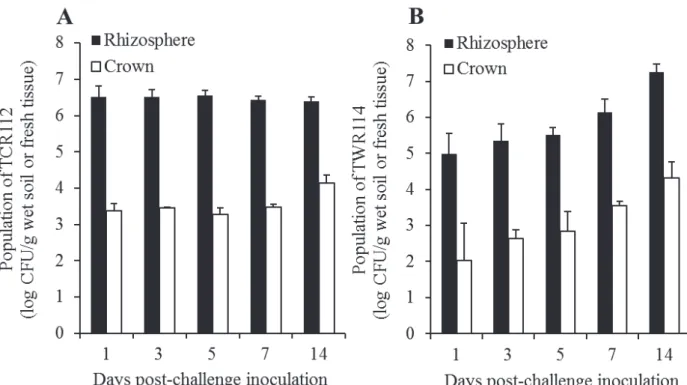
3.5. Colonization capacity of the isolates TCR112 and TWR114
The isolates TCR112 and TWR114 were successfully recovered from both the rhizosphere and crown of tomato plants during the growth period under the glasshouse conditions (Fig. 6). The colonization of the isolate TCR112 was relatively stable throughout the experiment, with a mean population of 6.5 log CFU/g wet soil and 3.5 log CFU/g fresh tissue in the rhizosphere and crown, respectively (Fig. 6A). In contrast, the isolate TWR114 showed an increased colonization throughout the experiment. At 1 dpi (4 days after bacterial treatment), the population densities of TWR114 steadily increased from 5.0 log CFU/g wet soil and 2.0 log CFU/g tissue to 7.3 log CFU/g wet soil and 4.3 log CFU/g tissue, respectively, in the rhizosphere and crown, respectively, at 14 dpi (Fig. 6B). TCR112- and TWR114-like colonies were not detected in both the rhizosphere and crown of untreated control tomato plants.
3.6. Evaluation of TCR112 and TWR114 in field experiments
In the field experiments, the mean initial pathogen population were 3.6 ± 0.2 (log CFU/g dry soil) and 3.0 ± 0.3 (log CFU/g dry soil) in 2016 and 2017, respectively (Fig. 7).
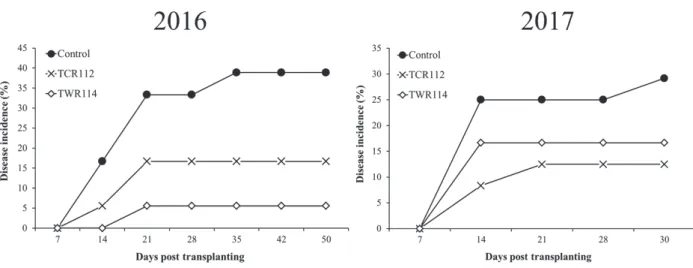
In two consecutive years, the wilt incidence was considerably suppressed in plots drenched treated with the biocontrol isolates at weekly intervals (Fig. 8). In the first year, the wilt incidence at the end of field experiment (50 days after transplanting) was reduced by 57.2% and 85.8% in plots drenched with TCR112 and TWR114, respectively (Fig. 8). Similarly, the wilt incidence at the end of field experiment in the second year (30 days after transplanting) was reduced by 57.2% and 35.3% in plot drenched with TCR112 and TWR114, respectively (Fig. 8).
At the end of the experiment in the first year, we enumerated the populations of both biocontrol isolates. Consequently, both isolates (TCR112 and TWR114) were successfully recovered from the rhizosphere (5.7 and 6.2 CFU/g wet soil, respectively) and crown (4.0 and 5.8 CFU/g fresh tissue, respectively) of the tomato plants (Fig. 9).
3.7. Characterization and identification of the biocontrol isolates TCR112 and TWR114 and the pathogen VT0801
The analysis of full-length 16S rRNA sequence of biocontrol isolates and the pathogen, indicated that TCR112 (approximately 1,230 bp) were 99.8% similar to those of R. pickettii (accession number: JOVL01000020) and TWR114 (approximately 1,200 bp) shared 99.3% similarity with Mitsuaria chitosanitabida (accession number: BCYP01000048), whereas VT0801 (approximately 880 bp) were shared 99.6% similarity with R. pseudosolanacearum (accession number: KC757037). To clarify the phylogenetic position of the biocontrol isolates and the pathogen, a phylogenetic tree was constructed on the basis of partial or complete 16S rRNA gene sequences (Figs. 10 and 11). As a result, the isolates TCR112,
TWR114, and VT0801 showed a clear distinction from the known type strains of R. pickettii ATCC 27511T, M. chitosanitabida 3001T, and R. pseudosolanacearum UQRS 461T, respectively (Figs. 10 and 11).
In order to construct a higher resolution phylogenetic tree for the isolate TCR112, an MLSA scheme using five housekeeping genes (gdhA, mutS, leuS, rplB, and gyrB) was applied. The lengths of the five protein-coding genes were: gdhA = 503 bp, mutS = 640 bp, leuS = 700 bp, rplB = 639 bp, and gyrB = 345 bp. The MLSA phylogenetic tree revealed that the isolate TCR112 have a clear distinction from its closest relative type strains of R. pickettii (Fig. 12).
The growth of TWR114 occurred at 15–37°C and 0–1.0% NaCl (w/v), being optimal at 25–30°C and 0.5% NaCl (w/v). The growth of TCR112 occurred at 15– 42°C and 0–1.0% NaCl (w/v), being optimal at 25–30°C and 0.5% NaCl (w/v). The TWR114 showed positive starch hydrolysis, whereas, it had negative urease and β-glucosidase activity. The TCR112 showed negative starch hydrolysis, while it showed positive urease and β-glucosidase activity. Both isolates did utilize sucrose, D-fructose, D-mannose, D-xylose, D-galactose, N-Acetyl-D-glucosamine, and L-Arabinose, but did not utilize capric acid (Table 7).
Based on the above characteristics, TWR114 was identified as a Mitsuaria sp. and TCR112 was identified as a Ralstonia sp., and VT0801 was identified as R. pseudosolanacearum.
● Ch ap te r 1 Primer s used f or the amplifica tion and s equencin g of p rotein-cod ing g en es in the non -path og eni c Ralstonia sp. TCR112. Gen e P ri m er Na m e Se que nc e ( 5’ -3 ’) Annealing T° Ref eren ce Te m p lat e size (bp) gdhA GdhAF GATGGAT G ACGGCCGCATCG 61 30 1056 GdhAR TGA A CGCCGCCGTCC G CAG gdhA486 -F † GCCGG A CGTG AA CACCAA C 50 220 639 gdhA1124 -R † GAGAAATCCTGCAC CCA CTCGAAA mutS mutS -RsF .1570 ACAGCGCCTTGAGCCGGTACA 62 220 758 mutSRsR1926 GCTGATCACCGGCCC GAACAT leuS leuS27 -F GGTCGAACAGCAGG C GCAGCAA 65 220 793 leuS819-R GGCGCAGAAGGTCA CGCCCA rplB rplB 39-F CCGCTCGA T GG TGAAG G TCGT 60 220 766 rplB 804-R CATG CTGG TCGTGCG C TTGTTG gyrB gy rB1 F GACAACGGCCGCGG S ATTCC 60 206 432 gy rB 2R CACGCCGTTGTTCAGGAASG Sequencin g primers
Table 2. Total number of bacteria successfully isolated from the rhizosphere soil of
Alliums and tomato plants.
Host plant Chinese chive Welsh onion Tomato
Table 3. Selected bacterial isolates used for the seedling bioassay and their disease
Isolate Antibacte rial activity † Sour ce ‡ Reduction in disease severity
(%)§ Closest hit (accession number)
Identity (%)
Trial 1#
Trial 2†
TTR103 + T 0.00 NT Paracoccus huijuniae (EU725799) 99.9
TTR108 + T 0.00 NT Pseudomonas sp. TCU-HL1 (CP015992) 99.1
TTR111 + T 47.9 NT Pseudomonas sp. TCU-HL1 (CP015992) 99.1
TTR115 + T 43.8 NT Pseudomonas brassicacearum subsp.
neoaurantiaca (EU391388) 99.1
TTR116 + T 0.00 NT Bacillus pseudomycoides (NVOR01000041) 99.5
TTR118 + T 0.00 NT P. huijuniae (EU725799) 99.8
TTR132 ++ T 0.00 NT Flavobacterium hibisci (KX263317) 99.2
TTR135 + T 0.00 NT Pseudomonas simiae (AJ936933) 92.1
TTR136 + T 0.00 NT Staphylococcus caprae (AB009935) 99.4
TTR139 + T 0.00 NT Cupriavidus oxalaticus (AF155567) 91.0
TTR144 ++ T 0.00 NT Paenibacillus cucumis (KU201962) 99.3
TTR163 + T 0.00 NT P. cucumis ( KU201962) 99.0
TTR168 + T 0.00 NT Arthrobacter oryzae (AB279889) 99.4
TTR171 + T 0.00 NT C. oxalaticus (AF155567) 91.0
TTR172 + T 0.00 NT C. oxalaticus (AF155567) 91.0
TTR174 + T 20.8 NT Pseudomonas umsongensis (NIWU01000003) 99.8
TTR178 + T 0.00 NT C. oxalaticus (AF155567) 91.0
TTR179 + T 10.4 NT P. umsongensis (NIWU01000003) 99.4
TTR188 + T 0.00 NT Streptomyces tanashiensis (AJ781362) 100
TTR191 + T 0.00 NT P. huijuniae (EU725799) 100
TTR192 + T 0.00 NT Streptomyces tuirus ( AB184690) 97.8
TTR197 +++ T 0.00 NT B. cenocepacia (JJOA01000042) 100
TTF101 ++ T 0.00 NT Burkholderia cenocepacia (JJOA01000042) 99.6
TTF102 ++ T 0.00 NT Burkholderia contaminans (LASD01000006) 99.7
TTF103 +++ T 0.00 NT B. cenocepacia (JJOA01000042) 99.2
TTF104 +++ T 0.00 NT B. cenocepacia (JJOA01000042) 99.5
TTF105 ++ T 0.00 NT B. cenocepacia (JJOA01000042) 100
TTF106 ++ T 0.00 NT B. contaminans (LASD01000006) 99.2
TTF107 ++ T 0.00 NT B. contaminans (LASD01000006) 99.2
TTF108 ++ T 0.00 NT Burkholderia puraquae (NBYX01000050) 100
TTF110 +++ T 0.00 NT B. cenocepacia (JJOA01000042) 99.8 TTF111 ++ T 0.00 NT B. contaminans (LASD01000006) 99.8 TTF112 ++ T 0.00 NT B. contaminans (LASD01000006) 99.8 TTF113 ++ T 0.00 NT B. contaminans (LASD01000006) 99.8 TTF114 +++ T 0.00 NT B. cenocepacia (JJOA01000042) 99.8 TTF115 +++ T 0.00 NT B. puraquae (NBYX01000050) 100