Dependency of preparatory inhibition of
corticospinal excitability on the sequence of
the to-be-signaled motor response
著者
鎌田 理之
内容記述
学位記番号:論保第5号, 指導教員:平岡浩一
博士学位論文
Dependency of preparatory inhibition of
corticospinal excitability on the sequence of the
to-be-signaled motor response
大阪府立大学大学院
総合リハビリテーション学研究科
博士後期課程
Contents
Abstract ··· 1
Overview Ⅰ. Preparation of voluntary movement ··· 4
Ⅱ. Modulation of corticospinal excitability preceding movement initiation ··· 4
Ⅲ. Dependency of preparatory suppression of corticospinal excitability on a to-be-signaled motor response ··· 5
Ⅳ. The first experiment ··· 5
Ⅴ. The second experiment ··· 6
Ⅵ. The difference of preparatory process about a to-be-signaled motor response between the first experiment and the second experiment ··· 7
Ⅶ.Summary ··· 9
Chapter 1: Corticospinal suppression during foreperiod is affected by advance information about the sequence of a to-be-signaled motor response Ⅰ. Introduction ··· 10
Ⅱ. Methods 1. Subjects ··· 12
2. Apparatus ··· 12
3. Transcranial magnetic stimulation ··· 12
4. Procedure ··· 13
5. Data analysis ··· 14
Ⅲ. Results 1. TMS intensity ··· 18
2. EMG activity and reaction time ··· 18
4. Motor-evoked potentials in the FDI muscle ··· 19
5. Motor-evoked potentials in the APB muscle ··· 19
Ⅳ. Discussion 1. Muscle specific preparation ··· 25
2. Event preparation of a to-be-signaled motor response ·· 25
3. Complexity of the sequence of a motor response ··· 26
4. The unique characteristic of the repetitive motor response during motor preparation ··· 27
Ⅴ. Summary ··· 28
Chapter 2: Muscle specificity of the effect of advance information about the sequence of a to-be-signaled motor response on corticospinal suppression during foreperiod Ⅰ. Introduction ··· 29
Ⅱ. Methods 1. Subjects ··· 31
2. Apparatus ··· 31
3. Transcranial magnetic stimulation ··· 32
4. Procedure ··· 32
5. Data analysis ··· 33
Ⅲ. Results 1. TMS intensity ··· 37
2. EMG bursts and reaction time ··· 37
3. Background EMG and MEP amplitude in the FDI muscle ···· 38
4. Background EMG and MEP amplitude in the ADM muscle ···· 38
Ⅳ. Discussion 1. Methodological considerations ··· 44
2. Preparation of the number of fingers and the number of finger transitions during the foreperiod ··· 44
4. Corticospinal excitability during foreperiod is affected by the
direction of to-be-signaled motor response ··· 46
5. Timing of determining the non-response finger ··· 47
6. Conflicting findings between the first and the second experiments ··· 48
Ⅴ. Summary ··· 51
Conclusion ··· 52
References ··· 53
Abstract 皮質脊髄下降路興奮性は警告信号から応答信号(response signal)までの期 間(foreperiod)に抑制され、この抑制の大きさは将来行うべき応答の運動方向 に依存する。この知見は foreperiod における皮質脊髄下降路興奮性が運動方向 の準備プロセスにおける皮質活動を反映することを示唆する。運動準備時の脳 活動は応答の運動系列と相関すると報告されている。従って、もし foreperiod の皮質脊髄下降路興奮性が準備プロセスにおける皮質活動を反映するならば、 その低下が将来行うべき応答の運動系列に依存すると考えられる。そこで第一 実験では、foreperiod の皮質脊髄下降路興奮性低下が予告された運動系列に影 響されるかについて検証した。選択反応時間課題において response signal 前 600ms に手がかり信号(precue signal)を提示し、対象者に単純応答(示指屈曲1 回)、反復応答(示指屈曲連続3回)、系列応答(示指、小指、中指順の屈曲)の3 つの運動系列のどの応答を行うかを予告した。その結果、response signal 前 300ms の 運 動 誘 発 電 位 (motor-evoked potential: MEP) 振 幅 は 母 指 外 転 筋 (abductor pollicis brevis: APB)では全ての予告条件で有意に減少したが、第 一背側骨間筋(first dorsal interosseous: FDI)では減少しなかった。EMG バー ストの結果から FDI は応答に参加する筋、APB は応答に参加しない筋であった。 これらより、foreperiod 中期の皮質脊髄下降路興奮性低下は被験筋が応答に参 加しない場合に生じると考えられた。他方、反復応答を予告した際の response signal 前 100ms の MEP 振幅減少は単純応答予告と比較して FDI で有意に小さく なり、系列応答予告と比較して FDI と APB の両方で有意に小さくなった。これ らより、foreperiod 後期の皮質脊髄下降路興奮性低下は予告された運動系列に 依存すると考えられた。
第一実験では、反復応答予告の際の response signal 前 100ms の MEP 振幅減 少は単純応答予告と比較して、FDI では有意に小さいが、APB では有意差を認め なかった。このことは、単純応答と反復応答が予告された運動系列の場合、 foreperiod の皮質脊髄下降路興奮性低下に対する予告された運動系列の影響が 筋特異的であることを示唆する。FDI は応答に参加する筋、APB は応答に参加し ない筋であった。そこで第二実験では、foreperiod の皮質脊髄下降路興奮性低
下に対する予告された運動系列の影響が被験筋の応答参加の有無に依存すると の仮説を設定し検証を行った。選択反応時間課題において response signal 前 600ms に提示した precue signal により、単純応答(示指屈曲1回、小指屈曲1 回)、反復応答(示指屈曲連続3回、小指屈曲連続3回)の4つの運動系列のどの 応答を行うかを予告した。その結果、response signal 前 100ms の MEP 振幅減少 は FDI と 小指外転筋のどちらにおいても 予告条 件間で有意差を認め ず、 foreperiod の皮質脊髄下降路興奮性低下が予告された運動系列に依存した第一 実験の結果とは異なった。従って、foreperiod の皮質脊髄下降路興奮性低下に 対する予告された運動系列の影響が被験筋の応答参加の有無に依存するとの仮 説は棄却された。 第一実験と第二実験における将来行うべき応答の準備過程の違いが 、 foreperiod の皮質脊髄下降路興奮性低下の運動系列依存性に影響した可能性が ある。第一実験では応答に参加する指の数や指変換数が運動系列(単純・反復・ 系列応答)間で異なるため、それらは予告前に決定されなかった。他方、第二実 験では応答に参加する指の数や指変換数は運動系列(単純・反復応答)間で同一 のため、それらは予告前から決定されている。従って、応答に参加する指の数 や指変換数が予告前に決定しているか否かが第一・第二実験間で異なる。この ことより、foreperiod において応答に参加する指の数や指変換数が準備される 場合、foreperiod の皮質脊髄下降路興奮性低下の運動系列依存性が生じる可能 性がある。また、第一実験では運動系列の第一反応は示指屈曲のみで構成され たため、第一反応に参加する指は予告前から決定された。他方、第二実験では 運動系列の第一反応は示指屈曲・小指屈曲のいずれかで構成されるため、第一 反応に参加する指は予告前には決定されていない。従って、第一反応に参加す る指が予告前に決定されるか否かが第一・第二実験間で異なる。このことより、 foreperiod 前に第一反応に参加する指が選択される場合、foreperiod の皮質脊 髄下降路興奮性低下の運動系列依存性が生じる可能性がある。 以上より、foreperiod 前に第一反応に参加する指が選択され る場合、 foreperiod 中期の皮質脊髄下降路興奮性低下は被験筋が応答に参加しない場合 に生じる。さらに、foreperiod 後期の皮質脊髄下降路興奮性低下は予告された 運動系列に依存するが、この依存性は foreperiod において応答に参加する指の
数や指変換数が準備される、もしくは foreperiod 前に第一反応に参加する指が 選択される場合に生じると結論する。
キーワード: 運動準備; foreperiod; 皮質脊髄下降路興奮性; 運動系列; 反応 時間課題
Key words: motor preparation; foreperiod; corticospinal excitability; motor sequence; reaction time task
Overview
Ⅰ. Preparation of voluntary movement
Various brain areas are active during preparation of voluntary movement. Previous studies in monkeys reported that parietal area1, 2,
premotor area3, 4, supplemental motor area5 and primary motor area6-8 were
associated with preparatory process of voluntary movement. Neuroimaging studies in humans showed increased neuronal activity during motor preparation in the contralateral parietal area, premotor area, supplemental motor area and primary motor area, as well as the basal ganglia and ipsilateral cerebellum9-11. The neural signals forming the prepared motor
program are transmitted to pyramidal neurons in primary motor area, and the programmed movement is finally executed via the corticospinal tract. Accordingly, corticospinal excitability is modulated by these neural activities during motor preparation.
Ⅱ . Modulation of corticospinal excitability preceding movement initiation
Several neurophysiological studies using transcranial magnetic stimulation (TMS) showed that amplitude of motor evoked potential (MEP) in the agonist muscle increased during the period between a response signal and onset of the muscle contraction12-18. On the other hand, if a precue
provides information of timing of the start cue, MEP amplitude decreases during the foreperiod (i.e., the phase between a warning signal and a response signal) 19-22. This preparatory suppression of corticospinal
excitability is just before a response signal. Accordingly, the neural activities of motor preparation must affected modulation of corticospinal suppression during the foreperiod more than that of premovement facilitation.
Ⅲ. Dependency of preparatory suppression of corticospinal excitability on a to-be-signaled motor response
It is not well understand which aspect of neural activities of motor preparation affects corticospinal suppression during the foreperiod. When a precue signal provides advance information about a feature of a to-be-signaled motor response, the response is able to be prepared specifically during the foreperiod. The previous study reported that, when a precue signal provided advance information about a finger (index or middle finger) or a hand (right or left hand) related with a to-be-signaled motor response, corticospinal excitability during the foreperiod was suppressed while this corticospinal suppression was not different among these types of advance information23. These findings indicate that corticospinal
suppression during the foreperiod reflects time preparation of a to-be-signaled motor response, rather than event preparatory response.
On the other hand, some previous studies reported that corticospinal excitability during the foreperiod was suppressed only in an agonist muscle21, 24. In addition, van Elswijk and colleagues reported that, when a
precue signal provided subjects advance information about the direction of a to-be-signaled motor response with the right thumb finger, corticospinal excitability during the foreperiod depended on the direction of a to-be-signaled motor response in an agonist muscle25. These findings
suggest that corticospinal suppression during the foreperiod may also reflect event preparation of a to-be-signaled motor response.
Ⅳ. The first experiment
The activities of several brain areas during a motor response are correlated with the sequence of the motor response26
. Accordingly, if corticospinal excitability during the foreperiod reflects event preparation of a to-be-signaled motor response, corticospinal suppression during the foreperiod may depend on the sequence of a to-be-signaled motor
response. In the first experiment (Chapter 1), I investigated whether corticospinal suppression during the foreperiod was affected by advance information about the sequence of a to-be-signaled motor response.
A warned choice reaction time task was performed. A precue signal appeared 600 ms before a response signal provided the sequence of one of three to-be-signaled motor responses, simple, repetitive or sequential motor responses. MEP amplitudes 100 ms before a response signal significantly decreased in both the first dorsal interosseous (FDI) and the abductor pollicis brevis (APB) muscle, but MEP amplitudes 300 ms before a response signal significantly decreased only in the APB muscle. The FDI muscle was related to a to-be-signaled motor response while the APB muscle was unrelated to a to-be-signaled motor response, because electromyographic (EMG) bursts during motor responses were present in the FDI muscle, and EMG bursts were almost absent in the APB muscle. These findings suggest that corticospinal suppression during the middle foreperiod is present when a to-be-signaled motor response does not involve the muscle tested. In addition, the decrease of MEP amplitudes 100 ms before a response signal when a repetitive motor response was a to-be-signaled motor response was smaller than that when a simple motor response was a to-be-signaled motor response in the FDI muscle, and was smaller than that when a sequential motor response was a to-be-signaled motor response Both in the FDI muscle and in the APB muscle. These findings suggest that corticospinal suppression during the late foreperiod is affected by advance information about the sequence of a to-be-signaled motor response.
Ⅴ. The second experiment
In the first experiment, the decrease of MEP amplitude 100 ms before a response signal by a precue signal indicating the repetitive motor response was significantly smaller than that by a precue signal indicating the simple motor response in the FDI muscle, but it was not significantly
different from that by a precue signal indicating the simple motor response in the APB muscle. These findings suggest that the effect of advance information about the sequence of a to-be-signaled motor response on corticospinal suppression during the foreperiod is muscle specific when a simple motor response or a repetitive motor response is a to-be-signaled motor response. The FDI muscle took part in the simple and repetitive motor responses and the APB muscle did not take part in these responses in the first experiment. Accordingly, I tested a hypothesis that the effect of advance information about the sequence of a to-be-signaled motor response on corticospinal suppression during the foreperiod depends on whether the to-be-signaled motor response involves the muscle tested when a simple motor response or a repetitive motor response was a to-be-signaled motor response in the second experiment (Chapter 2).
A warned choice reaction time task was performed in the second experiment. A precue signal appeared 600 ms before a response signal provided the sequence of one of four to-be-signaled motor responses, a simple motor response or a repetitive motor response with the index finger or with the little finger. The decrease of MEP amplitudes 100 ms before a response signal did not significantly differ among the four precued conditions both in the FDI muscle and the abductor digiti minimi (ADM) muscle, in contrast to the finding that corticospinal excitability during the foreperiod was affected by advance information about the sequence of a to-be-signaled motor response in the first experiment. Therefore, the hypothesis that the effect of advance information about the sequence of a to-be-signaled motor response on corticospinal suppression depends on whether the to-be-signaled response involves the muscle tested was rejected in the second experiment.
Ⅵ. The difference of preparatory process about a to-be-signaled motor response between the first experiment and the second experiment
The sequence of the motor response has two structures; the surface structure, such as sequence length or the number of fingers, and sequence-specific structure manifested by the relations between movements, such as repetitions or transitions26. In the first experiment, the number
of fingers and the number of finger transitions were different among simple, repetitive and sequential motor responses, and the sequence of a to-be-signaled motor response was not determined before a precue signal. On the other hand, the number of fingers and the number of finger transitions were same among simple and repetitive motor responses in the second experiment, and these of a to-be-signaled motor response were determined before a precue signal. Thus, choosing the number of fingers and the number of finger transitions of a to-be-signaled motor response processes during the foreperiod in the first experiment, but it does not in the second experiment.
In the first experiment, the initial element of the simple, repetitive and sequential motor responses was index finger flexion, and the initial element of a to-be-signaled motor response has been determined before a precue signal. On the other hand, the initial element of the simple and repetitive motor responses was index or little finger flexion, and the initial element of a to-be-signaled motor response has not been determined before a precue signal in the second experiment. Thus, the initial element of a to-be-signaled motor response is selected before the foreperiod in the first experiment, but it is not in the second experiment. Accordingly, corticospinal suppression during the foreperiod is affected by advance information about the sequence of a to-be-signaled motor response when choosing the number of fingers and the number of finger transitions of a to-be-signaled motor response is present during the foreperiod, or when the initial element of a to-be-signaled motor response is selected before the foreperiod.
Ⅶ. Summary
Corticospinal suppression during the middle foreperiod is present when a to-be-signaled motor response does not involve the muscle tested, and when the initial element of the to-be-signaled motor response is selected before the foreperiod. Corticospinal suppression during the late foreperiod is affected by advance information about the sequence of a to-be-signaled motor response when choosing the number of fingers and the number of finger transitions of a to-be-signaled motor response is present during the foreperiod, or when the initial element of a to-be-signaled motor response is selected before the foreperiod.
Chapter 1: Corticospinal suppression during the foreperiod is affected by advance information about the sequence of a to-be-signaled motor response
Ⅰ. Introduction
Voluntary movement is prepared in advance by the activation of various brain areas9-11, which change corticospinal excitability during
motor preparation. When the timing of a response signal was provided by a warning signal, corticospinal excitability is suppressed during the foreperiod (i.e., the interval between the warning signal and the response signal) 19-22. This preparatory suppression of corticospinal excitability may
be a braking mechanism which keeps the excitability below motor threshold, and to prevent the premature motor response22, 24, 27, 28
.
Corticospinal excitability during the foreperiod is suppressed not only by a warning signal, but also by a precue signal which provides advance information about features of a to-be-signaled motor response. When the precue signal provided advance information about the finger or the hand to perform a to-be-signaled motor response, corticospinal excitability during the foreperiod was suppressed and was not different among these features of advance information23. Accordingly, corticospinal suppression
during the foreperiod may not be affected by advance information about a to-be-signaled motor response.
However, two questions have been under debate about the effect of advance information on corticospinal suppression during the foreperiod. One question is if corticospinal suppression during the foreperiod depends on whether a to-be signaled motor response involves the tested muscle. When a precue signal provided advance information about the hand participating in a to-be-signaled motor response, corticospinal excitability during the foreperiod was facilitated in the muscle tested of the responded side, but it was suppressed in the muscle tested of the non-responded side29, 30. In
addition, suppressive change of corticospinal excitability during the foreperiod when the muscle tested was in a hand participating in a to-be-signaled motor response was larger than that when the muscle tested was not in the hand24, 31. On the other hand, when a precue signal provided
advance information about the direction of a to-be-signaled motor response by right thumb finger, corticospinal excitability during the foreperiod was suppressed in the tested muscles both related and unrelated to the to-be-signaled motor response in the responded side 25. Accordingly, it is
not clear if corticospinal suppression during the foreperiod depends on whether a to-be-signaled motor response involves the tested muscle.
Another question is whether corticospinal suppression during the foreperiod reflects event preparation of a to-be-signaled motor response. Previous study reported that, when subjects performed right thumb movement in choice reaction time task with a precue signal indicating the direction of the thumb movement, corticospinal suppression during the foreperiod was sensitive to the movement direction25. Motor sequence is defined by the
surface structure, such as the sequence length or the number of fingers, and by sequence-specific structure which is manifested by the relations between movements, such as repetitions or transitions26. The activities of
several brain areas during the response are correlated with these structures of motor sequence26. Accordingly, corticospinal suppression may
be also affected by advance information about the sequence of a to-be-signaled motor response, if corticospinal suppression during the foreperiod partially reflects event preparation. The purpose of the first experiment is to investigate if corticospinal suppression during the foreperiod depends on whether a to-be-signaled motor response involves the tested muscle, and to investigate the effect of advance information about the sequence of a to-be-signaled motor response.
Ⅱ. Methods
1. Subjects
Eleven healthy males, aged 24 to 35 years, were participated in the first experiment. They had no history of neurological disease. Ten out of 11 subjects were right-handed (the mean handedness score of 71) according to the Edinburgh Handedness inventory32. All subjects gave written informed
consent prior to this experiment. This experimental procedure was approved by the ethics committee of Osaka Prefecture University (Approval No. of research: 09-107).
2. Apparatus
The subjects were seated, with right hand placed on a flat table. The subject’s forearm was pronated, and the wrist was immobilized in the 0-degree extension position. The elbow was slightly flexed and the fingers were comfortably extended (Figure 1). Force transducers were placed on the table under the index finger, the middle finger, and the little finger to record the force pressing the table. An oscilloscope was placed in front of the subject. Surface electrodes recording EMG activity were placed on the belly of the FDI muscle and the APB muscle 1 cm apart. Ground electrodes were placed over the right processus styloideus and the head of the ulna. The EMG signals were amplified by an EMG amplifier (Nihon Kohden MEG-2100) with a band-pass filter from 15 Hz to 3 kHz.
3. Transcranial magnetic stimulation
TMS was delivered with a round coil (Nihon Kohden YM-133B) connected to a magnetic stimulator (Nihon Kohden SMN-1200). The coil had an outer diameter of 99 mm, and the maximum intensity of the coil was 0.96 T. First, the coil was placed over the vertex and moved little by little to search for a spot where MEP in the FDI muscle were largest. Then, the coil was
positioned at that spot and the TMS intensity was decreased trial by trial to assess the resting motor threshold of MEP in the FDI muscle. The resting motor threshold was defined as the lowest TMS intensity that produced MEP with amplitude of at least 50 μV in 50% of eight consecutive TMS. Before the experimental session, the TMS intensity at rest was adjusted to be 1.1 times the resting motor threshold. If the FDI-MEP evoked by TMS at the intensity did not stay between 0.1 and 1mV, the intensity was decreased to the intensity at which MEPs with 1 mV of the amplitude were constantly evoked.
4. Procedure
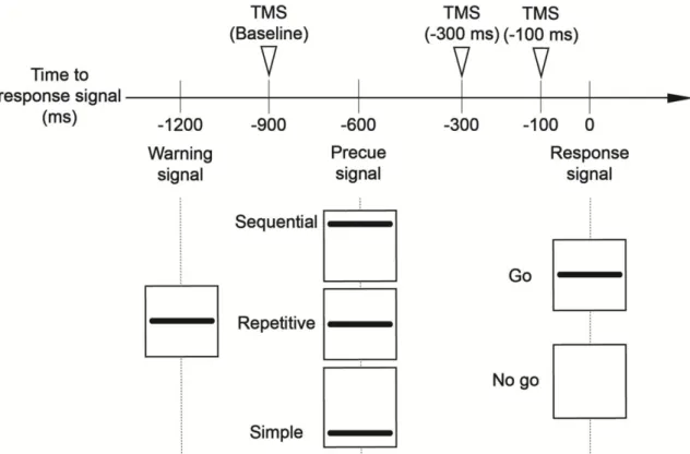
The subjects performed choice reaction time tasks. They were instructed to relax their muscles during and between the trials to prevent background muscle burst facilitating the corticospinal excitability, because MEP amplitude depended on background EMG activity of the target muscle. To make sure they really relaxed during and between trials, EMG signals were monitored online and checked visually by examiner. Three signals were presented visually at the horizontal line on the oscilloscope display (Figure 2). A warning signal indicated the start of a trial. A response signal appeared on the display 1200 ms after the warning signal. If the line of response signal was appeared at 80% probability (go trials), the subjects were instructed to perform the precued motor response as quickly and accurately as possible, or to withhold the response if the line was not (no go trials; 20% probability). These no go trials were adopted to help subjects preventing the premature motor response. A precue signal appeared 600 ms after the warning signal and was presented by one of the three lines in a trial. A precue signal appearing bottom of the display indicated that a simple motor response, to press the force transducer once with the index finger, was the to-be-signaled motor response. A precue signal appearing middle of the display indicated that a repetitive motor
response, to press the transducer with index finger three times continuously, was the to-be-signaled motor response. A precue signal appearing top of the display indicated that sequential motor response, to press the transducers sequentially with the index finger, little finger and middle finger, was the to-be-signaled motor response. This protocol was the same as the previous study25.
Each motor response had 40 trials and the experimental session consisted of total 120 trials. Each subject performed these motor responses randomly in the session. In 30 out of 40 trials of each motor response, a single TMS pulse was randomly delivered either 900 ms (baseline), 300 ms or 100 ms before response signal. In 10 out of 40 trials of the each motor response, the pulse was not delivered to measure the reaction time without TMS. Therefore, the experimental session consisted of 90 trials with TMS and 30 trials without TMS.
5. Data analysis
For the trials with TMS, the trials in which the EMG activity visually existed within 50 ms prior to the TMS pulse were excluded from the data analysis. Trials in which the onset of FDI EMG activity was earlier than the response signal, or in which a to-be-signaled motor response indicated by a precue signal was mistakenly performed, were excluded from the analysis. MEP amplitude was estimated on peak to peak basis. Background EMG amplitude was estimated from rectified EMG trace in the time window between 0 and 50 ms before TMS trigger. To control between subject variability, the MEP amplitude and background EMG amplitude were normalized to the mean MEP or EMG background amplitude at baseline respectively in each muscle. For the go trials with no TMS, reaction time, defined the time from the response signal to the onset of FDI EMG activity, were estimated. In the first experiment, it needed to be determined whether normalized MEP amplitudes or normalized background EMG amplitudes were significantly
different among the precued conditions in each TMS time and among TMS times in each precued condition, and whether the reaction times were significantly different among the precued conditions. Accordingly, multiple comparisons of simple main effects were carried out using Tukey’s multiple comparison tests. The significance level was set as 0.05 in these analyses. Data was presented as the mean values and standard error of the mean (mean± Standard Error).
Figure 1. Experimental setup. The right forearm and wrist were immobilized by a plastic splint and a Velcro strap. Force transducers were placed under the index, middle and little fingers. Surface electrodes (open circles) were placed on the FDI and the APB muscles.
Figure 2. Schematic representation of time course in an experimental trial. This Figure shows the timing of three signals (warning, precue, and response signal) and visual representation of them on the oscilloscope display. All signals appeared during 100 ms. Inverted triangles indicate the timing of a single-pulse TMS which were delivered at 900 ms (baseline), 300 ms and 100 ms before a response signal.
Ⅲ. Results
1. TMS intensity
The mean resting motor threshold corresponded 61.1±3.9% of maximum output of the magnetic stimulator. The TMS intensity for experimental sessions was 1.1 ± 0.0 times the resting motor threshold and corresponded 68.6±4.3 % of maximum output of the stimulator. 8.0% of go trials and 7.6% of no go trials were excluded from data analysis according to the exclusion criteria described in Data analysis section. Overall, 12.0% of all trials were excluded from the data analysis.
2. EMG activity and reaction time
Figure 3 shows a specimen record of the grand-averaged signals from the transducers and the rectified EMG of the FDI muscle and of the APB muscle during each motor response. The EMG bursts were present in the FDI muscle during each motor response, and the EMG bursts were almost absent in the APB muscle. Figure 4 shows the average reaction time for the simple, repetitive and sequential motor response in go trials without TMS. The average reaction time for the simple motor response was 230±15ms. The average reaction time for the repetitive motor response was 303±14ms, and that for the sequential motor response was 267±15ms. The multiple comparison test revealed that the average reaction time for the repetitive motor response was significantly longer than that for the simple motor response (p<0.05) and for the sequential motor response (p<0.05), and the reaction time for sequential motor response was longer than that for the simple motor response (p<0.05).
3. Background EMG amplitude
As shown in Figure 6, the normalized background EMG amplitudes tended to be constant throughout the time course. Multiple comparison tests
showed no significant change in the background EMG amplitudes between TMS time and between the motor responses in both muscles, suggesting that change of the MEP amplitudes observed in the first experiment was not influenced with the voluntary muscle contraction and background muscle activity.
4. Motor-evoked potentials in the FDI muscle
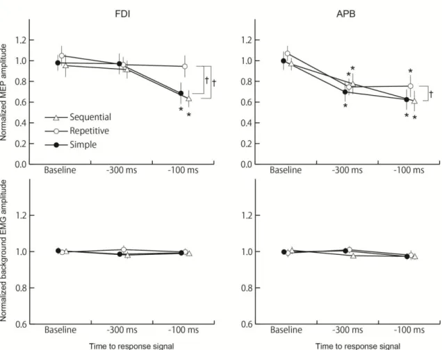
The mean MEP amplitude at baseline was 320±74μV in the FDI muscle. Figure 5 shows a specimen record of MEP of the FDI muscle or the APB muscle at baseline, 300 ms and 100 ms before a response signal when the simple, repetitive or sequential motor responses is a to-be-signaled motor response. In the FDI muscle, the MEP when the simple motor response was a to-be-signaled motor response and when the sequential motor response was a to-be-signaled motor response tended to decrease in proximity to a response signal. On the other hand, the MEP when the repetitive motor response was a to-be-signaled motor response seemed not to change through TMS time. The multiple comparison test revealed that the normalized MEP amplitudes 100 ms before a response signal significantly decreased when the precue signal indicated the simple motor response or the sequential motor response (p<0.05), while it did not significantly decreased when the precue signal indicated the repetitive motor response. In addition, the decrease of normalized MEP amplitude 100 ms before a response signal when the precue signal indicated the repetitive motor response was significantly smaller than that when the precue signal indicated the simple motor response or the sequential motor response (p<0.05) (Figure 6).
5. Motor-evoked potentials in the APB muscle
The mean MEP amplitude at baseline was 156±58μV in the APB muscle. In Figure 5, the MEP in the APB muscle tended to decrease in proximity to a response signal, and seemed to decrease earlier than the MEP in the FDI muscle. The normalized MEP amplitude 300 ms or 100 ms before a response
signal significantly decreased in all precued conditions (p<0.05) (Figure 6). In addition, multiple comparison test showed the decrease of the normalized MEP amplitude 100 ms before a response signal when the precue signal indicated the repetitive motor response was significantly smaller than that when the precue signal indicated the sequential motor response in the APB muscle (p<0.05).
Figure 3. Specimen record of the grand-averaged signals from the transducers in index, little and middle fingers, and the rectified EMGs in the FDI and the APB muscles between 100 ms before and 1000 ms after the FDI-EMG onset. Dashed vertical lines indicate the onset of EMG bursts in the FDI muscle.
Figure 4. The mean reaction time of simple, repetitive and sequential motor responses. Error bars indicate standard error. * Significant difference for the multiple comparison test; p<0.05.
Figure 5. Specimen record of MEP induced by a precue indicating the simple (―), the repetitive (╌╌) or sequential motor response (━) in the FDI muscle (upper subfigures) and the APB muscle (lower subfigures). Each MEP is between 20 ms before and 80 ms after TMS pulse which is delivered at 900 ms (baseline), 300 ms or 100 ms before a response signal. Each MEP indicates the average response of trials under each condition.
Figure 6. The mean normalized MEP and background EMG amplitudes as a function of TMS time period (Baseline, 300 ms and 100 ms before a response signal) and motor response (simple motor response, repetitive motor response and sequential motor response) in the FDI muscle and the APB muscle. The upper subfigures show mean MEP amplitudes and the lower present mean background EMG amplitudes. Error bars indicate standard error. Asterisk (*) indicates significant difference to the mean MEP amplitude at the baseline in each motor response. Dagger (†) indicates significant difference of mean MEP amplitudes between motor responses.
Ⅳ. Discussion
1. Muscle specific preparation
The MEP amplitude 100 ms before a response signal was significantly smaller than that at baseline both in the FDI muscle and the APB muscle, but the MEP amplitude 300 ms before a response signal decreased only in the APB muscle. In the first experiment, EMG bursts during motor responses were present in the FDI muscle, and they were almost absent in the APB muscle, indicating that the FDI muscle was the response related muscle, while the APB muscle was not. In the previous study, the MEP amplitude 100 ms before a response signal decreased by advance information about the thumb movement direction in both the APB and FDI muscles, but the MEP amplitude 300 ms before a response signal decreased only in the FDI muscle25
. The APB muscle was related to the thumb movement and the FDI muscle was not unrelated. These findings suggest that corticospinal suppression during the middle foreperiod is present when a to-be-signaled motor response does not involve the muscle tested.
2. Event preparation of a to-be-signaled motor response
The decrease of MEP amplitude 100 ms before a response signal when a repetitive motor response was a to-be-signaled motor response was smaller than that when a simple motor response or a sequential motor response was a to-be-signaled motor response in the FDI muscle, and was smaller than that when a sequential motor response was a to-be-signaled motor response in the APB muscle. In addition, MEP amplitude 100 ms before a response signal did not significantly decrease when a repetitive motor response was a to-be-signaled motor response only in the FDI muscle. van Elswijk and coauthors reported that the decrease of MEP amplitudes 300ms before a response signal did not depend on the movement direction of a to-be-signaled motor response, while the amplitudes 100 ms before a response signal
depended on the movement direction of the response25. These findings
indicate that corticospinal suppression during the late foreperiod is affected by advance information not only about the movement direction, but also about the sequence of a to-be-signaled motor response.
3. Complexity of the sequence of a motor response
The activities of several brain areas increase by the complexity of the sequence of a motor response. In a study using positron emission tomography study, regional cerebral blood flow (rCBF) in the ipsilateral sensorimotor cortex and the supplemental motor area was greater during complex sequential finger movements than during simple repetitive movement33.
In addition, increased rCBF in the ipsilateral premotor area, bilateral posterior parietal areas, and precuneus were related to the length of sequence34. In studies using electroencephalography, the amplitude of
Bereitschaftspotential and negative slope were larger in sequential finger movement than in simple finger movement35, 36. These findings suggest that
corticospinal suppression during the foreperiod may also depend on advance information about the complexity of the sequence of a to-be-signaled motor response.
In the first experiment, the sequence length of a repetitive motor response was longer than that of a simple motor response, and the number of fingers and finger transitions in a sequential motor response was set more than those in a repetitive motor response. Thus, most complex response among these responses applied in the first experiment must be a sequential motor response, followed in order by a repetitive motor response and a simple motor response. However, the reaction time for a repetitive motor response was significantly longer than that for a simple motor response or for a sequential motor response, suggesting that a repetitive motor response was most difficult among three motor responses during motor execution. Therefore, the modulation of corticospinal suppression induced
by a precue signal indicating a repetitive motor response is not affected by advance information about the complexity of the sequence of a to-be-signaled motor response, but may be affected by the difficulty of execution of a to-be-signaled motor response.
4. The unique characteristic of the repetitive motor response during motor preparation
Corticospinal excitability during the foreperiod was usually suppressed in previous studies19-22. On the other hand, corticospinal
excitability during the foreperiod was not suppressed when a repetitive motor response was a to-be-signaled motor response in the first experiment. Corticospinal excitability was facilitated by motor imagery of execution of a simple motor response37
. In addition, a previous study reported corticospinal excitability was also facilitated by motor imagery of execution of a sequential motor response, but it was not facilitated by motor imagery of execution of a repetitive motor response38. The neural
activity during motor imagery partly pertains to the neural activity of motor preparation39-41. These studies indicate that modulation of
corticospinal excitability is unique during motor preparation of a repetitive motor response. Accordingly, corticospinal suppression during the foreperiod when a repetitive motor response is a to-be-signaled motor response must reflect this unique characteristic of a repetitive motor response during motor preparation.
Ⅴ. Summary
Corticospinal suppression during the middle foreperiod is present when a to-be-signaled motor response does not involve the muscle tested. Corticospinal suppression during the late foreperiod is affected by advance information about the sequence of a to-be-signaled motor response.
Chapter 2: Muscle specificity of the effect of advance information about the sequence of a to-be-signaled motor response on corticospinal suppression during foreperiod
Ⅰ. Introduction
In the first experiment, the decrease of MEP amplitude 100 ms before a response signal induced by a precue signal indicating the repetitive motor response was smaller than that induced by a precue signal indicating the simple motor response and that induced by a precue signal indicating the sequential motor response in the FDI muscle, and was smaller than that induced by a precue signal indicating a sequential motor response in the APB muscle. These findings suggest that corticospinal suppression during the foreperiod is affected by advance information about the sequence of a to-be-signaled motor response. On the other hand, the decrease of the MEP amplitude 100 ms before a response signal induced by a precue signal indicating a repetitive motor response was not different from that induced by a precue signal indicating a simple motor response in the APB muscle. These findings suggest that the effect of advance information about the sequence of a to-be-signaled motor response on corticospinal suppression during the foreperiod was muscle specific when a simple motor response or a repetitive motor response is a to-be-signaled motor response.
In the first experiment, EMG bursts during a simple motor response and during a repetitive motor response were present in the FDI muscle, and were almost absent in the APB muscle, indicating that the FDI muscle took part in the simple and the repetitive motor responses and that the APB muscle did not take part in these motor responses. Therefore, I hypothesize that the effect of advance information about the sequence of a to-be-signaled motor response on corticospinal suppression during the foreperiod depends on whether the to-be-signaled response involves the muscle tested when a
simple motor response or a repetitive motor response is a to-be-signaled motor response. The second experiment was made for testing this hypothesis. On setting of the first experiment, the FDI muscle was always related to a to-be-signaled motor response because the initial element of the sequence of a to-be-signaled motor response was only index finger flexion. Accordingly, the tested muscle was set as both the muscle which was related to a to-be-signaled motor response and the muscle which was unrelated to a to-be-signaled motor response in the second experiment. The hypothesis is supported if corticospinal suppression during the foreperiod is affected by advance information about the sequence of a to-be-signaled motor response only when the to-be-signaled response involves muscle tested.
Ⅱ. Methods
1. Subjects
Eight males aged from 23 to 38 years participated in the second experiment. The subjects had no history of neurological disease. Seven out of 8 subjects were right-handed (the mean handedness score of 64.5) according to the Edinburgh Handedness inventory32. All subjects gave written
informed consent prior to the experiment, which was approved by the ethics committee of Osaka Prefecture University (Approval No. of research: 09-107).
2. Apparatus
The subject was seated, and his right forearm and hand were placed on a flat table. The forearm was pronated and the wrist joint was positioned in 0-degree extension, and the forearm and wrist were immobilized by a splint and Velcro strap (Figure 1). The fingers were comfortably extended. Force transducers were placed on the flat table under the index and little fingers to record the force of the finger tips pressing the surface of the table. The signals from the force transducers were amplified via strain amplifiers (DPM-712B, Kyowa Dengyo). An oscilloscope was placed in front of the subject. Surface electrodes recording EMG activity were placed on the belly of the FDI and the ADM muscles in the right hand, 1 cm apart. Ground electrodes were placed over the right processus styloideus and the head of the ulna. The EMG signals were amplified by an EMG amplifier (MEG-2100, Nihon Kohden) with a band-pass filter from 15 Hz to 3 kHz. The signals from the strain amplifiers and those from the EMG amplifier were converted to digital signals at a sampling rate of 5 kHz using an A/D converter (Unique Acquisition UAS3, Unique Medical) and the digital signals were stored in a personal computer.
3. Transcranial magnetic stimulation
TMS was delivered by a round coil (YM-133B, Nihon Kohden) connected to a magnetic stimulator (SMN-1200, Nihon Kohden). The coil had an outer diameter of 99 mm, and the maximum intensity of the coil was 0.96 T. The coil was placed over the vertex and moved little by little to find a hot spot where evoked MEP responses in the FDI muscle were largest. The coil was then positioned at the hot spot and the TMS intensity was decreased trial by trial to find the resting motor threshold of the MEP in the FDI muscle. The resting motor threshold was defined as the lowest stimulus intensity that produced the MEP with amplitude of at least 50 µV with 50% probability at rest. Before the experimental session, the TMS intensity at rest was adjusted to be 1.1 times the resting motor threshold. Then, if the FDI-MEP evoked by TMS at the intensity of 1.1 times the resting motor threshold exceeded 1 mV of amplitude, the intensity was decreased to the intensity at which MEPs with 1 mV of the amplitude were constantly evoked.
4. Procedure
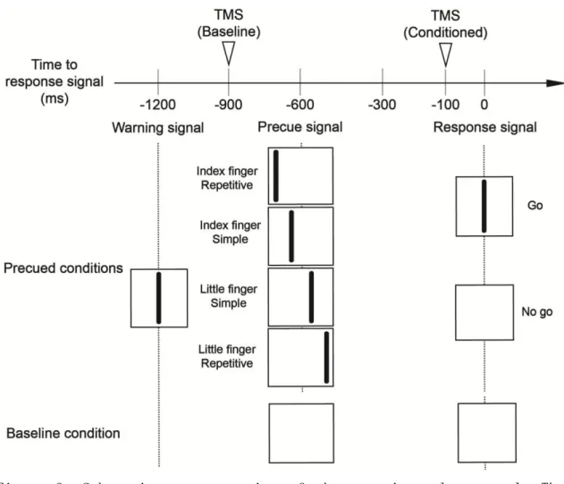
The subject performed a warned choice reaction time task with the right fingers. The subject was instructed to relax the muscles being recorded. Background EMG activities were visually monitored online. A warning signal, displayed as a vertical line, appeared on the middle of the oscilloscope display (Figure 2). A precue signal was then displayed as another vertical line on the display, appearing 600 ms after the warning signal. A precue signal appearing at the left medial side of the display indicated that a simple motor response pressing the rigid surface with the right index finger once was the to-be-signaled motor response. A precue signal appearing at the right medial side of the display indicated that a simple motor response pressing the rigid surface with the right little finger once was the to-be-signaled motor response. A precue signal appearing at the left lateral side of the display indicated that a
repetitive motor response pressing the rigid surface with the index finger three times was the to-be-signaled motor response. A precue signal appearing at the right lateral side of the display indicated that a repetitive motor response pressing the rigid surface with the little finger three times was the to-be-signaled motor response. A response signal appeared on the display 600 ms after the precue signal. The subject was instructed to perform the motor response indicated by the precue signal as quickly as possible in response to the response signal (go trial). The response signal was absent in 20% of the whole trials (no-go trial) to prevent premature motor responses. This protocol was the same as the previous study25.
Before the experimental session, subjects were trained to familiarize them with the precue-response pairs and to practice the complete muscle relaxation during trials. The experimental session consisted of total 74 trials with TMS and without TMS. Sixteen trials were conducted for each precue condition. In 10 of these 16 trials, TMS was randomly delivered 100 ms before the response signal. In the other 6 trials, TMS was not delivered in order to measure reaction time without TMS. In addition, ten trials were conducted for the baseline condition. In the baseline trials, TMS was delivered 300ms after the warning signal, and then precue and response signals were not applied to the subject.
5. Data analysis
The trials which EMG bursts were present during the foreperiod, defined as the period between a precue signal and a response signal, were excluded from the data analysis. The reaction times were estimated from the go trials without TMS. The MEP amplitude was estimated on a peak-to-peak basis. The EMG traces were rectified, and the background EMG amplitude was estimated from the rectified EMG traces in the time window between 0 and 50 ms before TMS onset. In the second experiment, it needed to be determined
whether MEP amplitudes or background EMG amplitudes were significantly different among the baseline and the precued conditions, and whether the reaction times were significantly different among the precued conditions. Accordingly, multiple a priori comparisons were carried out using Tukey’s multiple comparison tests. The alpha level was 0.05 for these statistical analyses. Data are presented as the mean values and standard error of the mean.
Figure 1. Experimental setup. The forearm and wrist were immobilized by a plastic splint with a Velcro strap. Force transducers were placed under the index and little fingers. Surface electrodes recording EMGs were placed over the belly of the FDI and ADM muscles.
Figure 2. Schematic representation of the experimental protocol. The vertical lines appeared on the oscilloscope display. A warning signal was delivered in the baseline and the precued conditions. A precue signal was delivered 600 ms after a warning signal in the precued conditions, and a response signal appeared 600 ms after the precue signal. Inverted triangles indicate the onset of TMS.
Ⅲ. Results
1. TMS intensity
Overall, 13.0% of the trials were excluded from the data analysis. The TMS intensity at the resting motor threshold was 65.4±5.3 % of maximum output in the FDI muscle. The TMS intensity for experimental sessions was 71.8 ±5.7 % of maximum output in the FDI muscle.
2. EMG bursts and reaction time
During the motor responses with the index finger, the main EMG burst was present during the simple motor response and the serial EMG bursts were present during the repetitive motor response in the FDI muscle (Figure 3). In contrast, the EMG burst was almost absent during these motor responses in the ADM muscle. These findings indicate that the FDI muscle took part in the simple and repetitive motor responses with index finger, and the ADM muscle did not take part in these motor responses with index finger. During the motor responses with the little finger, the EMG burst was absent in the FDI muscle. In contrast, the main EMG burst was present during the simple motor response and the serial EMG bursts were present during the repetitive motor response in the ADM muscle. These findings indicate that the FDI muscle did not take part in the simple and the repetitive motor responses with little finger, and the ADM muscle took part in the motor responses. The reaction time for the simple motor response with the index finger was 325± 33 ms, and that for the repetitive motor response with index finger was 291± 24 ms (Figure 4). The reaction time for the simple motor response with the little finger was 264± 33 ms, and that for the repetitive motor response with the little finger was 259±17 ms. The Multiple comparisons did not show significant difference of the reaction times among these precued conditions.
3. Background EMG and MEP amplitude in the FDI muscle
In the FDI muscle, the background EMG amplitude at the baseline condition was 46±7 μV. The background EMG amplitude before TMS trigger tended to be constant among the baseline and the precued conditions (Figure 5). The Multiple comparisons showed no significant difference of the background EMG amplitudes among the baseline and the precued conditions (Figure 6), suggesting that the MEP amplitudes observed in the FDI muscle was not facilitated by the voluntary muscle contraction and background muscle activity.
The MEP amplitude in the baseline condition was 382± 134 μV in the FDI muscle. The MEP amplitudes induced by a precue signal indicating the simple and the repetitive motor responses with the little finger seemed to be smaller than that in the baseline condition (Figure 5). The Multiple comparisons revealed that the MEP amplitude induced by a precue signal indicating the simple motor response with the little finger was smaller than that in the baseline condition (p<0.05), and revealed no significant difference of the MEP amplitudes among the precued conditions (Figure 6).
4. Background EMG and MEP amplitude in the ADM muscle
In the ADM muscle, the background EMG amplitude in the baseline condition was 85± 10 μV. The background EMG amplitude before TMS trigger tended to be constant among the baseline and the precued conditions (Figure 5). The Multiple comparisons showed no significant difference of the background EMG amplitudes among the baseline and the precued conditions (Figure 6), suggesting that the MEP amplitudes observed in the ADM muscle was not facilitated by the voluntary muscle contraction and background muscle activity.
The MEP amplitude in the baseline condition was 110± 27 μV in the ADM muscle. The MEP amplitudes in the precued conditions seemed to be
similar that in the baseline condition (Figure 5). The Multiple comparisons revealed no significant difference of the MEP amplitudes among the baseline and the precued conditions (Figure 6).
Figure 3. Specimen record of the grand-averaged signals from the transducers, and the rectified EMGs between 100 ms before and 1500 ms after EMG onset of the FDI muscle.
Figure 4. Mean reaction time for the simple or the repetitive motor response with the index finger or the little finger. Bars indicate the mean; error bars indicate the standard error.
Figure 5. Specimen record of averaged MEP responses. Traces show the MEP responses during the foreperiod in the baseline condition and the precued conditions. Each MEP response is between 50 ms before and 100 ms after TMS artifact.
Figure 6. Mean MEP amplitudes and background EMG amplitudes in the baseline and the precued conditions. The two upper graphs indicate the MEP amplitudes and the two lower graphs indicate background EMG amplitudes. Bars indicate mean; error bars indicate standard error. Asterisk indicates MEP amplitude in the baseline is significantly different from that in the precued condition (p<0.05).
Ⅳ. Discussion
In the second experiment, I investigated whether the effect of advance information about the sequence of a to-be-signaled motor response on corticospinal excitability during the foreperiod depends on involvement of the muscle tested for the to-be-signaled motor response.
1. Methodological considerations
MEP amplitude in the agonist muscle increases as closer to the onset of the muscle contraction when a subject responses to a start cue, called premovement facilitation12-18,42. Premovement facilitation may onset before
a response signal, if the subject prematurely responds. Reaction time of go trial during trials for the go/ no go reaction time task is longer than that for the simple reaction time task14, because the subject has to decide
whether producing response or not. Thus, go/ no go task was used for preventing the premature response, and the trials in which EMG onset of the FDI muscle initiated before a response signal were excluded from data analysis.
The MEP amplitude depends on background EMG activity43-45. I monitored
background EMG activity during and between trials online and the trials in which the EMG burst was present in the time window between 50 and 0 ms before TMS were visually excluded offline. Furthermore, the background EMG amplitudes were not significantly different between the baseline and the precued conditions. Thus, the MEP amplitudes must not be affected by background EMG activity.
2. Preparation of the number of fingers and the number of finger transitions during the foreperiod
In the second experiment, MEP amplitudes 100 ms before a response signal were not significantly different between the precued conditions both
in the FDI and ADM muscles. These findings rejected my hypothesis that the effect of advance information about the sequence of a to-be-signaled motor response on corticospinal suppression depends on whether the to-be-signaled motor response involves the muscle tested. This finding was conflicted with the findings in the first experiment. The sequence of the motor response has two structures; the surface structure, such as sequence length or the number of fingers, and sequence-specific structure manifested by the relations between movements, such as repetitions or transitions26.
In the first experiment, the number of fingers and the number of finger transitions were different among simple, repetitive and sequential motor responses, and the sequence of a to-be-signaled motor response was not determined before a precue signal. On the other hand, the number of fingers and the number of finger transitions were same among simple and repetitive motor responses in the second experiment, and these of a to-be-signaled motor response were determined before a precue signal. Thus, choosing the number of fingers and the number of finger transitions of a to-be-signaled motor response processes during the foreperiod in the first experiment, but it does not in the second experiment. Accordingly, corticospinal suppression during the foreperiod is affected by advance information about the sequence of a to-be-signaled motor response only when choosing the number of fingers and the number of finger transitions of a to-be-signaled motor response is present during the foreperiod.
3. Selecting the initial element during the foreperiod
In the first experiment, the initial element of the simple, repetitive and sequential motor responses was index finger flexion, and the initial element of a to-be-signaled motor response has been determined before a precue signal. On the other hand, the initial element of the simple and repetitive motor responses was index or little finger flexion, and the initial element of a to-be-signaled motor response has not been determined
before a precue signal in the second experiment. Accordingly, corticospinal suppression during the foreperiod is affected by advance information about the sequence of a to-be-signaled motor response only when the initial element of a to-be-signaled motor response is selected before the foreperiod.
4. Corticospinal excitability during foreperiod is affected by the direction of to-be-signaled motor response
Corticospinal excitability in the FDI muscle during the foreperiod was not significantly suppressed in the second experiment, but it was significantly suppressed in the first experiment, when a precue signal indicated that the simple motor response with the index finger was a to-be-signaled motor response. A previous study reported that corticospinal excitability during the foreperiod depends on the direction of a to-be-signaled motor response25. Thus, the direction of a
to-be-signaled motor response may be related to findings that corticospinal excitability in the FDI muscle during the foreperiod was suppressed in the first experiment and was not suppressed in the second experiment when a precue signal indicated that the simple motor response with the index finger was the to-be-signaled motor response. When the direction of a to-be-signaled motor response of the index finger was abduction, corticospinal excitability in the FDI muscle was suppressed during the foreperiod (Table) 24, 31, 46. On the other hand, when the direction of a
to-be-signaled motor response of the index finger was flexion, corticospinal excitability in the FDI muscle during foreperiod was various; facilitated in some studies29, 30 and suppressed in another study22. Thus, the
modulation of corticospinal excitability in the FDI muscle during the foreperiod is various when the direction of to-be-signaled motor response of the index finger is flexion.
to-be-signaled motor response, corticospinal excitability in the FDI muscle was suppressed during the foreperiod, if the finger participating to the to-be-signaled motor response had been already determined before the precue22. On the other hand, corticospinal excitability in the FDI muscle
was facilitated during the foreperiod if selection of the index finger for a to-be-signaled motor response was carried out after a precue 29, 30. Thus,
whether the finger participating to a to-be-signaled motor response has been determined before the foreperiod or not must be related to various modulation of corticospinal excitability in the FDI muscle during the foreperiod, when simple motor response by index finger flexion is the to-be-signaled motor response. However, the finger participating to a to-be-signaled motor response was determined after a precue signal in the first experiment because the finger participating to the simple motor response or repetitive motor response was different from the fingers participating to the sequential motor response, and then corticospinal excitability in the FDI muscle was suppressed during the foreperiod. In addition, choosing the finger participating to a to-be-signaled motor response was executed after a precue signal in the second experiment because the finger participating to the simple motor response or the repetitive motor response was the index finger or the little finger, and then corticospinal excitability in the FDI muscle was not suppressed during the foreperiod. Accordingly, I can not conclude that the period in which choosing the finger participating to a to-be-signaled motor response is the cause of various modulation of corticospinal excitability in the FDI muscle during the foreperiod between the first and second experiments.
5. Timing of determining the non-response finger
In the ADM muscle, any precued condition did not significantly decrease MEP amplitude 100 ms before a response signal. EMG bursts in the ADM muscle were present during little finger flexion and was not absent
during index finger flexion, indicating that the ADM muscle is involved in little finger flexion and is not involved in index finger flexion. MEP amplitude in the ADM muscle during the foreperiod when precue indicates advance information about a to-be-signaled motor response involving the target muscle has not been reported in previous studies. Accordingly, it is the original finding that MEP amplitude in the ADM muscle does not significantly decrease during the foreperiod when the muscle is involved in a to-be-signaled motor response. On the other hand, previous findings conflicted with the findings in the second experiment; MEP amplitude in the ADM muscle significantly decreased during the foreperiod when the muscle was not involved in a to-be-signaled motor response22, 30. In these
previous studies, the little finger had been determined not to take part in a to-be-signaled motor response before a precue signal. In the second experiment, the little finger had been determined not to take part in a to-be-signaled motor response during foreperiod. Thus, the difference between the previous studies and the second experiment is that whether involvement of the little finger for a to-be-signaled motor response has been determined before a precue signal or not. Accordingly, corticospinal excitability in the ADM muscle may not be suppressed during the foreperiod if involvement of the little finger for a to-be-signaled motor response has not been determined before a precue signal.
6. Conflicting findings between the first and the second experiments The effect of advance information about the sequence of a to-be-signaled motor response on corticospinal excitability during the foreperiod was not present in the second experiment, but was present in the first experiment. In addition, corticospinal excitability in the FDI muscle during the foreperiod was not significantly suppressed in the second experiment, but it was significantly suppressed in the first experiment, when a precue signal indicated that simple motor response with the index
finger was a to-be-signaled motor response. These findings suggest that some factors leads to the inconsistent findings between the first and second experiments. In the second experiment, the initial element of the simple and repetitive motor responses was the index finger movement or the little finger movement, and the initial element of a to-be-signaled motor response had not been selected before a precue signal. On the other hand, the initial element of the simple, repetitive and sequential motor responses was only the index finger movement, and the initial element of a to-be-signaled motor response had been selected before a precue signal in the first experiment. Accordingly, the findings in the first experiment must be limited to a specific condition that the initial element of a to-be-signaled motor response is selected before the foreperiod.
Ⅴ. Summary
A hypothesis that the effect of advance information about the sequence of a to-be-signaled motor response on corticospinal suppression during the foreperiod depends on whether the to-be-signaled response involves the muscle tested is rejected in the second experiment. Corticospinal suppression during the foreperiod is affected by advance information about the sequence of a to-be-signaled motor response, when choosing the number of fingers and the number of finger transitions of a to-be-signaled motor response is present during the foreperiod, or when the initial element of a to-be-signaled motor response is selected before the foreperiod.
Conclusion
Corticospinal suppression during the middle foreperiod is present when a to-be-signaled motor response does not involve the muscle tested, and when the initial element of the to-be-signaled motor response is selected before the foreperiod. Corticospinal suppression during the late foreperiod is affected by advance information about the sequence of a to-be-signaled motor response when choosing the number of fingers and the number of finger transitions of a to-be-signaled motor response is present during the foreperiod, or when the initial element of a to-be-signaled motor response is selected before the foreperiod.
References
1. Crammond DJ, Kalaska JF (1989) Neuronal activity in primate parietal cortex area 5 varies with intended movement direction during an instructed-delay period. Exp Brain Res, 76: 458-462.
2. Calton JL, Dickinson AR, Snyder LH (2002) Non-spatial, motor-specific activation in posterior parietal cortex. Nat Neurosci, 5: 580-588.
3. Riehle A, Requin J (1989) Monkey primary motor and premotor cortex: single-cell activity related to prior information about direction and extent of an intended movement. J Neurophysiol, 61:534–549.
4. Hoshi E, Tanji J (2000) Integration of target and body-part information in the premotor cortex when planning action. Nature, 408: 466-470.
5. Tanji J, Shima K (1994) Role for supplementary motor area cells in planning several movements ahead. Nature, 371: 413-416.
6. Tanji J, Evarts EV (1976) Anticipatory activity of motor cortex neurons in relation to direction of an intended movement. J Neurophysiol, 39: 1062-1068.
7. Bastian A, Schoner G, Riehle A (2003) Preshaping andcontinuous evolution of motor cortical representations during movement preparation. Eur J Neurosci, 18: 2047—2058.
8. Lu X, Ashe J (2005) Anticipatory activity in primary motor cortex codes memorized movement sequences. Neuron, 45: 967–973.