INTRODUCTION
Both hypofrontality and hyperfrontality have been reported in functional imaging studies of schizo-phrenia patients. These studies have used positron emission tomography (PET), single-photon emis-sion computed tomography (SPECT), and functional magnetic resonance imaging (fMRI) approaches.
Hypofrontality has frequently been observed in schizophrenia patients during performance of atten-tion and working memory tasks. Findings across studies have been inconsistent however, with some studies reporting reduced prefrontal cortex activa-tion (1-4). and others reporting unaltered or in-creased frontal activation (5-8). These discrepancies may be attributable to methodological differences in factors such as medication, task design, task per-formance and task requirements.
The Stroop task (9) is a useful test of selective at-tention and inhibition, and involves frontally medi-ated cognitive processes such as response inhibi-tion, interference resoluinhibi-tion, and behavioral conflict
ORIGINAL
Multi-channel near-infrared spectroscopy reveals reduced
prefrontal activation in schizophrenia patients during
performance of the kana Stroop task
Kyoko Taniguchi, Satsuki Sumitani, Yukina Watanabe, Mai Akiyama, and
Tetsuro Ohmori
Department of Psychiatry, Institute of Health Biosciences, the University of Tokushima Graduate School, Tokushima, Japan
Abstract : The purpose of the present study was to investigate the activity of frontal lobe of patients with schizophrenia during performance of two Japanese versions of the Stroop task (kana and kanji) by measuring changes in the concentration of oxygenated hemo-globin (oxyHb) with near-infrared spectroscopy (NIRS). Fourteen schizophrenia patients and 14 age- and gender-matched healthy control subjects participated in the study after giving consent. The relative changes of concentrations of oxyHb were measured by NIRS during performance of the Stroop task. Significant Stroop effects, as measured by the number of correct responses, were observed with both the kana and the kanji versions. Analysis of NIRS data revealed that the schizophrenia patients showed reduced activa-tion in the prefrontal cortex compared to healthy controls during performance of the kana Stroop task, and that both schizophrenia patients and healthy controls showed lack of activity in the prefrontal cortex during performance of the kanji Stroop task. The re-sults of the present study suggest the possibility that the kana Stroop task cause a greater Stroop effect than the kanji Stroop task, and schizophrenia patients show decreased pre-frontal vascular reactivity associated with the inhibition required during the perform-ance of the kana Stroop task. J. Med. Invest. 59 : 45-52, February, 2012
Keywords : near-infrared spectroscopy (NIRS), schizophrenia, Stroop task, hypofrontality, oxygenated hemo-globin (oxyHb)
Received for publication August 24, 2011 ; accepted September 15, 2011.
Address correspondence and reprint requests to Satsuki Sumitani, MD, PhD, Department of Psychiatry, Institute of Health Biosciences, the University of Tokushima Graduate School, 18 15 Kuramoto cho 3, Tokushima 770 8503, Japan and Fax : + 81 -88 - 633 - 7131.
resolution. Functional neuroimaging studies have found several areas of the frontal cortex that appear to be specifically activated during performance of the Stroop task.
The Stroop test has been used to compare the frontal activation patterns of schizophrenia patients with those of healthy subjects (3, 4, 8, 10-13). PET studies using the Stroop task have shown lower activities in the left paracingulate (10), left middle frontal gyri (11) or anterior cingulate region (3, 13) in schizophrenia patients. Studies with fMRI using the Stroop task have reported decreased or in-creased activation in various brain areas in schizo-phrenia patients compared with controls (4, 8). An-other fMRI study reported that the schizophrenia patients showed both decreased conflict- and error-related activity in the anterior cingulate cortex (12). The Stroop phenomenon is potentially interesting in Japanese linguistics because there exist differ-ences in the way phonogram kana and ideogram kanji are processed. Relatively little work has been done with the Stroop task in Japanese, though prior research has shown that there are differences in Stroop effects between kana and kanji. The previous study has reported that behavioral reaction time was longer in the kanji Stroop task than the kana Stroop task (14). It is necessary to study both kana and kanji of the Stroop task in the neuroimaging studies, too.
Near-infrared spectroscopy (NIRS) is an optical
imaging technique which allows non-invasive meas-urement of changes in the concentration of oxygen-ated (oxyHb) and deoxygenoxygen-ated (deoxyHb) hemo-globin in brain tissue (15). NIRS is therefore use-ful in assessing the brain function of healthy adults (16) and patients with psychiatric disorders, and has been used in several neuroimaging studies of schizophrenia patients (16-20).
In the present study, we used NIRS to examine differences between healthy subjects and schizo-phrenia patients during performance of the Stroop task. Two different writing systems are used in Japan. Japanese kana is a syllabic writing system in which each symbol represents a syllable, thus allowing direct phonetic reading, whereas Japanese kanji is a logographic writing system. A kana ver-sion and a kanji verver-sion of the Stroop task were used in the present study.
METHODS
Subjects
Fourteen patients with a DSM-IV-TR diagnosis of schizophrenia (age 29.8"5.7 years, mean"SD ; 3 males, 11 females), and 14 age-and sex-matched healthy control subjects (age 29.7"6.5 years, mean"SD ; 3 males, 11 females) were included (Table 1). The patients were outpatients or inpa-tients of the Department of Psychiatry of Tokushima
Table 1. The 2
!
2!
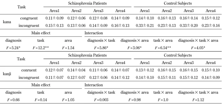
4 repeated ANOVA for changes in oxyHb concentration during performance of the kana Stroop task and the kanji Stroop task.Change in oxyHb concentration (mmol
!
mm)Task Schizophrenia Patients Control Subjects
Area1 Area2 Area3 Area4 Area1 Area2 Area3 Area4
kana congruent 0.11"0.09 0.12"0.06 0.12"0.08 0.14"0.09 0.14"0.10 0.16"0.13 0.16"0.14 0.15"0.12 incongruent 0.15"0.13 0.13"0.06 0.14"0.09 0.16"0.13 0.33"0.21 0.23"0.13 0.33"0.20 0.23"0.16
Main effect Interaction
diagnosis task area diagnosis
!
task diagnosis!
area task!
area diagnosis!
task!
areaF = 5.24* F = 12.2** F = 1.54 F = 5.86* F = 3.06* F = 6.54** F = 4.05*
Task Schizophrenia Patients Control Subjects
Area1 Area2 Area3 Area4 Area1 Area2 Area3 Area4
kanji conruent 0.12"0.07 0.14"0.04 0.11"0.06 0.14"0.07 0.13"0.12 0.16"0.15 0.16"0.15 0.15"0.10 incongruent 0.11"0.07 0.12"0.07 0.12"0.06 0.14"0.12 0.14"0.10 0.15"0.11 0.15"0.12 0.14"0.09
Main effect Interaction
diagnosis task area daignosis
!
task diagnosis!
area task!
area diagnosis!
task!
areaF = 0.66 F = 0.14 F = 1.05 F = 0.003 F = 0.98 F = 1.0 F =1.12
University Hospital. All subjects were right-handed, as assessed by the Edinburgh Handedness Inven-tory (21). The control subjects had no hisInven-tory of psychiatric or neurological disorder. All subjects completed the JART, which is a Japanese version of the NART (National Adult Reading Test), a widely used measure of premorbid IQ in English-speaking patients with dementia (22).
Schizophrenia symptoms were evaluated using the Positive and Negative Syndrome Scale (PANSS) (23). The patients had a mean total PANSS score of 46.4"7.9 (PANSS sub-scores : positive 10.9" 3.1 ; negative 11.5"2.5 ; global 24.0"4.8). All pa-tients were being treated with typical (n=2) or atypi-cal antipsychotic medication (n=9) or a combination of both (n=3) (mean chlorpromazine equivalent dose : 220 mg/day, range 100-400 mg/day) (24).
Written informed consent was obtained from all participants prior to study entry, and the study was approved by the Ethics Committee of Tokushima University Hospital.
Stroop task and procedure
The cognitive paradigm used in the present study was the Stroop task. The task was presented in a block design that consisted of rest periods and four test conditions (congruent and incongruent condi-tions in kana script, and then congruent and incon-gruent conditions in kanji script). The performance of each of the four task conditions was separated by a rest period. In the rest periods, subjects were instructed to look at a dot on the computer screen. The duration of each of the four task conditions and each rest period was 30 seconds. NIRS measure-ment was performed during the rest periods and all four task conditions. The task was presented in a block design, in which each block was separated by a rest period. Namely, the task consisted of the following sequence : rest, kana congruent condition, rest, kana incongruent condition, rest, kanji congru-ent condition, rest, and kanji incongrucongru-ent condition. During the Stroop task, subjects were asked to read as many words as possible on a computer screen displaying 100 words. In the Stroop task, subjects were randomly presented with the words ‘red’, ‘green’, ‘yellow,’ and ‘blue’ which were written in kana or kanji script and printed in red, green, yellow, or blue ink. In the congruent condition, word meaning was congruent with the color of the ink and subjects were instructed to read out the word. In the incongruent condition, the four words were printed in an incongruent color (e.g. the word
‘blue’ was printed in yellow ink). Subjects were in-structed to name, as quickly as possible, the color of the ink in which the words were printed. The investigator recorded the number of correct and incorrect verbal responses.
Near-Infrared Spectroscopy
NIRS measurement was performed using ETG4000, NIRS system with 24 optodes (Hitachi Medical Corporation, Tokyo, Japan), using two wavelengths of near-infrared light (695 and 830 nm). The absorption of light was measured, and changes in oxyHb and deoxyHb concentrations were calculated according to the Beer-Lambert law using the difference in absorption between the two wavelengths. The distance between the emission and the detector was 3.0 cm, and the machine took measurements at points located 2-3 cm beneath the scalp, i.e. on the surface of the cerebral cortices. The NIRS probes were placed symmetrically and bilaterally over the frontal region. Two plastic shells, to which 9 optodes were attached, were placed sym-metrically with one shell on the left forehead and one shell on the right forehead. The probes meas-ured the relative changes in oxyHb and deoxyHb concentrations at 12 measurement points within a 6
!
6-cm area of the left and right hemispheres, re-spectively. The lowest probes were positioned along the Fp1-Fp2 line in accordance with the Interna-tional 10/20 Electrode Placement System for elec-troencephalography. The distance from the midline to the most medial and the most lateral probes was 1.5 cm and 7.5 cm, respectively.Data analysis and statistics
The number of correct responses for each of the four task conditions was recorded for all subjects. Between-group non paired sample t-tests were performed for each of the four task conditions. Changes in oxy Hb concentration, which is the most sensitive indicator of changes in rCBF (25), were recorded from pre-task baseline. The peak change in oxyHb concentration in each task condition was recorded for each subject. Four areas were defined to allow investigation of the individual effects of the task parameters within different regions of NIRS measurement (Area 1 with channels 1,2,3,4,6, and 8 ; Area 2 with channels 5,7,9,10,11,and 12 ; Area 3 with channels 13,14,16,17,19, and 22 ; and Area 4 with channels 15,18,20,21,23,and 24) according to the method of Ehlis et al.(16) (Figure 1). The aver-age of the peak change in oxyHb concentration in
each area (Area1, Area2, Area3 and Area4) was cal-culated in each subject. Data concerning changes in OxyHb concentrations were analyzed with three way analysis of variance (ANOVA). The following variables were used : (i) “diagnosis” (control and schizophrenia), (ii) “task condition” (kana congru-ent and kana incongrucongru-ent, or kanji congrucongru-ent and kanji incongruent), and (iii) “area” (Areas 1, 2, 3, and 4). The Huynh-Feldt procedure was used to correct the degrees of freedom where necessary. Student’s t-test was used to compare behavioral and demographic data (age, education, JART, task performance). For the schizophrenia patient group, Spearman’s correlation coefficients were calculated to determine the relationship between changes in oxyHb concentration and the dose of antipsychotic medication, task performance, and PANSS score.
RESULTS
There was no significant difference between groups in the number of years of education (con-trols : 14.6"2.4 ; patients : 14.4"1.9 ; t=0.261, df = 26, p"0.05). However, a significant difference in the JART scores was observed between the two groups (controls : 103.6"6.9 ; patients : 95.9"9.6 ; t=2.419,
df =26 p!0.05). Stroop performance
There was no significant difference between groups in the performance of the kana congruent task (control : 64.14"15.0 ; patients : 63.0"9.2 ; t= 0.228, df =26, p"0.05), the kana incongruent task
( control : 36.79"8.5 ; patients : 31.43"8.0 ; t= 1.718, df =26, p"0.05), or the kanji congruent task ( control : 50.93"11.6 ; patients : 48.29"7.2 ; t= 0.725, df =26, p"0.05). In the kanji incongruent con-dition, however, controls gave more correct re-sponses than the schizophrenia patients(control : 37.71"5.7 ; patients : 32.21"6.8 ; t=2.326, df =26, p!
0.05). For both the control group and the schizo-phrenia patient group, within-subject comparisons using paired t-tests showed that the Stroop effect was significant in the kana condition (controls : t= 7.117, df =13, p!0.01 ; patients : t=9.77, df =13, p!
0.01), as indicated by a lower number of correct responses in the incongruent condition (controls : 36.8"8.5 ; patients : 31.4"8.0) than in the congru-ent condition (controls : 64.1"15.0 ; patients : 63.1" 9.2). A significant Stroop effect was also observed in the kanji Stroop task (controls : t=5.404, df =13 p!
0.01 ; patients : t=8.075, df =13, p!0.01), as indicated by a lower number of correct response in the in-congruent condition (controls : 37.8"5.7 ; patients : 32.2"6.8) than in the congruent condition (con-trols : 50.9"11.6 ; patients : 48.3"7.2).
NIRS data
No significant correlation was found between the JART score and the change in oxyHb concentration during any of the four task conditions for controls or schizophrenia patients(Spearman correlation, kana congruent : p=0.25 ; kana incongruent : p= 0.13 ; kanji congruent : p=0.84 ; kanji incongruent :
p=0.41), No significant correlation was found
be-tween the change in oxyHb concentration during any of the four task conditions and task performance
Figure 1. The optodes over the bilateral frontal region measured the relative changes in oxy Hb and deoxyHb concentrations at 12 measurement points within a 6
!
6 - cm area of the left and right hemispheres.(Spearman correlation, kana congruent : p=0.26 ; kana incongruent : p=0.28 ; kanji congruent : p= 0.95 ; kanji incongruent : p=0.68).
For the kana Stroop task, the 2
!
2!
4 repeated ANOVA for changes in oxyHb concentration re-vealed a significant main effect for the variable ‘diagnosis’ (F =5.243, df =1, p!0.05) and the vari-able ‘task’ (F = 12.228, df = 1, p!0.05).(Table 1, Figure 2).No significant main effect was found for the variable ‘area’(F = 1.544, df = 3, p"0.05). The post-hoc analysis using Bonferroni’s connection re-vealed that Schizophrenia patients showed a signifi-cantly less pronounced increase in oxyHb concentra-tion than controls (F =25.349, df =1, p!0.01). With respect to the ‘task’ there are significant differences between oxyHb concentrations in the kana congru-ent and in the kana incongrucongru-ent condition. (kana congruent!kana incongruent, F =20.329, df =1, p!0.01). The interactions ‘task
!
diagnosis’ (F =5.855,df =1, p!0.01), ‘area
!
diagnosis’ (F =3.063, df =3, p!0.05), ‘task
!
area’ (F =6.541, df =3, p!0.01), and ‘task!
area!
diagnosis’(F = 4.046, df = 3, p!0.05) were significant. The post-hoc analysis revealed that schizophrenia patients showed a significantly smaller increase in oxyHb concentration than the controls during the kana incongruent condition (F =33.249,df =1, p!0.01). The post-hoc analysis revealed that schizophrenia patients controls showed a signifi-cantly smaller increase in area1,2 and 3 than con-trols (area1 ; F = 9.505, df = 1, p!0.01, area2 ; F =
4.518, df = 1, p!0.05, area3 ; F = 12.799, df = 1, p!
0.01), but there were no significant differences be-tween controls and schizophrenia patients in area4 (F =1.648, df =1, p"0.05).
For the kanji Stroop task, the 2
!
2!
4 repeated ANOVA for changes in oxyHb concentration re-vealed no significant main effect for the variable ‘diagnosis’ (F =0.658, df =1, p"0.05) (Table 1). No significant main effect was found for the variable ‘task’(F =0.143, df =1, p"0.05) or ‘area’(F =1.048,df =2.356, p"0.05). The interactions ‘task
!
diagno-sis’ (F =0.003, df =1, p"0.05), ‘area!
diagnosis’(F= 0.975, df =2.356, p"0.05), ‘task!
area’ (F=0.996, df = 3, p"0.05), and ‘task!
area!
diagnosis’(F =1.116,df =3, p"0.05) were not significant.
In the schizophrenia patient group, no signifi-cant correlation was found between the change in oxyHb concentration and the dose of antipsychotic medication under any of the four task conditions (Spearman correlation, kana congruent : r= -0.101,
p"0.05 ; kana incongruent : r=0.779, p"0.05 ; kanji congruent : r=0.316, p"0.05 ; kanji incongruent : r= 0.348, p"0.05). No significant correlation was found between the PANSS score and the change in oxyHb concentration under any of the four task conditions for schizophrenia patients(Spearman correlation, kana congruent : r= -0.307, p"0.05 ; kana incongru-ent : r= -0.231, p"0.05 ; kanji congruent : r= -0.312,
p"0.05 ; kanji incongruent : r= -0.511, p"0.05).
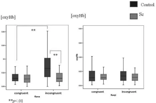
Figure 2. Left : the kana Stroop task. Right : the kanji Stroop task. Schizophrenia patients showed reduced activation in the pre-frontal cortex compared to healthy controls during performance of the kana version of the Stroop task, but not during performance of the kanji version of the Stroop task.
DISCUSSION
Regarding performance, there was no significant difference between schizophrenia patients and con-trols in the performance of the kana congruent, the kana incongruent, or kanji congruent tasks. Under the kanji incongruent condition, the controls gave more correct responses than the schizophrenia pa-tients. This result is compatible with those of pre-vious studies (26, 27), where a kanji Stroop task employing a mixture of congruent and incongruent tasks was used.
We examined activation patterns in the prefron-tal cortex during performance of a kana and a kanji version of the Stroop task. Ehlis et al. used the Stroop task in a NIRS study of the left frontal area, and demonstrated specific activation of left inferior-frontal regions in response to the Stroop interference condition (16). In the present study, increased activation was observed in the bilateral frontal cortex of healthy control subjects during per-formance of the kana Stroop task.
We also found that schizophrenia patients showed reduced activation in the prefrontal cortex compared with age- and sex-matched healthy controls during performance of the kana Stroop task. It has been suggested that differences in task performance may be a confounding factor in studies of schizophrenia. Several studies have reported that schizophrenia pa-tients perform poorly in the alphabet or kanji Stroop task in comparison to healthy controls (4, 21, 28). In the present study, we observed no significant differences between healthy subjects and schizo-phrenia patients in performance of the kana Stroop task, probably because the patients were mild cases with relatively high levels of intelligence who were receiving maintenance treatment. Our results sug-gest that there were significant differences between the bilateral frontal lobes of the schizophrenia pa-tients and those of the controls, even though no differences in task performance were observed.
Pervious neuroimaging studies have reported that schizophrenia patients show altered brain activation during performance of the Stroop task (3, 4, 13). Using a【15
O】H2O positron emission tomography
(PET) approach, Carter et al. demonstrated failure of activation of the anterior cingulate gyrus in schizo-phrenia patients during a single-trial Stroop task (13). In a PET study, Yücel et al. found that activa-tion occurred in both the limbic and the paralimbic anterior cingulate regions in healthy subjects during performance of the Stroop task, but that activation
only occurred in the paralimbic region in schizo-phrenia patients (3). A fMRI study by Weiss et al. reported reduced activation in the dorsolateral pfrontal, the anterior cingulate, and the parietal re-gions in schizophrenia patients compared to healthy controls during performance of a modified Stroop task (4). The results of the present NIRS study are consistent with those of the fMRI study of Weiss et
al. NIRS studies that have used alternative
atten-tion and executive tasks have reported hypofron-tality in schizophrenia patients (18, 20). The pre-sent results are also consistent with other NIRS studies using a different task. Ehlis et al. studied the left frontal area with NIRS and found significantly reduced activation in schizophrenia patients com-pared with healthy controls during performance of two verbal fluency tasks (18). Likewise, using 52-channel NIRS, Takizawa et al. demonstrated that schizophrenia patients had a slower and less pro-nounced increase in the bilateral prefrontal activa-tion than healthy controls during performance of a verbal fluency task (20). We found a significant dif-ference in OxyHb concentrations between schizo-phrenia patients and controls during performance of only kana but not kanji Stroop task. No differences in oxyHb concentration between control and patients during the performance of the kanji Stroop task are consistent with previous studies that used the kanji Stroop task (26, 27). The greater reduction of the number of correct answer in kana than kanji Stroop task suggests that the kana Stroop task may require more inhibition not to read kana letters. Schizophre-nia patients may have decreased prefrontal vascular reactivity associated with the inhibition required dur-ing the performance of the kana Stroop task.
The results of the present study should be inter-preted with caution. The sample size was small and there were an unequal number of male and fe-male subjects. Since men and women may have a different lateralization pattern, further studies are needed to examine differences of lateralization pat-tern between female and male subjects. The de-creased activation observed during the kana Stroop task in schizophrenia patients in the present study was therefore unlikely to have been due to a hy-perperfusion state at pre-task baseline. It is also possible that antipsychotic medication may have had an influence on our findings, although no sig-nificant correlation was found between the mean of the peak change in oxyHb concentration and an-tipsychotic dosages during any of the four Stroop task conditions. In the present study, a significant
difference in the JART scores was also observed between the two groups. It is also possible that the differences in intelligence estimated by JART may have influenced our findings, although no signifi-cant correlation was found between the mean of the peak change in oxyHb concentration and the JART score.
In contrast to the kana Stroop test, no increase in activation occurred during performance of the kanji incongruent task compared with the kanji congruent task in either controls or schizophrenia patients. Yamada et al. have established that the kanji script has a much stronger connection to se-mantics than to phonology (29), and thus less inter-ference occurs with use of the kanji Stroop task than with the kana version. It is possible that activation of increased blood flow is not necessary for perform-ance of the kanji incongruent Stroop task.
In summary, the change in oxyHb concentration during performance of the Stroop task was signifi-cantly less in schizophrenia patients than in healthy control subjects. The results of the present study suggest the existence of prefrontal dysfunction in patients with schizophrenia.
CONFLICT OF INTEREST
None of the authors have any conflicts of interest to declare.
ACKNOWLEDGEMENTS
This work was supported by a Grants-in-Aid for Scientific Research(C)No.20591410 from the Japa-nese Ministry of Education, Culture, Sports, Science and Technology.
REFERENCES
1. Callicott JH, Ramsey NF, Tallent K, Bertolino A, Knable MB, Coppola R, Goldberg T, van Gelderen P, Mattay VS, Frank JA, Moonen CT, Weinberger DR : Functional magnetic reso-nance imaging brain mapping in psychiatry : methodological issues illustrated in a study of working memory in schizophrenia. Neuropsy-chopharmacology 18 : 186-196, 1998
2. Curtis VA, Bullmore ET, Brammer MJ, Wright IC, Williams SC, Morris RG, Sharma TS,
Murray RM, McGuire PK : Attenuated frontal activation during a verbal fluency task in pa-tients with schizophrenia. Am J Psychiatry 155 : 1056-1063, 1998
3. Yücel M, Pantelis C, Stuart GW, Wood SJ, Maruff P, Velakoulis D, Pipingas A, Crowe SF, Tochon-Danguy HJ, Egan GF : Anterior cingu-late activation during Stroop task performance : a PET to MRI coregistration study of individual patients with schizophrenia. Am J Psychiatry 159 : 251-254, 2002
4. Weiss EM, Siedentopf C, Golaszewski S, Mottaghy FM, Hofer A, Kremser C, Felber S, Fleischhacker WW : Brain activation patterns during a selective attention test--a functional MRI study in healthy volunteers and unmedi-cated patients during an acute episode of schizo-phrenia. Psychiatry Research : Neuroimaging 154 : 31-40, 2007
5. Callicott JH, Bertolino A, Mattay VS, Langheim FJ, Duyn J, Coppola R, Goldberg TE, Weinberger DR : Physiologicaldysfunction of the dorsolateral prefrontal cortex in schizo-phrenia revisited. Cerebral Cortex 10 : 1078-1092, 2000
6. Manoach DS, Press DZ, Thangaraj V, Searl MM, Goff DC, Halpern E, Saper CB, Saper CB, Warach S : Schizophrenic subjects activate dor-solateral prefrontal cortex during a working memory task, as measured by fMRI. Society of Biological Psychiatry 45 : 1128-1137, 1999 7. Manoach DS, Gollub RL, Benson ES, Searl
MM, Goff DC, Halpern E, Saper CB, Rauch SL : Schizophrenic subjects show aberrant fMRI activation of dorsolateral prefrontal cor-tex and basal ganglia during working memory performance. Society of Biological Psychiatry 48 : 99-109, 2000
8. Weiss EM, Golaszewski S, Mottaghy FM, Hofer A, Hausmann A, Kemmler G, Kremser C, Brinkhoff C, Felber SR, Fleischhacker WW : Brain activation patterns during a selective at-tention test-a functional MRI study in healthy volunteers and patients with schizophrenia. Psychiatry Research 123 : 1-15, 2003
9. Stroop JR : Studies of interference in serial ver-bal reactions. Journal of Experimental Psy-cholgy 18 : 643-662, 1935
10. Yücel M, Brewer WJ, Harrison BJ, Fornito A, O’Keefe GJ, Olver J, Scott AM, Eagan GF, Velakoulis D, Pantelis C : Anterior cingulate activation in antipsychotic-naïve first-episode
schizophrenia. Acta Psychiatrica Scandinavica 115 : 155-118, 2007
11. Harrison BJ, Yücel M, Shaw M, Brewer WJ, Nathan PJ, Strother SC, Olver JS, Eagan GF, Velakoulis D, McGorry PD, Pantelis C : Dys-function of dorsolateral prefrontal cortex in antipsyachotic-naïve schizophreniform psycho-sis. Psychiatry Research : Neuroimaging 148 : 23-31, 2006
12. Kerns JG, Cohen JD, MacDonald III AW, Johnson MK, Stenger VA, Aizenstein H, Carter CS : Decreased conflict- and error-related ac-tivity in the anterior cingulate cortex in subjects with schizophrenia. Am I Psychiatry 162 : 1833-1839, 2005
13. Carter CS, Mintum M, Nicholos T, Cohen JD : Anterior cingulate gyrus dysfunction and selec-tive attention deficits in schizophrenia : [15O] H
2O
PET study during single-trial Stroop task per-formance. Am J Psychiatry 154 : 1670-1675, 1997 14. Morikawa Y : Stroop phenomena in the Japa-nese language : The case of ideographic char-acters(kanji) and syllabic characters(kana). Perceptual and Motor Skills 53 : 67-77, 1981 15. Jöbsis FF : Non-invasive, infrared monitoring
of cerebral and myocardial oxygen sufficiency and circulatory parameters. Science 198 : 1264-1267, 1977
16. Ehlis AC, Herrmann MJ, Wagener A, Fallgatter AJ : Multi-channel near-infrared spectroscopy detects specific inferior-frontal activation dur-ing incongruent Stroop trials. Biological Psy-chology 69 : 315-331, 2005
17. Shinba T, Nagano M, Kariya N, Ogawa K, Shinozaki T, Shimosato S, Hoshi Y : Near-infrared spectroscopy analysis of frontal lobe dysfunction in schizophrenia. Biological Psy-chiatry 55 : 154-164, 2004
18. Ehlis AC, Herrmann MJ, Plichta MM, Fallgatter AJ : Cortical activation during two verbal fluency tasks in schizophrenic patients and healthy controls as assessed by multi-channel near-infrared spectroscopy. Psychiatry Research : Neuroimaging 156 : 1-13, 2007 19. Hoshi Y, Shinba T, Sato C, Doi N : Resting
hy-pofrontality in schizophrenia : A study using near - infrared time - resolved spectroscopy. Schizophrenia Research 84 : 411-420, 2006 20. Takizawa R, Kasai K, Kawakubo Y, Marumo
K, Kawasaki S, Yamasue H, Fukuda M : Re-duced frontopolar activation during verbal flu-ency task in schizophrenia : A multi-channel
near-infrared spectroscopy study. Schizophre-nia Research 99 : 250-262, 2008
21. Oldfield RC : The assessment and analysis of handedness : the Edinburgh inventory. Neu-ropsychologia 9 : 97-113, 1971
22. Matsuoka K, Uno M, Kasai K, Koyama K, Kim Y : Estimation of premorbid IQ in indi-viduals with Alzheimer’s disease using Japa-nese ideographic script (Kanji) compound words : a Japanese version of NART. Psychiatry and Clinical Neurosciences 60 : 332-339, 2006 23. Kay SR, Fiszbein A, Opler LA : The positive and negative syndrome scale (PANSS) for schizo-phrenia. Schizophrenia Bulletin 13 : 261-276, 1987
24. American Psychiatric Association : Diagnostic and Statistical Manual of Mental Disorders, 4th Ed. American Psychiatric Press, Washington, DC. 1994
25. Hoshi Y, Oda I, Wada Y, Ito Y, Yamashita Y, Oda M, Ohta K, Yamada Y, Tamura M : Visu-ospatial imagery is a fruitful strategy for the digit span backward task : A study with near-infrared optical tomography. Cognitive Brain Research 9 : 339-342, 2000
26. Ikezawa K, Iwase M, Ishii R, Azechi M, Ganuet L, Ohi K, Yasuda Y, Iike N, Kurimoto R, Takahashi H, Nakahachi T, Sekiyama R, Yoshida T, Kazui H, Hashimoto R, Takeda M : Impaired regional hemodynamic response in schizophrenia during multiple prefrontal acti-vation tasks : A two-channel near-infrared spec-troscopy study. Schizophrenia Research 108 : 93-103, 2009
27. Azechi M, Iwase M, Ikezawa K, Takahashi H, Gaunet L, Kurimoto R, Nakahachi T, Ishii R, Fukumoto M, Ohi K, Yasuda Y, Kazui H, Hashimoto R, Takeda M : Discriminant analysis in schizophrenia and healthy subjects using prefrontal activation during frontal lobe tasks : A near-infrared spectroscopy. Schizophrenia Research 117 : 52-60, 2010
28. Matsuzawa D, Obata T, Shirayama Y, Nonaka H, Kanazawa Y, Yoshitome E, Takanashi J, Matsuda T, Shimizu E, Ikehira H, Iyo M, Hashimoto K : Negative correlation between brain Glutathione level and negative symptoms in schizophrenia : A 3T1H-MRS study. PLoS
ONE 3(4) : e1944, 2008
29. Yamada J, Kanamoto Y, Morita A : Japanese kanji as a semantically based orthography. Psy-chological Reports 84 : 637-642, 1999