-
12
-Effects of Zn
2+chelators, DTPA and TPEN, and ZnCl
2
on the cells treated
with hydrogen peroxide: a flow-cytometric study using rat thymocytes
Hiroko Matsui
a, Yoko Sakanashi
a, Yumiko Nishimura
b, Takuya Kawanai
c,
Yasuo Oyama
c,*, Shiro Ishida
d, Yoshiro Okano
da Faculty of Dental Science, Kyushu University, Fukuoka 812-8582, Japan b Okayama University Dental School, Okayama 700-8525, Japan
c Faculty of Integrated Arts and Sciences, The University of Tokushima, Tokushima 770-8502, Japan d Faculty of Pharmaceutical Sciences, Tokushima Bunri University, Tokushima 770-8512, Japan
* Corresponding author: Yasuo Oyama, Ph.D. E-mail: oyama@ias.tokushima-u.ac.jp
__________________________________________________________________________________________ ABSTRACT
Recently, we have revealed that trace Zn2+ partly attenuates Ca2+-dependent cell death induced by A23187, a calcium ionophore, in rat thymocytes. In this study, to see if Zn2+ attenuates the H
2O2-induced cell death that is also Ca2+-dependent, the effects of ZnCl
2 and chelators for Zn2+ have been examined by using a flow-cytometer with propidium iodide. The incubation with H2O2 increased the cell lethality. Simultaneous application of ZnCl2 greatly augmented the H2O2-induced increase in lethality. DTPA, a chelator for extracellular Zn2+, did not affect the increase in cell lethality by H
2O2. However, the H2O2-induced increase in cell lethality was greatly attenuated by TPEN, a chelator for extracellular and intracellular Zn2+. Taken together, it may be likely that intracellular Zn2+ modifies the H
2O2-induced cytotoxicity. However, it cannot be ruled out the possibility that TPEN chelates intracellular Fe2+, resulting in inhibiting the formation of hydroxyl radical from H2O2 that leads to an attenuation of H2O2 cytotoxicity.
Keywords: TPEN; DTPA; zinc; hydrogen peroxide; cytotoxicity
__________________________________________________________________________________________ 1. INTRODUCTION
Cell death induced by hydrogen peroxide (H2O2) in rat thymocytes is dependent on Ca2+ (Sakanashi et al., 2008). Thus, the application of H2O2 increases intracellular Ca2+ concentration and the removal of external Ca2+ significantly attenuates the H
2O2 -induced cell death (Okazaki et al., 1996; Nishizaki et al., 2003; Sakanashi et al., 2008). Sustained increase in intracellular Ca2+ concentration triggers either apoptotic or necrotic cell death (McConkey et al., 1989; Azmi et al., 1996; Berridge et al., 1998; Orrenius et al., 2003).
Recently, we have revealed that trace Zn2+ partly attenuates Ca2+-dependent cell death induced by A23187, a calcium ionophore, in rat thymocytes (Sakanashi et al., 2009). Zinc itself modifies or induces cell death in a concentration-dependent manner (MaCabe et al., 1993; Jiang et al., 1995; Iguchi et al., 1998; Kolenko et al., 2001;
Truong-Tran et al., 2001). Furthermore, zinc supplementation decreases oxidative stress induced by several types of compounds (DiSilvestro, 2000; Bao et al., 2008; Szuster-Ciesielska et al., 2009; Varghese et al., 2009). Therefore, in this study, to see if Zn2+ attenuates the H2O2-induced cell death, the effects of ZnCl2 and chelators for Zn2+, N,N,N',N'-tetrakis(2-pyridylmethyl)ethylenediamine (TPEN) and diethylenetriamine-N,N,N',N",N"- pentaacetic acid (DTPA), on the cell death of rat thymocytes induced by H2O2 have been examined by using a flow-cytometer with propidium iodide.
Rat thymocytes were used for present study because of following reasons. First, the cell membranes of thymocytes remain intact because single cells can be prepared without enzymatic treatment. Second, the process of cell death is extensively studied in murine thymocytes (McConkey et al., 1994; Tiso et al., 1995; Rinner et al., 1996; Winoto, 1997; Rennecke et al., 2000; Quaglino and
Ronchetti, 2001; Ortiz et al., 2001; Thompson et al., 2003).
2. MATERIALS AND METHODS
2.1. Chemicals
H2O2 was purchased from Sumitomo Chemical Co. (Osaka, Japan). So-called chelators for Zn2+, TPEN and DTPA, were obtained from Dojin Chemical Laboratory (Kumamoto, Japan). Propidium iodide was supplied from Molecular Probes Inc. (Eugene, Oregon, USA). Other chemicals (NaCl, CaCl2, MgCl2, KCl, glucose, HEPES, NaOH, and ZnCl2) were also purchased from Woko Pure Chemicals. 2.2. Animals and cell preparation
This study was approved by the Committee for Animal Experiments in the University of Tokushima (No. 05279 for Y. Oyama).
The procedure to prepare cell suspension was similar to that previously reported (Chikahisa and Oyama, 1992; Chikahisa et al., 1996). In brief, thymus glands dissected from ether-anesthetized rats were sliced at a thickness of 400-500 µm with razor under an ice-cold condition (1-4ºC). The slices were triturated by gently shaking in chilled Tyrode's solution (in mM: NaCl 150, KCl 5, CaCl2 2, MgCl2 1, glucose 5, HEPES 5, with an appropriate amount of NaOH to adjust pH to 7.3-7.4) to dissociate thymocytes. Thereafter, the Tyrode's solution containing the cells was passed through a mesh (a diameter of 10 µm) to prepare the cell suspension (about 5 ¥ 105 cells/ml). The beaker containing the cell suspension was water-bathed at 36ºC for 1 hr before the experiment.
2.3. Experimental protocol
H2O2 was added to cell suspension (2 ml cell suspension in 10 ml test tube). To examine the effects of ZnCl2, DTPA, and TPEN, the agents were respectively added to the suspension just before applying H2O2. The cell density was about 5 ¥ 105 cells/mL. The cells were incubated with respective agent and hydrogen peroxide at 36°C for 2 hr under room air condition. The data acquisition of fluorescence from 2 ¥ 103 cells by a flow cytometer required 10 sec at least.
2.4. Fluorescence measurements of cellular and membrane parameters
The method for measurement of cellular and membrane parameters, including forward scatter and side scatter, using a flow cytometer equipped with an argon laser (CytoACE-150, JASCO, Tokyo, Japan)
and fluorescent probe was similar to those previously described (Chikahisa and Oyama, 1992; Chikahisa et al., 1996). The fluorescence was analyzed by JASCO software (Ver.3XX, JASCO). As to chemicals used in this study, there was no fluorescence detected under our experimental condition.
To assess cell lethality, propidium iodide was added to cell suspension to achieve a final concentration of 5 µM. Since propidium stains dead cells, the measurement of propidium fluorescence from cells provides a clue to estimate the lethality. The fluorescence was measured at 2 min after the application of propidium iodide by a flow cytometer. Excitation wavelength for propidium was 488 nm and emission was detected at 600 ± 20 nm.
2.5. Statistics
Values were expressed as the mean ± standard deviation of 4 experiments. Statistical analysis was performed by using Tukey multivariate analysis. A P value of < 0.05 was considered significant.
3. RESULTS
3.1. Change of H2O2-induced increase in cell lethality
of rat thymocytes by ZnCl2, DTPA, or TPEN
There was no change in cell lethality of rat thymocytes after 2-3 h incubation with 10 µM ZnCl2, 10 µM DTPA, 10 µM TPEN, or 0.1 % DMSO as a solvent for TPEN. The application of 10 mM H2O2 time-dependently increased the population of cells exerting propidium fluorescence, presumably dead cells, indicating an increase in cell lethality. The cell lethality at 2 h after the start of H2O2 application ranged from 24.6 % to 44.0 %. Since prolonged incubation of cells with 10 mM H2O2 further increased the cell lethality, the values of cell lethality did not reach a steady state. DMSO at 0.1 % did not affect the H2O2-induced increase in cell lethality in the case of 2 h incubation.
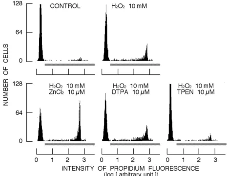
As shown in Fig. 1, the incubation with 10 mM H2O2 for 2 h increased the population of cells exerting propidium fluorescence. Simultaneous application of 10 µM ZnCl2 greatly augmented the H2O2-induced increase in the population (Fig. 1). DTPA at 10 µM, probably chelating extracellular Zn2+, did not affect the increase in cell lethality by 10 mM H2O2. However, the H2O2-induced increase in the population was greatly attenuated by 10 µM TPEN, probably chelating extracellular and intracellular Zn2+ (Fig. 1). It is noted that normal Tyrode's solution with rat thymocytes contains 200-300 nM Zn2+ derived from the cells (Sakanashi et al., 2009). Results are summarized in Fig. 2. ZnCl2 significantly augmented the cytotoxicity of H2O2
- 14
-while TPEN, but not DTPA, significantly attenuated it (Fig. 2). Thus, one may suggest that intracellular
Zn2+ has an essential role in H
2O2-induced increase in lethality.
Fig.1. Effects of ZnCl2, DTPA, and TPEN on H2O2-induced increase in population of cells exerting
propidium fluorescence. Each histogram was constructed from 2 ¥ 103 cells. Bar under the
histogram indicates the region of cells exerting propidium fluorescence, dead cells.
Fig. 2. Cell lethality of H2O2-treated cells in simultaneous presence of ZnCl2, DTPA, or TPEN.
Column and bar respectively indicate mean and standard deviation of four experiments. Symbol (##) near column indicates significant change (P < 0.01) to control group (CONTROL). Asterisks (* and **) show significant difference (P < 0.05 and P < 0.01, respectively) to the group of cells treated with H2O2 alone. It is noted that the difference
3.2. Change of H2O2-induced increase in population
of shrunken cells by ZnCl2, DTPA, or TPEN
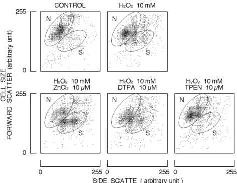
Cytogram (forward scatter versus side scatter) was also affected by the incubation of cells with 10 mM H2O2 for 2 h. As shown in Fig. 3, the population of area S increased in the presence of H2O2. Simultaneous application of 10 mM H2O2 and 10 µM ZnCl2 further increased the population of area S while 10 µM TPEN significantly attenuated the
H2O2-induced increase in the population (Fig. 3). DTPA at 10 µM did not significantly affect the population changed by 10 mM H2O2. Results are summarized in Fig. 4. ZnCl2 significantly augmented the H2O2-induced change in cytogram while TPEN significantly attenuated it. DTPA did not affect the H2O2-induced change. Therefore, it is likely that the cell shrinkage is well-associated with the increase in cell lethality.
Fig. 3. Effect of H2O2 on cytogram (forward scatter versus side scatter) of rat thymocytes in simultaneous
application of ZnCl2, DTPA, or TPEN. Each cytogram was constructed with 2 ¥ 103cells. Areas N and S
indicate normal size cells and shrunken cells, respectively.
Fig. 4. Proportion of shrunken cells in the cells treated with H2O2 in simultaneous presence of ZnCl2, DTPA,
or TPEN. Column and bar respectively indicate mean and standard deviation of four experiments. Symbol (##) near column indicates significant change (P < 0.01) to the control group (CONTROL). Asterisk (**) shows significant difference (P < 0.01) to the group of cells treated with H2O2 alone.
- 16
-4. DISCUSSION
DTPA is a chelator for extracellular Zn2+ while TPEN is one for both extracellular and intracellular Zn2+. Therefore, it is supposed that chelating intracellular Zn2+ is essential in attenuation of H
2O2 -induced cytotoxicity. The application of ZnCl2 increases intracellular Zn2+ concentration (Matsui et al., 2008). Thus, ZnCl2 may augment H2O2-induced cytotoxicity by increasing intracellular Zn2+ concentration. Therefore, it may be presumably suggested that intracellular Zn2+ augments H
2O2 -induced cytotoxicity. Although this suggestion well corresponds to the results in this study, it is challenged by previous findings. First, the cell death induced by H2O2 in rat thymocytes is significantly dependent on Ca2+ (Okazaki et al., 1996; Nishizaki et al., 2003; Sakanashi et al., 2008). Second, TPEN potentiates the Ca2+-dependent cytotoxicity of A23187 (Sakanashi et al., 2009). Thus, it is also supposed that the application of TPEN augments the cytotoxicity of H2O2 because the H2O2 cytotoxicity is Ca2+-dependent. However, the cytotoxicity of H2O2 is dependent on Fe2+, rather than Ca2+ (Walker and Shah, 1991; Hiraishi et al., 1993; Byler et al., 1994; Lomonosova et al., 1998). The combination of H2O2 and Fe2+ generates hydroxyl radical by Fenton reaction. Hydroxyl radical increases intracellular Ca2+ concentration (Dreher and Junod, 1995; Burlando and Viarengo, 2005). TPEN can chelate not only Zn2+ but also Fe2+. Therefore, it is supposed that TPEN can suppress the generation of hydroxyl radicals by chelating intracellular Fe2+,
resulting in attenuation of H2O2-induced cytotoxicity. Thus, the role of intracellular Zn2+ in H
2O2 cytotoxicity cannot be strongly addressed at present although TPEN is known to be a chelator for intracellular Zn2+.
The H2O2-induced increase in cell lethality was augmented by adding ZnCl2 (Figs. 1 and 2). In present study, we did not monitor the ZnCl2-induced change in intracellular Zn2+ concentration during H2O2 exposure. However, the addition of ZnCl2 may further elevate intracellular Zn2+ concentration since the application of H2O2 increases intracellular Zn2+ concentration (Hashimoto et al., 2009). Excess increase in intracellular Zn2+ concentration leads to cell death (Kim et al., 1999; Hamatake et al., 2000; Iitaka et al., 2001). In this aspect, further study will be necessary to elucidate the role of intracellular Zn2+ in the augmentation of H2O2 cytotoxicity.
As to the population of shrunken cells (Figs. 3 and 4), one of parameters during an early stage of apoptosis, TPEN almost completely suppressed the H2O2-induced increase in shrunken cell population while ZnCl2 greatly increased the population. Cell shrinkage is associated with an activation of Ca2+ -dependen K+ channels in rat thymocytes (Horimoto et al., 2006). Therefore, in the case of simultaneous presence of TPEN, it is unlikely that H2O2 increases intracellular Ca2+ concentration, as suggested above. On the other hand, the combination of ZnCl2 and H2O2 may further increase not only intracellular Zn2+ but also intracellular Ca2+ concentration. In this aspect, further studies on intracellular Ca2+ and Zn2+ concentrations will be required.
REFERENCES
Azmi, S., Dhawan, D., Singh, N., 1996. Calcium ionophore A 23187 induces apoptotic cell death in rat thymocytes. Cancer Lett. 107, 97-103.
Bao, B., Prasad, A.S., Beck, F.W., Snell, D., Suneja, A., Sarkar, F.H., Doshi, N., Fitzgerald, J.T., Swerdlow, P., 2008. Zinc supplementation decreases oxidative stress, incidence of infection, and generation of inflammatory cytokines in sickle cell disease patients. Transl. Res. .152, 67-80. Berridge, M.J., Bootman, M.D., Lipp, P., 1998.
Calcium – A life and death signal. Nature (London) 395, 645-648.
Burlando, B., Viarengo, A., 2005. Ca2+ is mobilized by hydroxyl radical but not by superoxide in RTH-149 cells: the oxidative switching-on of Ca2+ signaling. Cell Calcium. 38, 507-513.
Byler, R.M., Sherman, N.A., Wallner, J.S., Horwitz, L.D., 1994. Hydrogen peroxide cytotoxicity in cultured cardiac myocytes is iron dependent. Amer. J. Physiol. Heart. Circ. Physiol. 266, H121-H127.
Chikahisa, L., Oyama, Y., 1992. Tri-n-butyltin
increases intracellular Ca2+ in mouse thymocytes: a flow-cytometric study using fluorescent dyes for membrane potential and intracellular Ca2+. Pharmacol. Toxicol. 71, 190-195.
Chikahisa, L., Oyama, Y., Okazaki, E., Noda, K., 1996. Fluorescent estimation of H2O2-induced changes in cell viability and cellular nonprotein thiol level of dissociated rat thymocytes. Jpn. J. Pharmacol. 71, 299-305.
Di Silvestro, R.A., 2000. Zinc in relation to diabetes and oxidative disease. J Nutr. 130, 1509S-1511S. Dreher, D., Junod, A.F., 1995. Differential effects of
superoxide, hydrogen peroxide, and hydroxyl radical on intracellular calcium in human endothelial cells. J. Cell Physiol. 162, 147-153. Hamatake, M., Iguchi, K., Hirano, K., Ishida, R.,
2000. Zinc induces mixed types of cell death, necrosis, and apoptosis, in molt-4 cells. J. Biochem. 128, 933-939.
Hashimoto, E., Oyama, T.B., Oyama, K., Nishimura, Y., Oyama, T.M., Ueha-Ishibashi, T., Okano, Y., Oyama, Y.. 2009. Increase in intracellular Zn2+ concentration by thimerosal in rat thymocytes: Intracellular Zn2+ release induced by oxidative
stress. Toxicol. In Vitro. doi:10.1016/j.tiv.2009.05.020
Hiraishi, H., Terano, A., Razandi, M., Sugimoto, T., Harada, T., Ivey, K.J.. 1993. Role of iron and superoxide in mediating hydrogen peroxide injury to cultured rat gastric cells. Gastroenterol. 104, 780-788.
Horimoto, K., Nishimura, Y., Oyama, T.M., Onoda, K., Matsui, H., Oyama, T.B., Kanemaru, K., Masuda, T., Oyama, Y., 2006. Reciprocal effects of glucose on the process of cell death induced by calcium ionophore or H2O2 in rat lymphocytes. Toxicol. 225, 97-108.
Iguchi, K., Hamatake, M., Ishida, R., Usami, Y., Adachi, T., Yamamoto, H., Koshida, K., Uchibayashi, T., Hirano, K., 1998. Induction of necrosis by zinc in prostate carcinoma cells and identification of proteins increased in association with this induction. Eur. J. Biochem. 253, 766-770.
Iitaka, M., Kakinuma, S., Fujimaki, S., Oosuga, I., Fujita, T., Yamanaka, K., Wada, S., Katayama, S., 2001. Induction of apoptosis and necrosis by zinc in human thyroid cancer cell lines. J. Endocrinol. 169, 417-424.
Jiang, S., Chow, S.C., McCabe, M.J.Jr., Orrenius, S., 1995. Lack of Ca2+ involvement in thymocyte apoptosis induced by chelation of intracellular Zn2+. Lab. Invest. 73, 111-117.
Kim, E.Y., Koh, J.Y., Kim, Y.H., Sohn, S., Joe, E., Gwag, B.J., 1999. Zn2+ entry produces oxidative neuronal necrosis in cortical cell cultures. Eur. J. Neurosci. 11, 327-334.
Kolenko, V.M., Uzzo, R.G., Dulin, N., Hauzman, E., Bukowski, R., Finke, J.H., 2001. Mechanism of apoptosis induced by zinc deficiency in peripheral blood T-lymphocytes. Apoptosis 6, 419-429. Lomonosova, E.E., Kirsch, M., De Groot, H., 1998.
Calcium vs. iron-mediated processes in hydrogen peroxide toxicity to 1929 cells: Effects of glucose. Free Radic. Biol. Med. 25, 493-503.
Matsui, H., Sakanashi, Y., Oyama, T.M., Oyama, Y., Yokota, S., Ishida, S., Okano, Y., Oyama, T.B., Nishimura, Y., 2008. Imidazole antifungals, but not triazole antifungals, increase membrane Zn2+ permeability in rat thymocytes Possible contribution to their cytotoxicity. Toxicol. 248, 142-150.
McCabe, M.J.Jr., Jiang, S.A., Orrenius, S., 1993. Chelation of intracellular zinc triggers apoptosis in mature thymocytes. Lab. Invest. 69, 101-110. McConkey, D.J., Jondal, M., Orrenius, S., 1994. The
regulation of apoptosis in thymocytes. Biochem. Soc. Transac. 22, 606-610.
Nishizaki, N., Nakao, H., Umebayashi, C., Iwase, K., Tatsuishi, T., Satoh, M., Oyama, Y., 2003. Increase in number of annexin V-positive living cells of rat thymocytes by intracellular Pb2+. Environ. Toxicol. Pharmacol. 45, 45-51.
Okazaki, E., Chikahisa, L., Kanemaru, K., Oyama, Y., 1996. Flow cytometric analysis of the H2O2 -induced increase in intracellular Ca2+ concentration of rat thymocytes. Jpn. J. Pharmacol. 71, 273-280.
Orrenius, S., Zhivotovsky, B., Nicotera, P., 2003. Regulation of cell death: the calcium-apoptosis link. Nat. Rev. Mol. Cell. Biol. 4, 552-565.
Ortiz, R., Cortés, L., González-Márquez, H., Gómez, J.L., González, C., Cortés, E., 2001. Flow cytometric analysis of spontaneous and dexamethasone-induced apoptosis in thymocytes from severely malnourished rats. Br. J. Nutr. 86, 545-548.
Quaglino, D., Ronchetti, I.P., 2001. Cell death in the rat thymus: a minireview. Apoptosis 6, 389-401 Rennecke, J., Richter, K.H., Häussermann, S.,
Stempka, L., Strand, S., Stöhr, M., Marks, F., 2000. Biphasic effect of protein kinase C activators on spontaneous and glucocorticoid-induced apoptosis in primary mouse thymocytes. Biochim. Biophys. Acta. 1497, 289-296.
Rinner, I., Felsner, P., Hofer, D., Globerson, A., Schauenstein, K., 1996. Characterization of the spontaneous apoptosis of rat thymocytes in vitro. Int. Arch. Allergy Immunol. 111, 230-237.
Sakanashi, Y., Oyama, K., Matsui, H., Oyama, T.B., Oyama, T.M., Nishimura, Y., Sakai, H., Oyama, Y., 2008. Possible use of quercetin, an antioxidant, for protection of cells suffering from overload of intracellular Ca2+: a model experiment. Life Sci. 83,164-169.
Sakanashi, Y., Oyama, T.M., Matsuo, Y., Oyama, T.B., Nishimura, Y., Ishida, S., Imai, S., Okano, Y., Oyama, Y., 2009. Zn2+, derived from cell preparation, partly attenuates Ca2+-dependent cell death induced by A23187, calcium ionophore, in rat thymocytes. Toxicol. In Vitro. 23, 338-345. Szuster-Ciesielska, A., Plewka, K., Daniluk, J.,
Kandefer-Szersze_, M., 2009. Zinc supplementation attenuates ethanol- and acetaldehyde-induced liver stellate cell activation by inhibiting reactive oxygen species (ROS) production and by influencing intracellular signaling. Biochem. Pharmacol. 78, 301-314. Thompson, L.F., Vaughn, J.G., Laurent, A.B.,
Blackburn, M.R., Van De Wiele, C.J., 2003. Mechanism of apoptosis in developing thymocytes as revealed by adenosine deaminase-deficient fetal thymic organ cultures. Biochem. Pharmacol. 66, 1595-1599.
Tiso, M., Gangemi, R., Bargellesi-Severi, A., Pizzolitto, S., Fabbi, M., Risso, A., 1995. Spontaneous apoptosis in human thymocytes. Am. J. Pathol. 147, 434-444.
Truong-Tran, A.Q., Carter, J., Ruffin, R.E., Zalewski, P.D., 2001. The role of zinc in caspase activation and apoptotic cell death. Biometals 14, 315-330. Varghese, J., Faith, M., Jacob, M., 2009. Zinc
- 18
-prevents indomethacin-induced renal damage in rats by ameliorating oxidative stress and mitochondrial dysfunction. Eur. J. Pharmacol. 614, 114-121.
Walker, P.D., Shah, S.V., 1991. Hydrogen peroxide
cytotoxicity in LLC-PK1 cells: A role for iron. Kidney Internat. 40, 891-898.
Winoto, A., 1997. Genes involved in T-cell receptor-mediated apoptosis of thymocytes and T-cell hybridomas. Semi. Immunol. 9, 51-58.
Article history:
Received MS - 22 June 2009 Received revised MS - 26 June 2009 Accepted MS -28 June 2009