13
C/
15
N-Enriched l-Dopa as a Triple-Resonance NMR Probe
to Monitor Neurotransmitter Dopamine in the Brain and
Liver Extracts of Mice
Hisatsugu Yamada,*
[a]Tetsuro Kameda,
[b]Yu Kimura,
[b, c]Hirohiko Imai,
[d]Tetsuya Matsuda,
[d]Shinsuke Sando,
[e]Akio Toshimitsu,
[b, f]Yasuhiro Aoyama,*
[g]and Teruyuki Kondo*
[a, b]In an attempt to monitor mm-level trace constituents, we ap-plied here 1H-{13C-15N} triple-resonance nuclear magnetic reso-nance (NMR) to13C/15N-enriched l-Dopa as the inevitable pre-cursor of the neurotransmitter dopamine in the brain. The per-fect selectivity (to render endogenous components silent) and mm-level sensitivity (700 MHz spectrometer equipped with a cryogenic probe) of triple-resonance allowed the unambigu-ous and quantitative metabolic and pharmacokinetic analyses of administered l-Dopa/dopamine in the brain and liver of mice. The level of dopamine generated in the brain (within the range 7–76 mm, which covers the typical stimulated level of ~30 mm) could be clearly monitored ex vivo, but was slightly short of the detection limit of a 7T MR machine for small ani-mals. This work suggests that mm-level trace constituents are potential targets of ex vivo monitoring as long as they contain N atom(s) and their appropriate13C/15N-enrichment is syntheti-cally accessible.
Multiple-resonance NMR is a powerful technique,[1–4]by which particular protons in the sequence1H-13C-15N (1H-{13C-15N} triple resonance) or1H-13C (1H-{13C} double resonance) can be detect-ed highly selectively as a result of magnetic coherence transfer 1H!13C!15N!13C!1H or 1H!13C!1H. Although the applica-tion of multiple-resonance NMR (double resonance in many cases,[2,3] mostly dealing with main metabolic sources such as glucose, amino acids, and fatty acids, and triple resonance in some[4]) to metabolic analysis is by no means rare, little is
known, to the best of our knowledge, about its applicability to hormone-like trace (mm-level) constituents. In the present work, we applied triple-resonance NMR to 13C/15N-enriched l-Dopa (l-3,4-dihydroxyphenylalanine) as the inevitable precur-sor of neurotransmitter dopamine (2-(3,4-dihydroxyphenyl)-ethylamine) in the brain.
Dopamine plays important roles in motivation, reward, and motor control,[5]and problems with its metabolism can trigger several neurological/psychological disorders such as Parkin-son’s disease, schizophrenia, and depression.[6,7]Scheme 1 sum-marizes the l-Dopa-to-dopamine metabolism and its inhibition. The brain is not directly accessible by dopamine, which cannot pass through the blood–brain barrier (BBB). Instead, dopamine is generated in situ in the brain upon decarboxylation of its precursor, l-Dopa which is BBB-permeable, by the enzyme AAAD (aromatic l-amino acid decarboxylase). Dopamine thus generated in the brain undergoes rather rapid deactivation upon oxidative deamination by the enzyme MAO (monoamine oxidase, types A and B).
In clinical practice, l-Dopa as the precursor of dopamine is often administered together with inhibitors of enzymes AAAD[8] and MAO-A and MAO-B[9] as codrugs to maintain ap-propriate high concentrations of dopamine in the brain. An-other way to achieve high dopamine levels (~30 mm[10,11a, b]or ~2 mm[11c])[12]is to electrically stimulate the brain. In this work, we took 13C/15N-enriched l-Dopa as a triple-resonance probe to monitor dopamine in mice with a stimulated level of ~30 mm in the brain taken as a criterion to evaluate the
per-[a] Dr. H. Yamada,+Prof. Dr. T. Kondo
Advanced Biomedical Engineering Research Unit
Center for the Promotion of Interdisciplinary Education and Research Kyoto University, Katsura, Nishikyo-ku, Kyoto 615-8510 (Japan) E-mail: teruyuki@scl.kyoto-u.ac.jp
yamada.hisatsugu@tokushima-u.ac.jp
[b] T. Kameda, Dr. Y. Kimura, Prof. Dr. A. Toshimitsu, Prof. Dr. T. Kondo Department of Energy and Hydrocarbon Chemistry, Graduate School of Engineering, Kyoto University, Katsura, Nishikyo-ku, Kyoto 615-8510 (Japan) [c] Dr. Y. Kimura
Research and Educational Unit of Leaders for Integrated Medical System Center for the Promotion of Interdisciplinary Education and Research Kyoto University, Katsura, Nishikyo-ku, Kyoto 615-8510 (Japan) [d] Dr. H. Imai, Prof. Dr. T. Matsuda
Department of Systems Science, Graduate School of Informatics Kyoto University, Yoshida-honmachi, Sakyo-ku, Kyoto 606-8501 (Japan) [e] Prof. Dr. S. Sando
Department of Chemistry and Biotechnology, The University of Tokyo 7-3-1 Hongo, Bunkyo-ku, Tokyo 113-8656 (Japan)
[f] Prof. Dr. A. Toshimitsu
Division of Multidisciplinary Chemistry, Institute for Chemical Research Kyoto University, Gokanosho, Uji, Kyoto 611-0011 (Japan)
[g] Prof. Dr. Y. Aoyama
Kyoto University, Katsura, Nishikyo-ku, Kyoto 615-8510 (Japan) E-mail: aoyama.yasuhiro.78z@st.kyoto-u.ac.jp
[++] Present address: Department of Life Systems, Institute of Technology and
Science Graduate School, Tokushima University, Tokushima 770-8506 (Japan)
Supporting information for this article is available on the WWW under http://dx.doi.org/10.1002/open.201500196.
Ó 2015 The Authors. Published by Wiley-VCH Verlag GmbH & Co. KGaA. This is an open access article under the terms of the Creative Commons Attribution-NonCommercial-NoDerivs License, which permits use and distribution in any medium, provided the original work is properly cited, the use is non-commercial and no modifications or adaptations are made.
ChemistryOpen 2016, 5, 125 – 128 125 Ó 2016The Authors. Published by Wiley-VCH Verlag GmbH & Co. KGaA, Weinheim
formance of the present method. We use the selectivity and sensitivity of triple-resonance high resolution NMR with a cryo-genic probe to perform quantitative metabolic/pharmacokinet-ic analysis of l-Dopa/dopamine in the extracts of brain and liver of mice, showing that the stimulated dopamine level (~30 mm) in the brain can be detected ex vivo. This work also illustrates where we are on the path to direct in vivo MR spec-troscopic (MRS) monitoring of this neurotransmitter system.
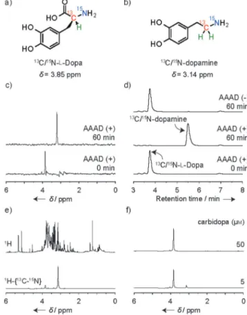
13C/15N-enriched l-Dopa (13C/15N-l-Dopa, Figure 1a) with a1 H-13C-15N sequence involving the asymmetric center was ob-tained starting from 13C/15N-glycine in an optical yield of 94% ee, as detailed in the Supporting Information.13C/15N-enriched dopamine (13C/15N-dopamine, Figure 1b) was also prepared from K13C15N. One-dimensional (1D) 1H-{13C-15N} triple-reso-nance spectra (13C-decoupled) of 13C/15N-l-Dopa and 13C/15 N-dopamine showed a single peak at 3.85 ppm or at 3.14 ppm for the methine proton (1H-13C-15N) of l-Dopa or the methylene protons (1H
2-13C-15N) of dopamine, respectively. The enzymatic decarboxylation of 13C/15N-l-Dopa (d= 3.85 ppm) to dopamine (d= 3.14 ppm) in 4-(2-hydroxyethyl)-1-piperazineethanesulfonic acid (HEPES) buffer containing the decarboxylation enzyme AAAD was completed in 1 h, as revealed by the triple-reso-nance analysis (Figure 1c) in accord with the results of high-performance liquid chromatography (HPLC) monitoring (Fig-ure 1d).
Decarboxylation and its inhibition in complex biological mix-tures such as liver lysate could also be readily monitored by triple-resonance. 13C/15N-l-Dopa (0.5 mm) in crude mouse liver lysate was incubated for 45 min. After workup, the mixture was subjected to NMR analysis. The conventional1H NMR
spec-trum (Figure 1e, top) was completely useless; all1H-containing molecules in the lysate represent their signals. The1H-{13C-15N} triple-resonance spectrum (Figure 1e, bottom) exhibited two signals at 3.85 ppm for l-Dopa and 3.14 ppm for dopamine in a ratio of 1:7 (12 % and 86% of l-Dopa used, respectively), indi-cating that most of the l-Dopa had undergone decarboxyla-tion by endogenous AAAD contained in the liver lysate to give dopamine. In the presence of carbidopa (BBB-impermeable), a potent AAAD inhibitor that is clinically used as a codrug to-gether with l-Dopa (referring to Scheme 1), the build-up of dopamine was effectively suppressed (~85%) even at [carbido-pa]= 5 mm (0.01 equiv of l-Dopa (0.5 mm)) (Figure 1 f, bottom) and completely suppressed at [carbidopa] =50 mm (0.1 equiv) (Figure 1 f, top). Triple resonance can thus completely suppress noise signals arising from endogenous components in complex biological mixtures to allow the unambiguous and quantitative metabolic analyses of13C/15N-enriched substrates therein.
In vitro and ex vivo spectra were obtained with a 700 MHz (16.4 T) NMR spectrometer equipped with a high-sensitivity
Scheme 1. l-Dopa-to-dopamine metabolism and its inhibition.
Figure 1. Structures of a)13C/15N-l-Dopa and b)13C/15N-dopamine and
analy-sis of the l-Dopa-to-dopamine conversion. Time course of the change in c)1H-{13C-15N} NMR spectra and d) HPLC profiles of13C/15N-l-Dopa (0.5 mm)
in 40 mm HEPES containing 80 mm NaCl, 100 mm pyridoxal phosphate, and AAAD (20 ng ml¢1), incubated at 378C for 0 or 60 min. The HPLC trace at the
top is for the control run in the absence of AAAD at 60 min. e) Conventional
1H (top) and1H-{13C-15N} (bottom) NMR spectra of a mouse liver lysate
con-taining13C/15N-l-Dopa (0.5 mm) in 2 mm Tris-HCl, 0.1 mm EDTA, 0.1 mm
2-mercaptoethanol, and 100 mm pyridoxal phosphate, incubated at 378C for 45 min. f) Inhibitory effects of carbidopa on the decarboxylation of13C/15
N-l-Dopa.1H-{13C-15N} NMR spectra obtained in the presence of carbidopa (5 or
50 mm) as an AAAD inhibitor, under the same conditions as in e). The1
H-{13C-15N} NMR spectra were obtained after 256 scans.
cryogenic probe after 256 scans (~7 min), where the detection limit, i.e., the lowest concentration to give S/N =3, of13C/15 N-dopamine lies at around 4 mm. Triple-resonance spectra of13C/ 15N-dopamine as phantom samples (500 mL) were also ob-tained with an MR machine for small animals operating at 7 T (300 MHz) without a cryogenic probe (Supporting Informa-tion); the detection limit after 3600 scans (1 h) turned out to be ~1 mm.
We proceeded to the l-Dopa/dopamine metabolic analysis in mice, focusing on the effects of inhibitors of decarboxylation (AAAD) and oxidation (MAO) enzymes. Mice (~16 g) were ad-ministered 13C/15N-l-Dopa (0.63 mmol kg¢1) with or without carbidopa (AAAD inhibitor) and MAO inhibitors [clorgyline and selegiline (BBB-permeable MAO-A and MAO-B inhibitors, re-spectively; see Scheme 1)]. After 1 h, brain and liver tissues were collected and, after workup, their triple-resonance spectra (256 scans) were obtained for doubly diluted (compared with the tissue weights) solutions, as shown in Figure 2, where the
signal intensities for the brain and liver samples are weight-normalized. In the absence of any inhibitors, a small amount of dopamine (3.14 ppm) was detected in the brain, while a much larger amount was found in the liver (bottom). l-Dopa with a signal at 4.19 ppm[13]was hardly detected in the liver or the brain. Most of the l-Dopa must have undergone wasteful de-carboxylation by endogenous AAAD in the liver, and any dopa-mine generated remained therein since it could not pass though the BBB to reach the brain. In the presence of the AAAD inhibitor carbidopa (63 mmol kg¢1), the dopamine level in the brain increased, but only slightly (middle), probably be-cause of its oxidative deamination by MAO to give dopal, which of course exhibited no 1H-13C-15N signals. Indeed, when the MAO inhibitors clorgyline (63 mmol kg¢1) and selegiline
(63 mmolkg¢1) were both present, there was a 5-fold increase in the dopamine level in the brain, and a substantial amount of l-Dopa that escaped decarboxylation remained in the liver (top).[14,15] The local concentrations of dopamine in the brain (~400 mg) were estimated by calibration using an authentic specimen to be 7 mm (in the absence of any inhibitors), 15 mm (with AAAD inhibitor), and 76 mm (with AAAD and MAO inhibi-tors). The inhibitor-dependent dopamine levels of 7–76 mm are consistent with those of 5–120 mm[9a]reported for rat based on HPLC analysis.
In this work, we investigated the usefulness of triple reso-nance for monitoring dopamine at a stimulated ~ 30 mm level. As shown above, a wide concentration range which includes this critical 30 mm could be easily accessed by a combination of l-Dopa and inhibitors. Selectivity and sensitivity are key issues in applying NMR to complex biological mixtures. In this context, the present work may be summarized as follows: 1) Triple resonance showed “perfect” selectivity. The probability of the natural occurrence of the sequence1H-13C-15N is as low as 0.011Õ0.0037 =0.00004 (0.004%), where 0.011 and 0.0037 are the natural abundance of13C and15N, respectively, and the mole-based selectivity factor for the 13C/15N-enriched target over endogenous components is 1/0.00004= 25000 (2.5 Õ104). Thus, endogenous components may effectively compete with the enriched target at, for example, 10 mm, only when they are present in unnaturally high concentrations of 10 Õ104mm= 0.1m. An implication of this observation is that selectivity is by no means a formidable issue to deal with for any mm-level trace constituents as long as they contain N-atom(s) and their appropriate double13C/15N-enrichment is synthetically accessi-ble. 2) This perfect, noiseless selectivity of triple resonance gives rise to a mm sensitivity [4 mm, 256 scans under less time-consuming (in minutes), one-dimensional (monitoring of 1H signals only) conditions]. This allows unambiguous and quanti-tative ex vivo metabolic/pharmacokinetic analyses of adminis-tered l-Dopa and its metabolite dopamine, i.e., ratiometric monitoring of their decay/build-up profiles, which clearly shows that the stimulated level of dopamine in the brain can be monitored ex vivo. 3) Unfortunately, however, the key dopa-mine level of 30 mm is short of the detection limit (~1 mm, 3600 scans) of the 7T MR machine (noncryogenic probe) for noninvasive, i.e., in vivo, monitoring.[16] However, the gap be-tween them is only a factor of ~ 30. This appears to be signifi-cant since an increase in sensitivity of this extent (~30-fold) may be achieved by combining an existing highest-field ma-chine and a high-sensitivity cryogenic probe equipped with a triple-resonance coil.[17]In-brain dopamine may then become a real target of direct in vivo MRS with which we can record the dopamine spectra in the brain.
In addition to a variety of techniques, based on HPLC,[18a] biological (enzyme-linked immunosorbent assay, ELISA) affinity (for the analysis of urine),[18b] microdialysis,[18c] electrochemical techniques,[18c]and chemical sensing,[18d] a couple of methods have recently been developed to monitor dopamine in the brain. One is positron emission tomography (PET) using 11C-raclopride, which competitively binds to the dopamine re-ceptor to enable the [dopamine]-dependent emission of
Figure 2. Effects of MAO inhibitors on the oxidative degradation of13C/15
N-dopamine in mice. Weight-normalized1H-{13C-15N} NMR spectra (256 scans)
for the extracts of brain (left) and liver (right) tissues of a mouse coadminis-tered with13C/15N-l-Dopa (0.63 mmolkg¢1) and carbidopa (63 mmol kg¢1) in
the absence (middle) or presence (top) of clorgyline (MAO-A inhibitor, 63 mmol kg¢1) and selegiline (MAO-B inhibitor, 63 mmolkg¢1). The
corre-sponding spectra in the absence of any inhibitors are shown at the bottom. The tissue extracts obtained were redissolved in D2O and subjected to NMR
analysis. The in-brain concentrations of dopamine were quantified via cali-bration and are shown.
gamma rays.[19] The other is MRI using a protein-engineered heme-based contrast agent which reversibly binds to dopa-mine, thereby changing the relaxivity, and thus gives [dopa-mine]-dependent images.[10]Both methods are highly sophisti-cated, but are indirect and involve complicated complexation processes. MRS is much simpler and can directly monitor the targets and their transformations with minimal noise signals which may arise from nonspecific binding, etc. Currently, the metabolic analysis of13C-glucose in the brain has received in-creasing attention.[3] The present work shows a way to detect mm-level trace constituents and has shed light on the issues to be overcome for in vivo imaging. Further work is now under-way along these lines with an ultimate goal of detection of hy-podopaminergy in related diseases.
Experimental Section
1) General methods, 2) preparation, 3) monitoring of the l-Dopa-to-dopamine conversion and subsequent dopamine oxidation, and 4) phantom MRS are included in the Supporting Information.
Acknowledgements
This work was supported by the Innovative Techno-Hub for Inte-grated Medical Bio-Imaging of the Project for Developing Innova-tion Systems, from the Ministry of EducaInnova-tion, Culture, Sports, Sci-ence and Technology, Japan (MEXT), and partly by Grant-in-Aid numbers 25350977, 15K01819, and 15H01403 from JSPS, Japan. H. Y. acknowledges financial support from the Daiwa Securities Health Foundation. T. K. acknowledges financial support from the Princess Takamatsu Cancer Research Fund, Magnetic Health Sci-ence Foundation, and SEI Group CSR Foundation. The authors thank Profs. Masahiro Shirakawa and Hidehito Tochio (Kyoto Uni-versity) for their support with the NMR measurements.
Keywords: dopamine · L-dopa · metabolic analysis · neurotransmitters · stable isotope enrichment · triple-resonance NMR
[1] a) L. E. Kay, M. Ikura, R. Tschudin, A. Bax, J. Magn. Reson. 1990, 89, 496 – 514; b) A. C. Wang, S. Grzesiek, R. Tschudin, P. J. Lodi, A. Bax, J. Biomol. NMR 1995, 5, 376– 382; c) H. Yamada, Y. Hasegawa, H. Imai, Y. Takayama, F. Sugihara, T. Matsuda, H. Tochio, M. Shirakawa, S. Sando, Y. Kimura, A. Toshimitsu, Y. Aoyama, T. Kondo, J. Am. Chem. Soc. 2015, 137, 799–806. [2] a) T. W-M. Fan, A. N. Lane, Prog. Nucl. Magn. Reson. Spectrosc. 2008, 52, 69–117 and references therein; b) E. Chikayama, M. Suto, T. Nishihara, K. Shinozaki, T. Hirayama, J. Kikuchi, PLoS ONE 2008, 3, e3805 and refer-ences therein.
[3] a) P. C. M. van Zijl, A. S. Chesnick, D. Despres, C. T. W. Moonen, J. Ruiz-Ca-bello, P. van Gelderen, Magn. Reson. Med. 1993, 30, 544– 551; b) R. A. de Graaf, G. M. I. Chowdhury, K. L. Behar, Anal. Chem. 2014, 86, 5032 –5038. [4] a) W. C. Hutton, J. J. Likos, J. K. Gard, J. R. Garbow, J. Labelled Compd. Ra-diopharm. 1998, 41, 87– 95; b) H. Yamada, K. Mizusawa, R. Igarashi, H. Tochio, M. Shirakawa, Y. Tabata, Y. Kimura, T. Kondo, Y. Aoyama, S. Sando, ACS Chem. Biol. 2012, 7, 535 –542; c) R. Ueki, K. Yamaguchi, H. Nonaka, S. Sando, J. Am. Chem. Soc. 2012, 134, 12398 – 12401.
[5] E. S. Bromberg-Martin, M. Matsumoto, O. Hikosaka, Neuron 2010, 68, 815– 834.
[6] J. Lotharius, P. Brundin, Nat. Rev. Neurosci. 2002, 3, 932 –942.
[7] M. B. H. Youdim, D. Edmondson, K. F. Tipton, Nat. Rev. Neurosci. 2006, 7, 295– 309.
[8] A. C. Whitfield, B. T. Moore, R. N. Daniels, ACS Chem. Neurosci. 2014, 5, 1192–1197.
[9] a) N. T. Buu, M. Angers, Biochem. Pharmacol. 1987, 36, 1731– 1735; b) P. Riederer, L. Lachenmayer, G. Laux, Curr. Med. Chem. 2004, 11, 2033 – 2043.
[10] T. Lee, L. X. Cai, V. S. Lelyveld, A. Hai, A. Jasanoff, Science 2014, 344, 533– 535.
[11] a) A. G. Ewing, J. G. Bigelow, R. M. Wightman, Science 1983, 221, 169– 171; b) L. C. Nicolaysen, M. Ikeda, J. B. Justice, D. B. Neill, Brain Res. 1988, 460, 50– 59; c) P. A. Garris, M. Kilpatrick, M. A. Bunin, D. Michael, Q. D. Walker, R. M. Wightman, Nature 1999, 398, 67–69.
[12] The reported values for rat are ~35 mm (electrochemical),[11a]a)~30 mm
(electrochemical),[11b] b)~27 mm (MRI),[10] and ~ 2 mm
(electrochemi-cal).[11c]
[13] The workup procedure involves treatment with 10% trichloroacetic acid (TCA).13C/15N-l-Dopa was independently shown to be stable when
treated with 10% TCA under the same conditions and exhibited me-thine proton resonance at 4.19 ppm, which is shifted downfield by 0.34 ppm (probably as a result of changes in the protonation state) from that (3.85 ppm) for TCA-untreated l-Dopa.
[14] The liver spectrum (Figure 2, top, right) shows a notable amount of dopamine (3.14 ppm), indicating that 0.1 equiv of carbidopa is not enough to completely suppress in vivo decarboxylation of l-Dopa in the liver, although enzymatic in vitro decarboxylation of l-Dopa can be completely inhibited by 0.1 equiv of carbidopa (Figure 1 f).
[15] Interestingly, the l-Dopa level in the brain is higher with carbidopa and MAO inhibitors than with carbidopa alone.
[16] The sensitivity of MR machines for small animals with a wider bore di-ameter, a longer sample-to-coil distance, and more pronounced inho-mogeneity of the magnetic field is known to be considerably lower than that of spectrometers dealing with solution samples in 5 mm tubes. Under these circumstances, recent attention has been directed to specific signal amplification techniques such as hyperpolarization and CEST (chemical exchange saturation transfer). For example, a) A. Viale, S. Aime, Curr. Opin. Chem. Biol. 2010, 14, 90–96 and references therein; b) A. R. Lippert, K. R. Keshari, J. Kurhanewicz, C. J. Chang, J. Am. Chem. Soc. 2011, 133, 3776 –3779; c) S. Viswanathan, Z. Kovacs, K. N. Green, S. J. Ratnakar, A. D. Sherry, Chem. Rev. 2010, 110, 2960 –3018 and references therein.
[17] The commercially available, highest-field MR machine for small animals operates at 21T (Bruker, Billerica, MA, USA). The NMR sensitivity increas-es in proportion to B03/2, where B0is the static magnetic field. A
cryo-genic probe is known to enhance sensitivity by a factor of ~ 5 com-pared with noncryogenic probes. Thus, a 21 T (900 MHz) machine with a cryogenic probe is expected to be more sensitive than a 300 MHz (7T) machine without a cryogenic probe by a factor of (21/7)3/2Õ5=26.
[18] a) G. Cannazza, A. Di Stefano, B. Mosciatti, D. Braghiroli, M. Baraldi, F. Pinnen, P. Sozio, C. Benatti, C. Parenti, J. Pharm. Biomed. Anal. 2005, 36, 1079 –1084; b) M. Nichkova, P. M. Wynveen, D. T. Marc, H. Huisman, G. H. Kellermann, J. Neurochem. 2013, 125, 724–735; c) D. L. Robinson, A. Hermans, A. T. Seipel, R. M. Wightman, Chem. Rev. 2008, 108, 2554– 2584 and references therein; d) M. Maue, T. Schrader, Angew. Chem. Int. Ed. 2005, 44, 2265–2270; Angew. Chem. 2005, 117, 2305–2310. [19] I. K. Goerendt, C. Messa, A. D. Lawrence, P. M. Grasby, P. Piccini, D. J.
Brooks, Brain 2003, 126, 312 –325.
Received: October 13, 2015
Published online on December 30, 2015