ORIGINAL
Effects of thrombus suction therapy on myocardial blood flow
disorders in males with acute inferior myocardial infarction
Yuji Ozaki
1,3, Masahiro Nomura
2, Toru Nakayama
1, Tatsuro Ogata
3, Kimiko Nakayasu
2,
Yutaka Nakaya
4, and Susumu Ito
3 1Department of Internal Medicine, Kochi Red Cross Hospital;2
Faculty of Integrated Art and Sciences, Department of Human and Social Sciences,The University of Tokushima ;3
Department of Digestive and Cardiovascular Medicine, and4
Department of Nutrition and Metabolism, Institute of Health Biosciences, The University of Tokushima Graduate School, Tokushima, Japan
Abstract : Several studies have reported that the use of a distal protection device decreases the incidence of slow-flow and/or no-reflow in patients with myocardial infarctions. In the present study, we investigated the influence of a RESCUE/Thrombuster system and a PercuSurge GuardWire catheter on coronary microcirculation disorders in patients with acute myocardial infarction using the natriuretic polypeptide (ANP), the brain natriuretic peptide (BNP), and
99mTc-tetrofosmin myocardial scintigraphy (TF). The group consisted of a 77patients with
initial inferior myocardial infarction who had undergone emergency coronary angioplasty. The patients were randomly divided into : Group D (n=28), in which a direct stent alone was inserted, Group R/T (n=25), in which a stent was inserted after RESCUE system or a Thrombuster system was performed, and Group P (n=24), in which a stent was inserted after thrombus suction using a PercuSurge GuardWire catheter. Patients with coronary slow-flow/no-reflow were 3, 2 and 0cases in Group D, Group R / T and Group P, respectively. In the present study, patients with good-reflow were enrolled in order to investigate the coronary microcirculation disorder in patients with visually similar coronary blood flow obtained in coronary angiography after percutaneous coronary reperfusion therapy. TF myocardial scintigraphy was performed 10±3 days after admission. Bull’s eye images were divided into 8 sections, and each section was evaluated in 4 grades. The grade of each segment was regarded as the defect score. The results were compared with the database prepared based on bull’s eye maps from 50 healthy adults in our hospital, and count areas of−2×SD (standard deviation) or less were calculated
as the extent score (%), reflecting the area in which myocardial blood flow was decreased. The extent and severity scores in Groups P and R/T were significantly lower than those in Group D. Coronary angiography at the chronic stage (6 months after surgery) showed the patency of the responsible vascular lesion in all patients. However, the ANP, BNP, cardiac index, and pulmonary capillary wedge pressure (PCWP) were significantly improved in Groups R/T and P, compared to Group D (p<0.01). These results suggest that the use of a RESCUE/ Thrombuster system and a PercuSurge GuardWire catheter system in patients with acute inferior wall infarction improves coronary microcirculation disorders and acute- to chronic-phase cardiac function. J. Med. Invest. 53 : 167-173, February, 2006
Keywords : acute myocardial infarction; coronary microcirculation; thrombus suction therapy, TF myocardial scintigraphy
Received for publication October 20, 2005 ; accepted January 23, 2006.
Address correspondence and reprint requests to Yuji Ozaki,
M.D., Department of Digestive and Cardiovascular Medicine, Institute of Health Biosciences, The University of Tokushima Graduate School, Tokushima, Japan and Fax : +81-88-633-9235.
The Journal of Medical Investigation Vol. 53 2006
INTRODUCTION
A thrombus of the coronary artery exists in many lesions in acute myocardial infarctions, and a throm-boembolism may occur in a distal area during treatment of the infarction. A peripheral embolism or no-reflow phenomena also sometimes occurs(1-7). Therefore, the presence of a thrombus in the coronary artery is important with respect to myo-cardial salvage (8-10). In addition, in many patients with marked thrombosis, treatment by catheter intervention is difficult. Several studies have re-ported that thrombus suction therapy decreases the incidence of slow flow or no-reflow of the nary artery (11-13), but the question of whether coro-nary microcirculation disorders improve after throm-bus suction therapy in patients in whom good-reflow of Thrombolysis in Myocardial Infarction (TIMI) grade 3 has been achieved, using myocar-dial scintigraphy has not been investigated.
A purpose of the present study is to investigate the coronary microcirculation disorder in patients with visually similar coronary blood flow obtained in coronary angiography after percutaneous coro-nary reperfusion therapy. Therefore, the issue of whether thrombus suction therapy improves car-diac load and coronary microcirculation disorders in males with acute inferior myocardial infarction was studied, using the atrial natriuretic polypep-tide (ANP), the brain natriuretic peppolypep-tide (BNP), and nuclear medical parameters, such as the ex-tent and severity scores of99m
Tc-tetrofosmin myo-cardial scintigraphy (TF).
PATIENTS AND METHODS
1) Patients
The study group consisted of 77 males with an initial acute inferior myocardial infarction (age : 65.5±12.1 years) in whom reperfusion therapy was
successful at the Emergency Center of the Kochi Red Cross Hospital, Kochi City, Japan, between September 2000 and December 2004.
We employed the following criteria for acute myocardial infarction : chest pain persisting for 30 minutes or more ; more than 1-mm increases in ST at 2 or more leads on standard 12-lead electro-cardiograms ; a plasma creatinine (CK) level more than 2 times higher than the normal value ; abnor-malities in left ventricular wall motion on echocar-diography ; and an interval of 6 hours or less from
the development of chest pain. Patients who had undergone fibrinolytic treatment with tissue plas-minogen activator (t-PA) or urokinase before ad-mission, those with cardiogenic shock, and those in whom aspirin or ticlopidine was contraindicated were excluded from the study. Patients with coro-nary no-reflow/slow flow and chronic-stage resteno-sis were excluded. Written informed consent was obtained from all patients before the study.
Each of the subjects were assigned to one of three groups by the envelope method with respect to the procedures for coronary reperfusion ther-apy : Group D, in which only a direct stent was used (n=28), Group R/T, in which a stent was in-serted after thrombus suction using a RESCUE system (Boston Scientific Scimed, Inc., Maple Grove, Minnesota, USA) or a Thrombuster system (Kaneka Medix Corp, Japan)(n=25), or Group P, in which a stent was inserted after thrombus suction using a PercuSurge GuardWire catheter (Medtronic AVE, Santa Rosa, California, USA) (n=24). A MultiLink Trister!stent was used in all patients. Restenosis was not observed for 6 months after reperfusion, except in 12 cases.
2) Coronary angiography and percutaneous coro-nary reperfusion therapy
Prior to coronary reperfusion therapy, 162 mg of aspirin and 200 mg of ticlopidine hydrochloride were administered, along with an intra-arterial ad-ministration of heparin at 10,000 units, as antiplate-let therapy.
Intervention for the coronary artery responsible for myocardial infarction was performed by punc-ture of the femoral artery using a 7Fr guiding cathe-ter. Coronary angiography was performed using a MODEL CAS 210-A device (TOSHIBA Co., Ltd., Japan) and a polygraph (RMC-2000, NIHON KOHDEN Co., Ltd., Tokyo, Japan).
Three or more cardiologists evaluated the suc-cess or failure of the acute-stage coronary reperfu-sion ; in patients in whom the lereperfu-sion was evaluated as good-reflow on coronary angiography, reperfu-sion was considered successful. Similarly, 3 or more cardiologists evaluated chronic-stage resteno-sis ; patients with 50% or more stenoresteno-sis on coro-nary angiography according to the classification described by the American Heart Association were regarded as having restenosis. For the analy-sis of coronary angiography findings, the diameter and minimum lumen diameter (MLD) of the infarct-related artery was measured using the direct
cali-Y. Ozaki, et al. Coronary myocardial blood flow after PTCA
per method before and after reperfusion therapy and 6 months after the onset of acute myocardial infarction.
For antiplatelet therapy after coronary reperfu-sion therapy, heparin at 20,000 units/day was ad-ministered intra-venously, and aspirin at 162 mg/ day and ticlopidine hydrochloride at 200 mg/day were orally administered in each group.
3) Measurement of ANP and BNP
We measured the levels of ANP and BNP imme-diately after reperfusion and 6 months after reper-fusion. Venous blood through the cubital vein was collected after the end of percutaneous translumi-nal coronary angioplasty (PTCA). In the chronic stage, blood was collected through the cubital vein after fasting early in the morning on the day be-fore catheterization. ANP was measured by a ra-dioimmunoassay (RIA), and BNP by a sandwich immunoradiometric assay (IRMA).
4) Evaluation of hemodynamics using left ventric-ulography and a Swan-Gantz catheter
Left ventriculography was performed 1 and 6 months after reperfusion therapy. The left ven-tricular systolic volume index (ESVI), end-diastolic volume index (EDVI), and ejection frac-tion (EF) were obtained from 30 - degree right an-terior oblique left ventriculograms according to Simpson’s method. Using a Swan-Ganz catheter, we evaluated the hemodynamics immediately after reperfusion therapy and 6 months after the onset of myocardial infarction based on heart rate (HR), pulmonary capillary wedge pressure (PCWP), pulmonary arterial pressure (PAP), right ventricu-lar pressure, right atrial pressure, cardiac output (CO), cardiac index (CI), and left ventricular end-diastolic pressure (LVEDP).
5) Myocardial scintigraphy with 99m
Tc-tetrofosmin
Myocardial scintigraphy with99m
Tc-tetrofosmin was performed on all patients 10±3 days after
reper-fusion therapy, at rest after fasting. Single photon emission computed tomography (SPECT) was per-formed 15 minutes after an intravenous injection of 600 MBq of99m
Tc-tetrofosmin through the cubital vein. A 3-head rotating gamma camera (GCA, 7200 A, Toshiba, Japan) with a low-energy all-purpose col-limator was used for the imaging. The collection time was 30 seconds per frame, and 360 - degree data were taken by 72 image stops (5°step ; 24 directions×3 heads) for SPECT imaging. A data
processing super computer (GMS, 5000A, Toshiba, Japan) was used for analysis of the SPECT data.
From a bull’s eye map, an area counting 2×
stan-dard deviation (SD) or less in comparison to a da-tabase prepared from 50 normal adults in our hos-pital (25 men, 25 women) was calculated. The area of reduced99m
Tc-tetrofosmin uptake (extent score) and severity of deficiency (severity score) were cal-culated. That is, the extent score (%) and severity score reflect an area in which myocardial blood flow is decreased using a bull’s eye map prepared from SPECT transverse images.
6) Statistical analysis
All values are expressed as the mean±standard
deviation (SD), and statistical analyses were per-formed using StatView 5.0 software (SAS Institute, Inc., U.S.A.). The values were compared between two groups using an unpaired t-test and chi square test. P<0.05 was regarded as significant.
RESULTS
1) Patient characteristics
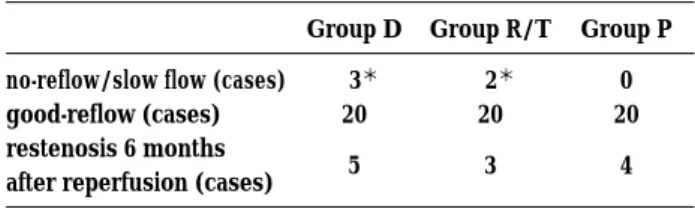
The incidence of coronary circulation disorder in each group was shown in Table 1. Patients with coronary no-reflow/slow-flow were 3, 2 and 0 cases in Group D, Group R/T, and Group P, respec-tively. The incidences of no-reflow/slow-flow in Group D and Group R/T were significantly higher than Group P (p<0.05).
Table 2 shows the characteristics of coronary good-flow patients in the present study. There were no significant differences in age, number of affected vessels, presence of concurrent diseases, such as ischemic heart disease, hypertension, and diabetes, lipid levels, fasting blood sugar, blood pressure, combination therapy with ACE inhibi-tors, interval to obtaining peak CK, post reference vessel diameter, or post minimal diameter among Group D, Group R/T, and Group P. However, the
Table 1. Incidence of coronary circulation disorder in each group
Group D Group R/T Group P no-reflow/slow flow (cases)
good-reflow (cases) restenosis 6 months after reperfusion (cases)
3* 20 5 2* 20 3 0 20 4 *p<0.05 vs Group P
peak CK levels in Group P and Group R/T were significantly lower than that in Group D (p<0.01).
2) Comparison of ANP and BNP concentrations on admission and six months after reperfusion ther-apy
Table 3 shows the serum levels of ANP and BNP on admission (at the onset of acute myocar-dial infarction) and 6 months after reperfusion ther-apy. There were no significant differences in the level of ANP on admission among Groups D, R/T, and P. However, 6 months after admission, the lev-els of ANP in Group P and Group R/T were sig-nificantly lower than that in Group D (p<0.05).
The level of BNP on admission in Group R/T was significantly higher than the values in Groups
D and P (p<0.01). However, 6 months after admis-sion, the levels of BNP in Groups P and R/T were significantly lower than that in Group D (p<0.01).
3) Myocardial scintigraphy with99m
Tc-tetrofosmin in Groups D, P, and R/T.
Fig. 1 shows an extent score map for a 64-year-old subject in Group D. In this patient, reperfusion therapy with a stent was performed, regarding the right coronary artery #3 as a responsible coronary lesion. The area of the defect involved the inferior wall, the anterior wall septum, and the posterior lateral wall. His extent score was 49.2, and the se-verity score was 34.4.
Fig. 2 shows an extent score map for a 72-year-old subjects in Group P. In this patient, distal pro-tection with a PercuSurge GuardWire catheter and reperfusion therapy were performed, regard-ing the right coronary artery #3 as a responsible coronary lesion. The defect area involved the infe-rior wall and the anteinfe-rior wall septum. His extent score was 23.5, and the severity score was 12.5.
Fig. 3 shows an extent score map for a 68-year-old subject in Group R/T. In this patient, distal protection with the RESCUE system and reperfu-sion therapy were performed, regarding the right coronary artery #3 as a responsible coronary lesion. The defect area involved the inferior wall. His extent score was 30.6, and his severity score
Table 2. Patient charactaristics
D group R/T group P group
age (yrs)
number of lesion branch ischemic heart disease hypertension smoking diabetes mellitus hyperlipidemia T-cho(mg/dl) TG(mg/dl) HDL-cho(mg/dl) FBS (mg/dl) ACE-Inhibitor sBP(mmHg) dBP(mmHg) peak CK (IU/l) time to peak CK (hrs) Post ref. (acute phase) Post MLD (acute phase)
66 ± 13 1.8 ± 0.8 4 /20 (20%) 10/20 (50%) 13/20 (65%) 8 /20 (40%) 13/20 (65%) 182±32 139 ± 83 48 ± 8 125 ± 32 15/20 (75%) 115 ± 22 75 ± 15 3151 ± 2467 14 ± 6 3.4 ± 0.4 3.1 ± 0.2 68 ± 9 1.5 ± 0.5 3 /20 (15%) 9 /20 (45%) 10/20 (50%) 9 /20 (45%) 11/20 (55%) 190 ± 49 115 ± 62 53 ± 12 157 ± 84 14/20 (70%) 123 ± 32 72 ± 12 1956 ± 2036* 12 ± 6 3.3 ± 0.3 3.2 ± 0.2 60 ± 18 1.7 ± 0.8 4 /20 (20%) 10/20 (50%) 11/20 (55%) 9 /20 (45%) 13/20 (65%) 187 ± 30 142 ± 100 44 ± 6 132 ± 55 14/20 (70%) 118 ± 18 76 ± 8 2010 ± 1278* 13 ± 6 3.6 ± 0.3 3.3 ± 0.3
*p<0.01 difference between D group.
T-cho, total cholesterol ; T-G, triglyceride ; HDL-cho, high-density lipoprotein cholesterol ; FBS, fasting blood sugar ; ACE, an-giotensin converting enzyme ; sBP, systolic blood pressure ; dBP, diastolic blood pressure ; CK, creatine kinase ; post ref., post ref-erence vessel diameter ; post MLD, post minimal lumen diameter
Table 3. Serum levels of ANP and BNP on admission and 6 months after reperfusion
D group R/T group P group ANP (pg/ml) admission 6 months 85±72 53±30 86±118 25±15** 78±64 30±28** BNP (pg/ml) admission 6 months 58±59 133±127 121±191* 62±50* 65±95 82±76*
*p<0.01, **p<0.05 difference between D group.
ANP, A type natriuretic peptide; BNP, B type natriuretic peptide
Y. Ozaki, et al. Coronary myocardial blood flow after PTCA
was 15.6.
4) Comparison of scintigraphic scores among Groups D, P, and R/ T.
As shown in Table 4, the extent and severity scores were compared among Groups D, P, and R/ T. The extent and severity scores in Groups P and R/T were significantly lower than those in Group D.
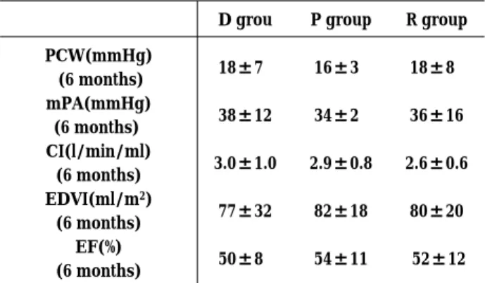
5) Pressure study and ejection fraction in the chronic stage
Table 5 shows the pressure study and EF on heart catheterization 6 months after the onset of myo-cardial infarction. No significant differences were found in PCWP, mean PAP, CI, EDVI, or EF among the 3 groups.
DISCUSSION
It has been reported that thrombus suction ther-apy decreases the incidence of slow flow or no-reflow during coronary artery perfusion therapy in patients with acute myocardial infarctions (14). However, the question of whether thrombus suc-tion therapy improves coronary microcirculasuc-tion disorders by myocardial scintigraphy in patients in whom good-reflow is achieved after thrombus suc-tion therapy has not been investigated. In the pre-sent study, there was no difference between Groups D and R/T in the incidence of slow-flow/ no-reflow, but the improvement of microcircula-tion at the chronic stage in good-reflow patient of Group R/T was more frequent than that of Group D. Moreover, thrombus suction therapy with a RESCUE system, a Thrombuster system, or a PercuSurge GuardWire catheter significantly im-proved ANP, BNP, and peak CK levels and myo-cardial scintigraphic parameters in comparison to direct stenting, indicating the usefulness of these therapies on myocardial coronary microcirculation disorders.
The use of direct stent therapy has decreased in treatment of acute myocardinal infarctions. How-ever, it continues to be performed in some
hospi-Fig. 1. Extent score map in a 64-year-old man in Group D.
Fig. 2. Extent score map in a 72-year-old man in Group P.
Fig. 3. Extent score map in a 68-year-old man in Group R/T.
Table 4. Comparison of scintiphotogrhic scores among D, R/T and P groups
D group R/T group P group extent score severity score 42±9 36±7 30±6* 25±3* 31±5** 26±4*
*p<0.01, **p<0.05 difference between D group.
Table 5. Pressure study and ejection fraction on heart cath-erization 6 months after the on set of myocardial infarction
D grou P group R group PCW(mmHg) (6 months) mPA(mmHg) (6 months) CI(l/min/ml) (6 months) EDVI(ml/m2) (6 months) EF(%) (6 months) 18±7 38±12 3.0±1.0 77±32 50±8 16±3 34±2 2.9±0.8 82±18 54±11 18±8 36±16 2.6±0.6 80±20 52±12 PCWP, pulmonary capillary wedge pressure ; mPA, mean pul-monary artery pressure;C.I., cardiac index;EDVI, end-diastolic volume index ; EF, ejection fruction
tals. We previously reported differences in cardiac function of the chronic stage between POBA and stent therapy in acute myocardial infarctions (15). Therefore, patients who had received direct stent therapy were enrolled in this study in order to standardize the control subjects.
For percutaneous transluminal coronary an-gioplasty for acute myocardial infarction, TIMI grade 3 blood flow in the early stage is required, for to a good prognosis (16-18). However, coro-nary reperfusion at the site of the lesion, where many thrombi are present, causes peripheral em-bolism related to rupture of the thrombus or the no-reflow phenomena (19). According to previous reports, the combination of distal protection with a balloon and thrombus suction therapy in patients with acute myocardial infarction resulted in high-grade TIMI blood flow, suggesting the usefulness of this combination therapy in marked thrombotic lesions (11-13).
In the present study, myocardial scintigraphic findings as well as the ANP and BNP levels were significantly improved in patients in whom a Per-cuSurge GuardWire catheter or a RESCUE system/ Thrombuster system was employed as a protec-tive device for preventing peripheral embolisms, even among patients with good-reflow. Thus, among patients in whom direct stent therapy with-out distal protection achieved TIMI grade 3 blood flow, and coronary angiography confirmed good circulatory reconstruction, a significant improve-ment was achieved in patients in whom thrombus suction therapy with a RESCUE system/Throm-buster system or a PercuSurge GuardWire cathe-ter was performed, suggesting that the combina-tion of distal proteccombina-tion with a balloon and throm-bus suction therapy is effective regardless of the presence of a visually recognizable thrombus.
However, there were no significant differences in cardiac function (EF) among Groups D, R/T, and P 6 months after the onset of the myocardial infarction. Possible reasons for the discrepancy between EF and ANP/BNP levels are as follows. First, EF values were almost normal or slightly de-creased in all patients because of successful reper-fusion therapy, second, ANP and BNP are more sensitive markers of cardiac function than EF, third, ANP and BNP affect the left ventricular dia-stolic function.
Significant changes were found in myocardial scintigraphic findings and ANP, BNP levels in pa-tients in whom visually high-grade TIMI blood
flow was achieved. The following mechanism is as-sumed. Platelets are usually activated after percu-taneous transluminal coronary angioplasty (20). However, the removal of a peripheral microthrom-bus using a distal protection device inhibits plate-let activation enhancement after percutaneous transluminal coronary angioplasty.
In the present study, we investigated only male with inferior myocardial infarction, because myo-cardial infarction is less frequent in woman, and because most women with a myocardial infarction are older and suffer a severe infarction. Because of these gender-related differences, the study was limited to males. Furthermore, we focused on acute inferior myocardial infarction, because a thrombosis is involved in right coronary infarction in many cases (21). In the future, left coronary le-sions should be investigated.
In conclusion, it has been recently reported that the use of drug-eluting stents is associated with the reduction of coronary artery restenosis compared with the bare thin-strut stent (22-24). These re-sults suggest that thrombus suction therapy prior to stenting may improve coronary microcircula-tion disorders in the acute to chronic stages in males with inferior myocardial infarction.
REFERENCES
1. Kragel AH, Gertz SD, Roberts WC : Mor-phologic comparison of frequency and types of acute lesions in the major epicardial coro-nary arteries in unstable angina pectoris, sud-den coronary death and acute myocardial in-farction. Am Coll Cardiol 18 : 801-808, 1991 2. Fuster V, Badimon L, Badimon JJ, Chesebro
JH : The pathogenesis of coronary artery dis-ease and the acute coronary syndromes (1). N Engl J Med 326 : 242-250, 1992
3. Dorros G, Cowley MJ, Simpson J, Bentivoglio LG, Block PC, Bourassa M, Detre K, Gosselin AJ, Gruntzig AR, Kelsey SF, Kent KM, Mock MB, Mullin SM, Myler RK, Passamani ER, Stertzer SH, Williams DO : Percutaneous trans-luminal coronary angioplasty : report of com-plications from the National Heart, Lung, and Blood Institute PTCA Registry. Circulation 67 : 723-730, 1983
4. Grube E, Gerckens U, Yeung AC, Rowold S, Kirchhof N, Sedgewick J, Yadav JS, Stertzer S: Prevention of distal embolization during
Y. Ozaki, et al. Coronary myocardial blood flow after PTCA
coronary angioplasty in saphenous vein grafts and native vessels using porous filter protec-tion. Circulation 104 : 2436-2441, 2001
5. Braunwald E : Unstable angina. A classification: Circulation 80 : 410-414, 1989
6. Topol EJ, Yadav JS : Recognition of the impor-tance of embolization in atherosclerotic vas-cular disease. Circulation 101 : 570-580, 2000 7. Erbel R, Heusch G : Coronary
microemboliza-tion. J Am Coll Cardiol 36 : 22-24, 2000
8. Kloner RA, Ganote CE, Jennings RB : The
“no-reflow” phenomenon after temporary coro-nary occlusion in the dog. J Clin Invest 54 : 1496-1508, 1974
9. Rezkalla SH, Kloner RA : No-reflow phenome-non. Circulation 105 : 656-662, 2002
10. Reffelmann T, Kloner RA : The “no-reflow” phenomenon : basic science and clinical cor-relates. Heart 87 : 162-168, 2002
11. Yip HK, Wu CJ, Chang HW, Fang CY, Yang CH, Chen SM, Hung WC, Chen CJ, Cheng CI, Hsieh YK : Effect of the PercuSurge GuardWire device on the integrity of microvasculature and clinical outcomes during primary transra-dial coronary intervention in acute myocartransra-dial infarction. Am J Cardiol 92 : 1331-1335, 2003 12. Rogers C, Huynh R, Seifert PA, Chevalier B,
Schofer J, Edelman ER, Toegel G, Kuchela A, Woupio A, Kuntz RE, Macon ND : Embolic protection with filtering or occlusion balloons during saphenous vein graft stenting retrieves identical volumes and sizes of particulate de-bris. Circulation 109 : 1735-1740, 2004
13. Bonderman D, Teml A, Jakowitsch J, Adlbrecht C, Gyongyosi M, Sperker W, Lass H, Mosgoeller W, Glogar DH, Probst P, Maurer G, Nemerson Y, Lang IM : Coronary no-reflow is caused by shedding of active tissue factor from dis-sected atherosclerotic plaque. Blood 99 : 2794-2800, 2002
14. Stone GW, Webb J, Cox DA, Brodie BR, Qureshi M, Kalynych A, Turco M, Schultheiss HP, Dulas D, Rutherford BD, Antoniucci D, Krucoff MW, Gibbons RJ, Jones D, Lansky AJ, Mehran R : Enhanced Myocardial Efficacy and Recov-ery by Aspiration of Liberated Debris (EMER-ALD) Investigators. Distal microcirculatory pro-tection during percutaneous coronary inter-vention in acute ST-segment elevation myo-cardial infarction : a randomized controlled trial. JAMA 293 : 1063-1072, 2005
15. Nakayama T, Nomura M, Fujinaga H, Ikefuji H, Kimura M, Chikamori K, Nakaya Y, Ito S : Does coronary artery stenting for acute myo-cardial infarction improve left ventricular over-loading at the chronic stage? Jpn Heart J 45 : 217-29, 2004
16. An international randomized trial comparing four thrombolytic strategies for acute myocar-dial infarction. The GUSTO investigators. N Engl J Med 329 : 673-682, 1993
17. Lee KL, Woodlief LH, Topol EJ, Weaver WD, Betriu A, Col J, Simoons M, Aylward P, Van de Werf F, Califf RM : Predictors of 30-day mortality in the era of reperfusion for acute myocardial infarction. Results from an inter-national trial of 41,021 patients. GUSTO-I In-vestigators. Circulation 91 : 1659-1668, 1995 18. ISIS-2 (Second International Study of Infarct
Survival) Collaborative Group. Randomised trial of intravenous streptokinase, oral aspirin, both, or neither among 17,187 cases of sus-pected acute myocardial infarction. Lancet 2 (8607) : 349-360, 1988
19. Mawson JB, Gavin JB, Herdson PB : A com-parison of the effects of ischemia and of selective occlusion of capillaries, pre-capillaries and terminal arterioles on coronary reperfu-sion. Pathology 14 : 369-373, 1982
20. Topol EJ, Byzova TV, Plow EF : Platelet GPIIb-IIIa blockers. Lancet 353(9148) : 227-231, 1999 21. Nagata Y, Usuda K, Uchiyama A, Uchikoshi M, Sekiguchi Y, Kato H, Miwa A, Ishikawa T : Characteristics of the pathological images of coronary artery thrombi according to the infarct-related coronary artery in acute myo-cardial infarction : Circ J 68 : 308-314, 2004 22. Pache J, Dibra A, Mehilli J, Dirschinger J,
Schomig A, Kastrati A : Drug-eluting stents compared with thin-strut bare stents for the reduction of restenosis : a prospective, ran-domized trial. Eur Heart J, 2005 Feb 28 ; [Epub ahead of print]
23. Katritsis DG, Karvouni E, Ioannidis JP : Meta-analysis comparing drug-eluting stents with bare metal stents. Am J Cardiol 95 : 640-3, 2005 24. Chieffo A, Stankovic G, Bonizzoni E, Tsagalou E, Iakovou I, Montorfano M, Airoldi F, Michev I, Sangiorgi MG, Carlino M, Vitrella G, Colombo A : Early and mid-term results of drug-eluting stent implantation in unprotected left main. Circulation 111 : 791-5, 2005