http://internmed.jp
【 CASE REPORT 】
Four Cases of Desquamative Esophagitis Occurring after
Hematopoietic Stem Cell Transplantation
Masaya Iwamuro
1, Daisuke Ennishi
2, Ken-ichi Matsuoka
2, Takehiro Tanaka
3,
Shotaro Okanoue
1, Yuka Obayashi
1, Hiroyuki Sakae
1,
Yoshiro Kawahara
4and Hiroyuki Okada
1Abstract:
We herein report four patients with desquamative esophagitis that developed one to nine days after periph-eral blood stem cell transplantation (PBSCT). Three patients underwent allogeneic PBSCT for leukemia, and the other underwent autologous PBSCT for pineoblastoma. Esophagogastroduodenoscopy revealed mucosal sloughing and fresh blood in the esophagus. Fasting and intravenous proton pump inhibitor therapy in addi-tion to blood transfusion improved the esophageal lesions within five to seven days in three patients. These cases indicate that desquamative esophagitis can occur in patients who receive hematopoietic stem cell trans-plantation. Although blood transfusions may be required, it can be resolved within seven days.
Key words:desquamative esophagitis, sloughing esophagitis, esophagitis dissecans superficialis, peripheral blood stem cell transplantation
(Intern Med 59: 3015-3022, 2020) (DOI: 10.2169/internalmedicine.4977-20)
Introduction
Desquamative esophagitis, also known as sloughing esophagitis or esophagitis dissecans superficialis, is a benign condition characterized by sheets of sloughed squamous tis-sue in the esophagus (1-3). While a rare disorder, cases of desquamative esophagitis have been reported to be associ-ated with skin diseases (e.g., pemphigus vulgaris and lichen planus), medication intake (e.g., bisphosphonates, non-steroidal anti-inflammatory drugs, and direct oral anticoagu-lants), esophageal injury (e.g., hot beverages, endoscopic dilatation, and sclerotherapy), and underlying esophageal disorders (e.g., eosinophilic esophagitis) (4-10). In contrast, few cases of desquamative esophagitis occurring after stem cell transplantation have been reported (11-13).
We herein report four cases of desquamative esophagitis that developed after peripheral blood stem cell
transplanta-tion (PBSCT). Of note, although the esophageal mucosa sloughing remained unchanged in one patient despite treat-ment, esophagogastroduodenoscopy performed five to seven days later revealed the improvement of desquamative esoph-agitis in the other three patients.
Case Reports
The patient characteristics of the four cases of desquama-tive esophagitis after PBSCT are shown in Table 1. The medications used by the patients during the period from myeloablative conditioning therapy to the diagnosis of desquamative esophagitis are listed in Table 2.
Case 1
An 18-year-old Japanese woman underwent autologous PBSCT for refractory pineoblastoma. She received seven cy-cles of chemotherapy with ifosfamide, cisplatin, and
1Department of Gastroenterology and Hepatology, Okayama University Graduate School of Medicine, Dentistry, and Pharmaceutical Sciences, Japan,2Department of Hematology and Oncology, Okayama University Graduate School of Medicine, Dentistry and Pharmaceutical Sciences, Japan,3Department of Pathology, Okayama University Hospital, Japan and4Department of Practical Gastrointestinal Endoscopy, Okayama
Uni-Intern Med 59: 3015-3022, 2020 DOI: 10.2169/internalmedicine.4977-20
Table 1. Characteristics of Patients who Developed Desquamative Esophagitis after Hematopoietic Stem Cell Transplantation. Case Age (years) Sex Underlying disease SCT Days from SCT to
the diagnosis of DE Outcome of DE 1 18 F Pineoblastoma Autologous PBSCT 1 DE was healed on day
7 after the diagnosis 2 29 M B-cell acute lymphoblastic leukemia Allogeneic PBSCT from an HLA-haploidentical related donor
6 DE was healed on day 7 after the diagnosis 3 21 M T-cell acute lymphoblastic leukemia Allogeneic PBSCT from an HLA-haploidentical related donor
9 DE was healed on day 5 after the diagnosis 4 16 M Acute myeloid
leukemia
Allogeneic PBSCT from an HLA-1-locus mis-matched related donor
5 DE persisted on day 7 after the diagnosis
SCT: stem cell transplantation, DE: desquamative esophagitis, PBSCT: peripheral blood stem cell transplantation
Table 2. The Medications Used by the Patients During the Period from Myeloablative Conditioning Therapy to the Diagnosis of Desquamative Esophagitis.
Antacid Antibiotic Antiemetic Immunosuppressive agent Chemotherapeutic agent Others 1 Esomeprazole, lansoprazole Acyclovir, cefepime, doripenem, fluconazole, levofloxacin, micafungin Aprepitant, fosaprepitant, granisetron, metoclopramide, prochlorperazine, ramosetron Hydrocortisone Cyclophosphamide, melphalan, nogitecan Acetaminophen, diphenhydramine, famotidine, furosemide, glycyrrhizin, haptoglobin, hydroxyzine, morphine, sodium bicarbonate, ursodeoxycholic acid,
zonisamide 2 Lansoprazole, omeprazole, rabeprazole, sodium alginate Acyclovir, fluconazole, letermovir, meropenem, micafungin, sulfamethoxazole/ trimethoprim, valacyclovir, vancomycin, voriconazole Aprepitant, granisetron, metoclopramide, olanzapine Cyclophosphamide, hydrocortisone, mycophenolate mofetil, tacrolimus
Fludarabine, mesna Acetaminophen, calcium gluconate, clostridium butyricum, dalteparin sodium,
d-chlorpheniramine, furosemide, lenograstim, magnesium sulfate, potassium
chloride, potassium hydrogen phosphate, sodium bicarbonate, ursodeoxycholic acid
3 Omeprazole, vonoprazan Acyclovir, cefepime, letermovir, levofloxacin, meropenem, micafungin, pentamidine, sulfamethoxazole/ trimethoprim, valacyclovir, voriconazole Aprepitant, granisetron, prochlorperazine Cyclophosphamide, dexamethasone, hydrocortisone, mycophenolate mofetil, tacrolimus Fludarabine, melphalan, mesna Acetaminophen, alfacalcidol, carbazochrome, clostridium butyricum, dalteparin sodium, d-chlorpheniramine, filgrastim, furosemide, lenograstim,
levothyroxine, magnesium oxide, magnesium sulfate, midodrine, morphine, oxycodone, potassium chloride, potassium hydrogen phosphate, ramelteon, sodium bicarbonate, thiamazole, ursodeoxycholic
acid, zolpidem 4 Lansoprazole, omeprazole, vonoprazan Acyclovir, caspofungin, itraconazole, letermovir, levofloxacin, meropenem, pentamidine, sulfamethoxazole/ trimethoprim, tazobactam/ piperacillin, teicoplanin, valacyclovir, vancomycin Aprepitant, domperidone, granisetron, metoclopramide, prochlorperazine dexamethasone, hydrocortisone, methotrexate, tacrolimus Gemtuzumab ozogamicin, cyclophosphamide, mesna Acetaminophen, brotizolam, carbazochrome, clostridium butyricum, dalteparin sodium, d-chlorpheniramine, fentanyl, furosemide, haptoglobin, herbal
medicine Choreito, hydroxyzine, lenograstim, loperamide, magnesium sulfate, mosapride, nicardipine, potassium
chloride, sodium bicarbonate, tranexamic acid, ursodeoxycholic acid
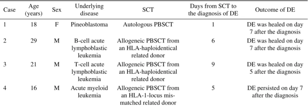
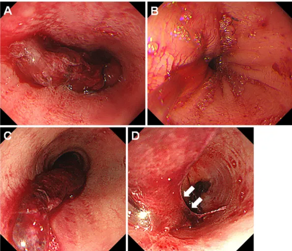
Figure 1. Endoscopic images of case 1. An 18-year-old woman developed desquamative esophagitis one day after PBSCT (A-C). She was treated with fasting, intravenous administration of lansoprazole, and red blood cell transfusion. Esophagogastroduodenoscopy performed seven days later showed improvement of the esophageal lesion (D, E). PBSCT: peripheral blood stem cell transplantation
Figure 2. Pathological examination findings of case 1. The biopsy specimen from the sloughed esophageal mucosa re-vealed necrotic tissue and degenerative squamous cells.
etoposide before PBSCT. After conditioning with a myeloablative regimen consisting of topotecan, melphalan, and cyclophosphamide, seven days before PBSCT, the pa-tient developed febrile neutropenia and was treated with ce-fepime followed by doripenem. The administration of acy-clovir was also started seven days before PBSCT. On the day of PBSCT, she manifested oral mucositis and diarrhea, and morphine was administered. Since she vomited blood a day after undergoing PBSCT, we performed esophagogastro-duodenoscopy, which revealed extensive sloughing of the esophageal mucosa and fresh blood (Fig. 1A-C). We
there-tive squamous cells (Fig. 2). Immunostaining for herpes simplex virus was negative. Concomitantly, her white blood cells were 40/μL, hemoglobin was 6.5 g/dL, and platelets were 57,000/μL. She was treated with fasting and intrave-nous lansoprazole along with red blood cell transfusions. Esophagogastroduodenoscopy performed seven days later showed a healed esophageal mucosa (Fig. 1D, E).
Case 2
A 29-year-old Japanese man underwent haploidentical PBSCT for B-cell acute lymphoblastic leukemia. Although the leukemia was initially treated with intensive induction polychemotherapy, remission was not achieved. Thus, the patient received two cycles of inotuzumab ozogamicin and subsequently underwent PBSCT. Myeloablative conditioning was performed with 12 Gy of total body irradiation and fludarabine. The administration of dalteparin sodium was started the day before PBSCT to prevent sinusoidal obstruc-tion syndrome. Valacyclovir and letermovir administraobstruc-tion was also initiated the day before and after PBSCT, respec-tively. After PBSCT, the patient received a two-day infusion of cyclophosphamide, followed by the administration of my-cophenolate mofetil and tacrolimus. Six days after PBSCT, he developed melena and hematochezia. His white blood cell count was 20/μL, hemoglobin level was 5.6 g/dL, and platelet count was 14,000/μL. Esophagogastroduodenoscopy showed a huge bloodlike lump in the middle to lower esophagus. In the middle part of the esophagus, the bloody
Intern Med 59: 3015-3022, 2020 DOI: 10.2169/internalmedicine.4977-20
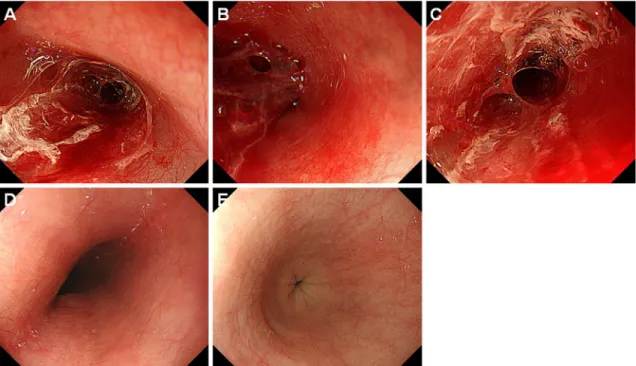
Figure 3. Endoscopic images of case 2. A 29-year-old man who underwent haploidentical PBSCT developed desquamative esophagitis on day 6 (A, B). A bloody mass was observed at the longitudinal border in the middle part of the esophagus (A, arrows), and squamous mucosa was found in the lower end of the lesion (B, arrows). Treatment with fasting, administration of omeprazole and sodium alginate, and red blood cell and platelet transfusion was initiated. However, the sloughed esophageal mucosa remained unchanged four days later (C, D). Improvement of the esophageal lesions was con-firmed by esophagogastroduodenoscopy performed seven days after the diagnosis (E, F). Half of the mucosa of the middle esophagus was reddish, which we considered to be vestiges of the desquamated area (E, arrows). Damaged esophageal mucosa was also partly observed in the reddish area (F, ar-row). PBSCT: peripheral blood stem cell transplantation
squamous mucosa was seen in the lower end of the lesion (Fig. 3B, arrows). We therefore suspected that a hematoma had formed after desquamation of the esophageal mucosa.
The patient was treated with fasting and intravenous ome-prazole therapy, and per-oral sodium alginate. The admini-stration of dalteparin sodium was stopped. Red blood cells and platelets were also transfused. Esophagogastroduodeno-scopy performed four days after treatment showed persis-tence of mucosal sloughing, hematoma, and bleeding (Fig. 3C, D). Although the esophageal lesions mostly healed
seven days after the diagnosis of desquamative esophagitis, half of the mucosa of the middle esophagus was reddish, which we considered to be vestiges of the desquamated area (Fig. 3E, arrows). Damaged esophageal mucosa was partly observed in the reddish area (Fig. 3F, arrow).
Case 3
A Japanese boy was diagnosed with T-cell acute lym-phoblastic leukemia at 5 years of age and underwent haploi-dentical PBSCT at 7 years of age. However, the leukemia
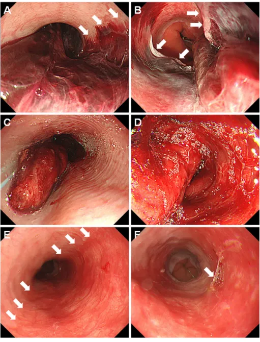
Figure 4. Endoscopic images of case 3. On day 9 after haploidentical PBSCT, a 21-year-old man developed desquamative esophagitis (A, B). Esophagogastroduodenoscopy performed five days later showed epithelialized esophageal mucosa (C, D). PBSCT: peripheral blood stem cell transplantation
recurred, and he received multiple chemotherapies. Thus, a second haploidentical PBSCT procedure was planned at 21 years of age. Myeloablative conditioning was performed with total body irradiation, fludarabine, and melphalan. In-travenous injection of dalteparin sodium was started six days before PBSCT to prevent sinusoidal obstruction syndrome. Valacyclovir and letermovir administrations were also initi-ated seven days before and on the day of PBSCT, respec-tively. After PBSCT, mycophenolate mofetil and tacrolimus were administered on day 5. On day 9, he developed he-matemesis. Esophagogastroduodenoscopy results revealed fresh blood and peeled esophageal mucosa (Fig. 4A, B). Desquamative esophagitis was treated with fasting, intrave-nous omeprazole therapy, and discontinuation of dalteparin sodium. Since his white blood cell count was 30/μL, hemo-globin was 8.9 g/dL, and platelet count was 24,000/μL, platelet transfusion was performed. Esophagogastroduodeno-scopy performed five days later showed epithelialized esophageal mucosa (Fig. 4C, D).
Case 4
A 15-year-old Japanese boy was diagnosed with acute myelomonocytic leukemia and received chemotherapy with daunorubicin and cytarabine, followed by consolidation and
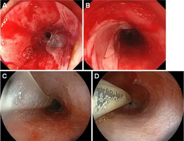
ozogamicin was administered. Subsequently, he underwent allogeneic PBSCT from his human leukocyte antigen (HLA)-1-locus mismatched mother. Myeloablative condi-tioning was performed with total body irradiation and cyclo-phosphamide. Dalteparin sodium injection was administered once five days before PBSCT. Tacrolimus was administered a day before PBSCT, and methotrexate was administered on day 3 after PBSCT. Valacyclovir and letermovir administra-tions were also initiated seven days before and on the day of PBSCT, respectively. On day 5, the patient developed he-matemesis, and esophagogastroduodenoscopy showed a bloody mass in the esophagus (Fig. 5A, B). Because the en-doscopic images (Fig. 3C, D) and the patient’s clinical course resembled that of case 2, we diagnosed the patient with desquamative esophagitis.
His white blood cell count was 10/μL, hemoglobin was 9.1 g/dL, and platelet count was 14,000/μL. Subsequently, red blood cells and platelets were transfused. Although the esophagitis was treated with fasting and intravenous omepra-zole, esophagogastroduodenoscopy performed seven days af-ter the diagnosis revealed that the clump of blood-like sub-stance was unchanged (Fig. 5C). The lower end of the le-sion was partly membranous (Fig. 5D, arrows), which we considered to be peeled-off esophageal mucosa. Because his
Intern Med 59: 3015-3022, 2020 DOI: 10.2169/internalmedicine.4977-20
Figure 5. Endoscopic images of case 4. A 16-year-old boy who underwent HLA-1-locus mismatched PBSCT developed desquamative esophagitis on day 5 (A, B). Esophagogastroduodenoscopy per-formed seven days after the diagnosis revealed that the sloughed esophageal mucosa was unchanged (C, D). The lower end of the lesion was partly membranous (D, arrows), which we considered to be peeled-off esophageal mucosa. PBSCT: peripheral blood stem cell transplantation, HLA-1: human leucocyte antigen-1
Discussion
We herein present the reports of four cases of desquama-tive esophagitis that occurred one to nine days after PBSCT. McDonald et al. reported that, among 63 patients with chronic graft-versus-host disease who were examined after allogeneic bone marrow transplantation, 8 had esophageal involvement (13). Although an endoscopic examination was not performed in one patient, all other patients developed desquamative esophagitis. The mean duration until the esophageal symptom onset was 220 days after transplanta-tion (range, 69-632 days). Other previous reports of desqua-mative esophagitis after hematopoietic stem cell transplanta-tion have described it as a manifestatransplanta-tion of chronic graft-versus-host disease (11, 12). However, in our four patients, desquamative esophagitis was diagnosed within nine days after PBSCT, indicating that esophageal mucosal sloughing can occur in the acute phase in patients who undergo hema-topoietic stem cell transplant.
The typical endoscopic appearance of desquamative esophagitis includes sheets of partially sloughed, crumpled,
or detached mucosa and linear strips of desquamated mu-cosal layers (1-4). In the present study, all of the patients showed blood adhesion in the esophagus and sloughed mu-cosa. Thrombocytopenia after hematopoietic stem cell trans-plantation and dalteparin sodium therapy, which was con-tinuously administered in cases 2 and 3, may have caused the bleeding from the desquamated esophageal wall, which resulted in hematemesis, melena, and/or hematochezia. In addition, a hematoma (Fig. 3A, B) and a clump of blood were observed in case 2 (Fig. 3C) and case 4 (Fig. 5A, C). We speculated that, as described above, hematoma formation had occurred beneath the desquamated esophageal mucosa. The clump of blood might have been composed of rolled up sloughed mucosa and blood. However, because we did not perform a pathological analysis of the hematoma or the clump of blood, the mechanisms underlying the occurrence of these lesions have not been elucidated.
Several hypotheses may be able to explain the pathogene-sis of desquamative esophagitis in the present four patients: graft-versus-host disease, infection, radiation, intensive treat-ment sequences, and medication-induced mucosal damage. In general, the esophagus is infrequently affected by acute
graft-versus-host disease. Furthermore, no patient manifested symptoms of acute graft-versus-host disease, such as skin rashes, diarrhea, or high levels of liver enzymes. However, an aggressive alloimmune reaction may have been involved in the development of desquamative esophagitis in cases 2-4, as these patients underwent HLA-mismatched PBSCT.
Viruses, such as cytomegalovirus and herpes simplex vi-rus, can affect the esophagus, particularly in immunocom-promised critically ill patients. Patients in the acute phase after PBSCT are susceptible to viral infection even under prophylactic antibiotic treatment. Thus, although antiviral drugs had been administered to all our patients prior to PBSCT, viral infection may have induced esophageal dam-age. Radiation therapy also causes inflammation, edema, erythema, and erosion of the mucosal surface of the esopha-gus. Therefore, it may lead to desquamative esophagitis. Nevertheless, radiation-induced mucosal damage is unlikely to be the common etiology for desquamative esophagitis in our patients because case 1 did not undergo total body irra-diation. Among those who underwent allogeneic transplant, all three were young and had recurrent/refractory leukemia; therefore, intensive pretransplant treatments and HLA-mismatched PBSCTs with myeloablative conditioning were performed. Such intensive treatment sequences might have affected the esophageal tissue.
Other drugs administered before symptom onset could also be possible causes of medication-related toxicity and/or esophageal infection, which resulted in esophageal mucosal sloughing. Mycophenolate mofetil has been reported to in-duce gastrointestinal tract abnormalities, such as circumfer-ential inflammation, ulcers, and esophageal stricture (14-16). However, only two patients received mycophenolate mofetil, whereas the other two did not take the drug. As shown in Table 2, the common drugs administered to all four patients were mesna, acyclovir, aprepitant, granisetron, cyclophos-phamide, hydrocortisone, furosemide, sodium bicarbonate, and ursodeoxycholic acid. Some of these drugs are known to cause adverse gastrointestinal manifestations (17-19). Thus, one of these drugs or their combination might have caused the desquamative esophagitis in our patients. Further investigations will be required to determine the etiology of desquamative esophagitis that develops in the acute phase after PBSCT.
It is noteworthy that three of the four patients recovered from desquamative esophagitis within seven days of the di-agnosis. Although the infusion of red blood cells and/or platelets was required for the treatment of anemia and thrombocytopenia, patients were treated with fasting and the intravenous administration of proton pump inhibitors. These results indicate that desquamative esophagitis occurring in the acute phase of PBSCT is only transient damage to the superficial esophageal mucosa and should be treated conser-vatively.
nation with blood transfusion helped ameliorate the sloughed esophageal mucosa in three of the four patients. Although the exact pathogenesis is uncertain, we believe that understanding the unique manifestation and outcome of this esophageal lesion will help hematologists and gastroen-terologists manage adverse events in patients undergoing he-matopoietic stem cell transplantation.
The authors state that they have no Conflict of Interest (COI).
References
1. Akhondi H. Sloughing esophagitis: a not so common entity. Int J Biomed Sci 10: 282-286, 2014.
2. Purdy JK, Appelman HD, McKenna BJ. Sloughing esophagitis is associated with chronic debilitation and medications that injure the esophageal mucosa. Mod Pathol 25: 767-775, 2012.
3. Carmack SW, Vemulapalli R, Spechler SJ, Genta RM. Esophagitis dissecans superficialis (“sloughing esophagitis”): a clinicopa-thologic study of 12 cases. Am J Surg Pathol 33: 1789-1794, 2009.
4. Shah R, Thoguluva V, Bansal N, Manocha D. Esophageal disse-cans: a rare life-threatening presentation of recurrent pemphigus vulgaris. Am J Emerg Med 33: e1-e2, 2015.
5. Ma H, Chen M, Liu J, Li Y, Li J. Serious stomatitis and esophagi-tis: a peculiar mucous reaction induced by pegylated liposomal doxorubicin. An Bras Dermatol 90: 209-211, 2015.
6. Shah SA, Cho M, Chaptini L, Parikh N. Sloughing esophagitis: an atypical cause of food impaction. ACG Case Rep J 3: e85, 2016. 7. Dumas-Campagna M, Bouchard S, Soucy G, Bouin M.
IgG4-related esophageal disease presenting as esophagitis dissecans su-perficialis with chronic strictures. J Clin Med Res 6: 295-298, 2014.
8. Hart PA, Romano RC, Moreira RK, Ravi K, Sweetser S. Esoph-agitis dissecans superficialis: clinical, endoscopic, and histologic features. Dig Dis Sci 60: 2049-2057, 2015.
9. Hokama A, Yamamoto Y, Taira K, et al. Esophagitis dissecans su-perficialis and autoimmune bullous dermatoses: a review. World J Gastrointest Endosc 2: 252-256, 2010.
10. Nidimusili AJ, Dhadam GC, Shaheen K. Sloughing esophageal mucosa. QJM 107: 77-78, 2014.
11. Trabulo D, Ferreira S, Lage P, Rego RL, Teixeira G, Pereira AD. Esophageal stenosis with sloughing esophagitis: a curious manifes-tation of graft-vs-host disease. World J Gastroenterol 21: 9217-9222, 2015.
12. Nakshabendi IM, Maldonado ME, Coppola D, Mamel JJ. Esopha-geal cast: a manifestation of graft-versus-host disease. Dig Dis 18: 103-105, 2000.
13. McDonald GB, Sullivan KM, Schuffler MD, Shulman HM, Thomas ED. Esophageal abnormalities in chronic graft-versus-host disease in humans. Gastroenterology 80: 914-921, 1981.
14. Matin T, Zhao Y, Goyal J, Weber F. Mycophenolate induced GI mucosal injury - case report and review of the literature. MOJ Clin Med Case Rep 8: 96-98, 2018.
15. Liu K, Kia L. Mycophenolate mofetil-induced esophagitis. Clin Gastroenterol Hepatol 17: e139, 2019.
16. Delacruz V, Weppler D, Island E, et al. Mycophenolate mofetil-related gastrointestinal mucosal injury in multivisceral transplanta-tion. Transplant Proc 42: 82-84, 2010.
17. Blijlevens NM. Cytotoxic treatment-induced gastrointestinal symp-toms. Curr Opin Support Palliat Care 1: 16-22, 2007.
Intern Med 59: 3015-3022, 2020 DOI: 10.2169/internalmedicine.4977-20
10: e02903-e02918, 2019.
19. Xie JH, Fan ST, Nie SP, et al. Lactobacillus plantarum NCU116 attenuates cyclophosphamide-induced intestinal mucosal injury, metabolism and intestinal microbiota disorders in mice. Food Funct 7: 1584-1592, 2016.
The Internal Medicine is an Open Access journal distributed under the Creative Commons Attribution-NonCommercial-NoDerivatives 4.0 International License. To view the details of this license, please visit (https://creativecommons.org/licenses/ by-nc-nd/4.0/).
Ⓒ 2020 The Japanese Society of Internal Medicine