Journal of the American Heart Association
ORIGINAL RESEARCH
Significance of Exercise-Related Ventricular
Arrhythmias in Patients With Brugada
Syndrome
Hiroshi Morita, MD, PhD ; Saori T. Asada, MD, PhD; Masakazu Miyamoto, MD; Yoshimasa Morimoto, MD;
Tomonari Kimura, MD, PhD; Tomofumi Mizuno, MD; Koji Nakagawa, MD, PhD; Atsuyuki Watanabe, MD, PhD; Nobuhiro Nishii, MD, PhD; Hiroshi Ito, MD, PhD
BACKGROUND: Sinus tachycardia during exercise attenuates ST-segment elevation in patients with Brugada syndrome, whereas ST-segment augmentation after an exercise test is a high-risk sign. Some patients have premature ventricular contractions (PVCs) related to exercise, but the significance of exercise-related PVCs in patients with Brugada syndrome is still unknown. The objective of this study was to determine the significance of exercise-related PVCs for predicting occurrence of ventricular fibrillation (VF) in patients with Brugada syndrome.
METHODS AND RESULTS: The subjects were 307 patients with Brugada syndrome who performed a treadmill exercise test. We evaluated the occurrence of PVCs at rest, during exercise and at the peak of exercise, and during recovery after exercise (0–5 minutes). We followed the patients for 92±68 months and evaluated the occurrence of VF. PVCs occurred in 82 patients (27%) at the time of treadmill exercise test: PVCs appeared at rest in 14 patients (4%), during exercise in 60 patients (20%), immediately after exercise (0–1.5 minutes) in 28 patients (9%), early after exercise (1.5–3 minutes) in 18 patients (6%), and late after exercise (3–5 minutes) in 12 patients (4%). Thirty patients experienced VF during follow-up. Multivariable analysis includ-ing symptoms, spontaneous type 1 ECG, and PVCs in the early recovery phase showed that these factors were independently associated with VF events during follow-up.
CONCLUSIONS: PVCs early after an exercise test are associated with future occurrence of VF events. Rebound of vagal nerve activity at the early recovery phase would promote ST-segment augmentation and PVCs in high-risk patients with Brugada syndrome.
Key Words: Brugada syndrome ■ exercise test ■ premature ventricular contractions ■ sudden death ■ ventricular fibrillation
V
entricular fibrillation (VF) frequently occurs atnight in association with parasympathetic nerve activation during sleep in patients with Brugada
syndrome.1,2 Occurrence of VF is rare during exercise2
since the activated sympathetic nerve suppresses ar-rhythmogenesis in patents with Brugada syndrome. Sinus tachycardia during exercise usually reduces
ST-segment elevation in right precordial leads,3 but
some patients have augmentation of ST-segment el-evation in the early recovery phase after an exercise
test.4 ST-segment augmentation after an exercise test
can predict lethal arrhythmic events during follow-up. ST-segment augmentation can be caused by with-drawal of sympathetic nerve activation and rebound of
parasympathetic nerve activity after exercise.5 A rise
in body temperature during exercise is also a possible
cause of augmentation of ST-segment elevation.6
Although occurrence of VF during exercise is rare in patients with Brugada syndrome, ventricular tach-yarrhythmias as well as supraventricular arrhythmias
Correspondence to: Hiroshi Morita, MD, PhD, Department of Cardiovascular Therapeutics, Okayama University Graduate School of Medicine, Dentistry and Pharmaceutical Sciences, 2-5-1 Shikata-Cho, Okayama City, Okayama 700-8558, Japan. E-mail: hmorita@cc.okayama-u.ac.jp
Supplementary Material for this article is available at https://www.ahajo urnals.org/doi/suppl/ 10.1161/JAHA.120.016907 For Sources of Funding and Disclosures, see page 10.
© 2020 The Authors. Published on behalf of the American Heart Association, Inc., by Wiley. This is an open access article under the terms of the Creative Commons Attribution-NonCommercial-NoDerivs License, which permits use and distribution in any medium, provided the original work is properly cited, the use is non-commercial and no modifications or adaptations are made.
JAHA is available at: www.ahajournals.org/journal/jaha
sometimes occur in association with exercise4,7–9.10 Moreover, monomorphic ventricular tachycardia (VT) can occur in some patients with Brugada syndrome
during exercise.11–13 Detection of ventricular
arrhyth-mias can be a high-risk sign in Brugada syndrome14,15;
however, little is known about the significance of ar-rhythmias, especially ventricular tachyarar-rhythmias, as-sociated with exercise.
The objective of this study was to clarify the inci-dence and significance of exercise-related arrhythmias, especially ventricular tachyarrhythmias, in patients with Brugada syndrome.
METHODS
The data that support the findings of this study are available from the corresponding author on reason-able request.
Subjects of the Study
The subjects of this study comprised 307 patients with Brugada-type ECG who visited our hospital. The mean age of the patients was 45±12 years (range, 17–79 years)
and men were predominant (299 men; 97%). At the first visit to the hospital, 219 patients were asymptomatic, 75 had syncope, and 13 had experienced aborted car-diac arrest. The diagnosis of Brugada syndrome was made according to the criteria of J-Wave-Syndromes
Expert Consensus Statements.16 Spontaneous type
1 was defined as the appearance of coved-type ST-segment elevation (J point ≥0.2 mV) without a sodium channel blocker. There were no patients from the same family. We followed all of the patients at our outpatient clinic.
This study was approved by the Ethics Committee on Human Research and Epidemiology of Okayama University, and analysis of the SCN5A gene was per-formed in 117 patients, in compliance with guidelines for human genome studies of the Ethics Committee of Okayama University.
ECG Evaluations
All of the 307 patients performed a treadmill exercise test (TMT). All ECGs were recorded without the use of any cardiac drugs. We explained the methods, re-quirements, and risks of the exercise test and obtained informed consent before the TMT. A repeat exercise test was performed in 140 patients at ≥1 month after the first exercise test. The protocol of the exercise test was the Bruce protocol with symptom-limited or sub-maximal exercise (up to 90% of maximum predicted heart rate by age). A 12-lead ECG with leads V1 and V2 located at the third intercostal spaces was recorded at rest, at every stage during exercise, at peak exer-cise, and every 1 minute during the recovery phase to 5 minutes and 20 seconds after exercise with a low-pass filter of 0 to 25 Hz. We evaluated heart rate, blood pressure, ST-segment levels as the J point in leads V1 and V2, and occurrence of arrhythmias in each ECG recording phase. ST-segment elevation was meas-ured as the calculated mean of 3 successive beats. ST-segment augmentation after exercise was defined as ST-segment amplitude increase ≥0.05 mV in one of leads V1 and V2 in the early recovery phase of the
exercise test (2 and 3 minutes after peak exercise).4
To classify the morphologies of the exercise-related premature ventricular contractions (PVCs), we defined morphologies of PVCs as superior axis (0° to −90° and +180° to +270°) and inferior axis (0° to +180°) in limb leads and right bundle-branch block (RBBB) type (volt-age of R wave ≥ volt(volt-age of S wave in lead V1) and left bundle-branch block (LBBB) type (voltage of R wave <voltage of S wave in lead V1). ECG patterns were re-viewed blindly by 3 cardiologists (HM, TK, and SA).
Statistical Analysis
Continuous data are expressed as means±SD values.
Fisher’s exact test or the χ2 test was used for categorical
CLINICAL PERSPECTIVE
What Is New?
• The significance of exercise test–induced pre-mature ventricular contractions has not been recognized in patients with Brugada syndrome.
• Patients with premature ventricular contractions during early recovery phase after an exercise test frequently experienced ventricular fibrilla-tion during follow-up.
What Are the Clinical Implications?
• Premature ventricular contractions during the early recovery phase (1.5–3 minutes) after an exercise test by vagal rebound can identify the high-risk patients with Brugada syndrome.
• Exercise tests are safe because they do not induce ventricular fibrillation in patients with Brugada syndrome.
• Exercise tests can be a useful risk stratifica-tion tool for predicting future occurrence of ventricular fibrillation in patients with Brugada syndrome.
Nonstandard Abbreviation and Acronym
TMT treadmill exercise testvariables. Comparison of 2 groups was made with the Student t test or a nonparametric test for paired and unpaired data, as appropriate. Arrhythmic events were defined as occurrence of VT/VF, aborted cardiac arrest, sudden death, or appropriate therapy of an implantable cardioverter defibrillator (ICD). Time from the initial hos-pital visit to the first arrhythmic event during follow-up was analyzed with Cox’s proportional hazards model. Hazard ratios and CIs are presented for results of uni-variable analysis. For multiuni-variable analysis, we selected significant parameters (symptoms [history of syncope or VF], spontaneous type 1 ECG and exercise-related PVCs at the first exercise test) to assess predictors of VF events during follow-up. Survival curves were plotted by the Kaplan–Meier method and analyzed by the log-rank test. We also assessed the results of univariable analysis of exercise-related PVCs in all exercise tests including re-peated exercise tests to determine the relation between PVCs and all VF events and to calculate the odds ratios (ORs). Significance was defined as P<0.05. All statistical analyses were performed with the use of JMP version 14.2 (SAS Institute, Inc., Cary, NC). All authors had full access to and take full responsibility for the integrity of the data.
RESULTS
Characteristics of Patients and TMT
Table 1 shows the baseline characteristics of the pa-tients. Spontaneous type 1 ECG appeared in 219 patients (71%). Fifty patients had a family history of sudden death before 45 years of age. SCN5A mutation was found in 14 of the 117 patients in whom genetic tests were performed.
The median of maximum exercise was stage 4 (minimum, stage 1; maximum, stage 7). Heart rate
increased until the peak of exercise and gradually de-creased after the exercise test (Figure 1A). Systolic blood pressure rose during exercise and became high-est at the peak of the exercise and during the early recovery phase from exercise, and then it gradually decreased during the late recovery phase (Figure 1B). Diastolic blood pressure increased slightly at the peak of exercise.
ST-segment level at the J point in leads V1 and V2 decreased during exercise and became minimum at the peak of exercise, and then it gradually recov-ered to the baseline level after exercise (Figure 1C). Augmentation of ST-segment elevation in leads V1 and V2 appeared during the recovery phase after exer-cise in 66 patients (Figure S1A through S1C). Patients who had ST-segment augmentation after exercise had higher ST-segment level at baseline and throughout the exercise test than did patients without ST-segment augmentation after exercise (Figure S1C). Horizontal ST-segment depression (≤0.1 mV) in inferolateral leads was observed in 10 patients, and coronary artery dis-ease was found in 1 of those patients (Figure S2). After percutaneous coronary stenting to the right and left descending branches of the coronary arteries, that pa-tient still had spontaneous type 1 ECG but did not have ST-segment depression after an exercise test. None of the other 9 patients with ST-segment depression after exercise had ischemic heart disease or ventricular hy-pertrophy, and we considered that ECG change was false positive for ischemia.
We implanted an ICD in 70 patients (28 asymptom-atic patients, 29 patients with syncope, and 13 patients with VF). Thirty patients experienced lethal arrhythmic events during 92±68 months’ follow-up (event ratio, 1.3%/year; asymptomatic patients, n=7; patients with syncope, n=12; patients with VF, n=11). The arrhythmic events were appropriate ICD therapies in 21 patients,
Table 1. Baseline Characteristics of Patients
Overall Event+ Event− P Value*
Total Patients (%) 307 30 277
Age y 45±12 44±12 45±12 0.4668
Male Patients (%) 299 (97.4%) 29 (96.7%) 270 (97.5%) 0.5651
FH of SD <45 y Patients (%) 50 (16.3%) 5 (16.7%) 45 (16.2%) 1.0000
SCN5A mutation Patients (%) 14/117 (12.0%) 4/25 (16.0%) 10/92 (10.9%) 0.4944
Spontaneous type 1 Patients (%) 219 (71.3%) 27 (90.0%) 192 (69.3%) 0.0182
Symptom at the first visit
Asymptomatic Patients (%) 219 (71.3%) 7 (23.3%) 212 (76.5%) <0.0001
Symptomatic Patients (%) 88 (28.7%) 23 (76.7%) 65 (30.7%)
Syncope Patients (%) 75 (24.4%) 12 (40.0%) 63 (22.7%)
VF Patients (%) 13 (4.2%) 11 (36.7%) 2 (7.2%)
Event Patients (%) 30 (9.8%) 30 (100.0%) 0 (0%) …
FH indicates family history; SD, sudden death; and VF, ventricular fibrillation. *Comparison of patients with events and without events.
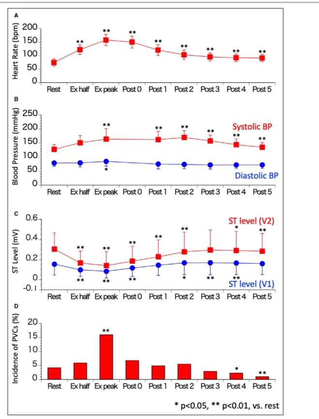
Figure 1. Changes in heart rate, blood pressure, ST level and incidence of PVC in association with exercise.
A, Heart rate. Heart rate increased after starting exercise and gradually recovered during the recovery
phase. B, Blood pressure. Systolic blood pressure increased during and early after exercise and
recovered to baseline during the late recovery phase. C, ST-segment level. ST-segment level decreased
during exercise and then gradually recovered to baseline level after exercise. D, Incidence of premature
ventricular contractions (PVCs). PVCs significantly increased at the peak of exercise. PVCs decreased during the late recovery phase compared with those at rest. *P<0.05, **P<0.01, vs rest. BP indicates blood pressure; and Ex, exercise test.
VT/VF in 8 patients, and sudden death in 1 patient. Patients who experienced VF events during follow-up more frequently had spontaneous type 1 ECG and were already symptomatic (Table 1). Other clinical fac-tors, including age, sex, family history, and SCN5A mutation, were not different between patients with and those without VF events. There were no differences in occurrence of ST-segment augmentation or depres-sion after exercise between patients with and without VF.
Exercise-Related Ventricular Arrhythmias
at the Time of the First TMT
Exercise-related PVCs were observed in 82 patients (26.7%) in the first TMT. PVCs appeared before ex-ercise in 14 patients and during or after exex-ercise in 81 patients (26.4%). PVCs occurred during exercise in 60 patients and in the recovery phase in 44 pa-tients. PVCs occurred during or after exercise in 13 patients with PVCs before exercise. Twenty-three patients had PVCs during both the exercise and the recovery phase. PVCs significantly increased at the peak of exercise (n=48) and immediately recov-ered to baseline after exercise (Figure 1D, Table 2). The frequency of PVCs was significantly reduced at 4 minutes (7 patients; P=0.0346) and 5 minutes (3 patients; P=0.0043) of the recovery phase compared with that before exercise (14 patients).
PVCs at 2 minutes of the recovery phase (2.0– 3.0 minutes) occurred more frequently in patients with ST-segment augmentation than in patients without ST-segment augmentation (PVCs at 2 minutes of re-covery: 7/66 patients with ST augmentation [10.6%], versus 10/241 patients without ST-segment augmen-tation [4.1%]; P=0.0425; Figure S1D). PVCs did not occur in association with ST-segment depression.
Patients with VF events more frequently had exer-cise-related PVCs, especially PVCs during 1.0–2.0 min-utes and 2.0–3.0 min1.0–2.0 min-utes of the recovery phase, than did patients without VF events (Figure 2, Table 2). Furthermore, the frequencies of PVCs in the first half of 1 minute of the recovery phase (1.0–1.5 minutes) were not different between the 2 groups (P=0.2689), but PVCs in the second half of 1 minute of the recov-ery phase (1.5–2.0 minutes) appeared more frequently in patients with VF events (P=0.0291). Patients with VF events more frequently had PVCs in the first half and second half of 2 minutes of the recovery phase than did patients without VF events (P=0.0290 and
P=0.0051, respectively). Thus, the frequency of PVCs
during 1.5 to 3.0 minutes of the recovery phase was significantly higher in patients with VF events than in patients without VF events (Table 2).
Various morphologies of PVCs were observed, and 24 patients had multifocal PVCs (Figure 3, Table 3).
Eleven patients had couplet of PVCs (n=9) or triplet of PVCs (n=2) (Figure 3E), but exercise did not induce VT/ VF. Couplet or triplet of PVCs and morphology of PVCs were not different between patients with and without VF during follow-up. PVCs with LBBB morphology, suggesting RV origin, frequently appeared during 1.5 to 3.0 minutes of the recovery phase (LBBB type, n=16; RBBB type, n=6; P<0.001).
Risk Factors Predicting VF Events During
Follow-Up
Univariable analysis of baseline clinical characteristics showed that existence of spontaneous type 1 ECG and symptoms were associated with VF events during follow-up (Table 4, Figure 4A and 4B). Age, sex, family history, and SCN5A mutation were not associated with VF events. Among ECG changes and arrhythmias as-sociated with exercise, occurrence of PVCs during 2.0 to
Table 2. Occurrences of Arrhythmias in Association With Exercise Test
Overall Event+ Event−
P Value* 307 30 277 PVC (first TMT), n 82 (26.7 13 (43.3) 69 (24.9) 0.0305 Rest 14 (4.2) 1 (3.3) 13 (4.7) 0.7349 During exercise 18 (5.9) 1 (3.3) 17 (6.1) 0.5353 Peak exercise 8 (15.6) 6 (20.0) 42 (15.2) 0.4890 Post 0 min 21 (6.8) 4 (13.3) 17 (6.1) 0.1387 Post 1 min 15 (4.9) 4 (13.3) 11 (4.0) 0.0241 Post 2 min 17 (5.5) 5 (16.7) 12 (4.3) 0.0051 Post 3 min 9 (2.9) 0 (0) 9 (3.3) 0.1371 Post 4 min 7 (2.3) 0 (0) 7 (3.6) 0.3792 Post 5 min 3 (1.0) 0 (0) 3 (1.1) 0.5674 Post 0–1.5 min 28 (9.1) 5 (16.7) 23 (8.3) 0.1313 Post 1.5–3 min 18 (5.9) 6 (20.0) 12 (4.3) 0.0005 Post (1.5–3 min)−(4–5 min) 14 (4.6) 6 (20.0) 8 (2.9) <0.0001 PVCs ≥ cpl 12 (3.9) 2 (6.7) 10 (3.6) 0.4127 PAC, n (%) 52 (16.9) 3 (10.0) 49 (17.7) 0.2869 PAT/PSVT, n (%) 4 (1.3 0 (0) 4 (1.4) 0.5083 PAF, n (%) 0 (0) 0 (0) 0 (0) 1.0000 SSS, n (%) 1 (0.3) 0 (0) 1 (0.4) 0.7421 Paroxysmal AVB, n (%) 1 (0.3) 0 (0) 1 (0.4) 0.7421 Complete RBBB, n (%) 1 (0.3) 0 (0) 1 (0.4) 0.7421 ST-segment depression, n (%) 8 (2.6) 1 (3.3) 7 (2.5) 0.7926 ST-segment augmentation, n (%) 66 (21.5) 5 (16.7) 61 (22.0) 0.4984 AVB indicates atrioventricular block; cpl, couplet; PAC, premature atrial contraction; PAF, paroxysmal atrial fibrillation; PSVT, paroxysmal supraventricular tachycardia; PVC, premature ventricular contraction; RBBB, right bundle-branch block; and TMT, treadmill exercise test.
*Comparison of patients with events and without events.
3.0 minutes of the recovery phase was associated with VF events. Occurrence of PVCs during the early recovery phase (1.5–3.0 minutes) showed a better correlation with VF events (Figure 4C). Sensitivity, specificity, and posi-tive and negaposi-tive predicposi-tive values of PVCs during the early recovery phase for occurrence of VF events were
20%, 96%, 33%, and 83%, respectively. Patients with VF events did not have PVCs after 4 minutes of the late recovery phase, and thus patients who had PVCs in the early recovery phase but did not have PVCs in the late recovery phase were at high risk for VF events during follow-up (Figure 4D). Morphology of PVCs and multifocal
Figure 2. Occurrence of premature ventricular contractions (PVCs) associated with exercise in patients with and those without VF events during follow-up.
PVCs significantly increased at the peak of exercise in both patients with and those without VF events during follow-up. The frequencies of PVCs at 1 and 2 minutes of the recovery phase were significantly higher in patients with VF events than in patients without VF events. PVCs did not appear after 3 minutes of the recovery phase in patients with VF events. Ex indicates exercise test.
Figure 3. Morphologies of exercise-related premature ventricular contractions (PVCs).
A, A PVC with left bundle-branch (LBBB) type and inferior axis (n=44). B, A PVC with LBBB and superior axis (n=33). C, A PVC with
right bundle-branch block (RBBB) and inferior axis (n=9). D, A PVC with RBBB and superior axis (n=40). E, Occurrence of couplets of
PVCs during the recovery phase (circles).
PVCs were not associated with VF events. Multivariable analysis including symptoms, spontaneous type 1 ECG, and PVCs in the early recovery phase (1.5–3.0 minutes) showed that these factors were independently associ-ated with VF events (Table 5).
Impact of PVCs in Repeat Exercise Tests
on Overall VF Events
Repeat exercise tests were performed in 140 patients (2–15 times, average 2.9 times). Repeat exercise test added exercise-related PVCs in 24 additional patients out of 116 patients who did not have PVCs at the first exercise test (Table S1). The morphologies of PVCs were LBBB in 75 patients and RBBB in 45 patients (Table 3). Thirty-three patients who experienced VF previously or during follow-up (overall VF events) more frequently had exercise-related PVCs around 2 minutes of the recovery phase than did patients who never experienced VF at the end of follow-up (OR of PVCs during 2.0–3.0 minutes of exercise, 3.84; CI, 1.37–10.71; P=0.0102; Figure S3). Exercise-related PVCs including those in repeat exercise tests showed that the PVCs during 1.5 to 3.5 minutes of the recovery phase were associated with overall VF epi-sodes (OR of PVCs during 1.5–3.5 minutes of the recov-ery phase, 3.61; CI, 1.39–9.40; P=0.0084). Patients who experienced VF did not have PVCs after 4 minutes of the recovery phase. Thus, excluding patients with PVCs after 4 minutes of the recovery phase from patients with PVCs during 1.5 to 3.5 minutes of the recovery phase was strongly associated with overall VF episodes (OR, 5.41; CI, 1.98–14.74; P=0.0010) (Table S2).
Exercise-Related Supraventricular
Arrhythmias and Bradyarrhythmias
Supraventricular arrhythmias occurred in association with the first exercise test in 52 patients (52 patients with premature atrial complexes and 4 patients with paroxysmal atrial tachycardia or supraventricular tachycardia). Exercise revealed sinus node dysfunc-tion, paroxysmal atrioventricular block, and transient
complete RBBB were observed in 1 patient each (Table 2).
Repeat TMT induced premature atrial contractions in 18 additional patients. New occurrence of supraven-tricular tachycardia and atrial fibrillation during repeat exercise tests were observed in 1 and 2 additional pa-tients, respectively. Sick sinus syndrome and RBBB during repeat TMT tests were observed in 2 and 1 ad-ditional patients, respectively.
Supraventricular arrhythmias, bradyarrhythmias, and exercise-related ECG changes, such as ST changes, were not associated with VF events (Table 4, Table S1).
DISCUSSION
New Observations
Exercise-induced PVCs were observed in 26% of the patients with Brugada syndrome. In most of the pa-tients, PVCs increased at the peak of exercise, then gradually decreased after exercise. However, in pa-tients at high risk for VF, occurrence of PVCs showed a bimodal increment in association with the exercise test: PVCs increased at the peak of exercise and around 2 minutes of the recovery phase. Occurrence of PVCs in the early recovery phase (1.5–3.0 minutes) was associated with VF events during follow-up. Exercise test should be a part of routine examina-tion for risk stratificaexamina-tion in patients with Brugada syndrome.
Exercise Test and Brugada Syndrome
Occurrence of lethal arrhythmias during exercise is very rare in patients with Brugada syndrome since cat-echolamine release attributable to acute sympathetic activation and parasympathetic withdrawal by exer-cise attenuate ST-segment elevation and inhibit the
occurrence of VF.17,18 Thus, there is no strict limitation
on competitive sports even in symptomatic patients
with Brugada syndrome.19 Although an exercise test
Table 3. Morphologies of PVCs Associated With Initial and Repeat Exercise Test
PVC Morphology
PVC at Initial TMT PVC at Repeat TMT
Overall Recovery 1.5–3 min Overall Recovery 1.5–3.5 min
LBBB type 60 16 75 23 LBBB+inferior axis 36 9 44 15 LBBB+superior axis 34 9 43 12 RBBB type 33 6 45 9 RBBB+inferior axis 8 0 9 1 RBBB+superior axis 28 6 40 9 Multifocal 11 4 14 6
LBBB indicates left bundle-branch block; PVC, premature ventricular contraction; RBBB, right bundle-branch block; and TMT, treadmill exercise test.
is a safe examination for patients with Brugada syn-drome, various ECG changes in an exercise test have
been reported8,10,20,21: ST-segment augmentation at
the early recovery phase,4,20 QRS widening,3 delayed
heart rate recovery,21 and arrhythmias. Some of these
ECG changes are associated with VF. Makimoto et al4
reported that augmentation of ST-segment elevation
was observed in 37% of patients with Brugada syn-drome in the early recovery phase after the exercise. ST-segment augmentation after exercise was associ-ated with VF events during follow-up in both sympto-matic and asymptosympto-matic patients. During the recovery phase after exercise, the occurrences of sympathetic withdrawal and parasympathetic rebound and rise in body temperature can cause augmentation of
ST-segment elevation.5,22
Some studies have also shown that arrhythmias can occur in association with exercise, but the signif-icance of these arrhythmias has not been evaluated. Description of the occurrence of supraventricular ar-rhythmias associated with exercise test is very limited: One study showed the occurrence of premature atrial contractions in one patient and atrial fibrillation in 1 of
46 patients with Brugada syndrome.10 The occurrence
of ventricular tachyarrythmias during an exercise test is also rare. Previous studies showed that monomorphic
VT occurred during an exercise test,9,11–13,21 but there
have been no reports of VF induction during an exer-cise test. However, PVCs during or after an exerexer-cise test do not seem to be rare. The incidences of PVCs associated with exercise in patients with Brugada
syn-drome have been reported to be 0% to 33%,4,7,10,23 but
the impact of PVCs during exercise on the pathogene-sis of Brugada syndrome has not been reported.
The present study showed that PVCs increased at the peak of exercise in overall patients. These PVCs at the peak of exercise were not associated with cardiac events, and PVCs at that phase might therefore not be directly associated with arrhythmogenicity in Brugada syndrome. PVCs significantly increased in the early re-covery phase of exercise (1.5–3.0 minutes) in patients with VF during follow-up compared with those in pa-tients without VF. Although the occurrence of PVCs in that phase was not frequent, PVCs occurring in only about 20% of the patients with future VF events, these PVCs might be directly associated with arrhythmoge-nicity in Brugada syndrome. Since ECG type and ST-segment level fluctuate in association with occurrence
of VF24,25 and arrhythmogenic substrate will progress in
some patients,26 occurrence of exercise-related PVCs
could show day-by-day variation. Then repeat exer-cise test should be important to detect PVCs for risk stratification. The predominance of LBBB-type PVCs in this phase, usually indicating right ventricular origin, is compatible with the observation that the arrhyth-mogenic substrate of Brugada syndrome exists in the
right ventricle.27 Analysis of PVC morphologies might
indicate a critical area in which ventricular arrhythmias are initiated in patients with Brugada syndrome.
The occurrence of PVCs in the recovery phase corresponded to the occurrence of ST-segment level augmentation after exercise. The mecha-nism of PVCs in this phase might coincide with the
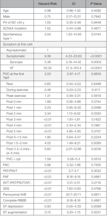
Table 4. Risk for VF of Baseline Characteristics and Exercise-Related Arrhythmias—Univariable Analysis
Hazard Risk CI P Value
Age 0.98 0.96–1.02 0.4085 Male 0.75 0.17–13.31 0.7842 FH of SD <45 y 1.03 0.35–2.49 0.9448 SCN5A mutation 1.52 0.44–3.99 0.4671 Spontaneous type 1 3.54 1.25–14.83 0.0144
Symptom at first visit
Asymptomatic 1 … … Symptomatic 9.36 4.23–23.63 <0.0001 Syncope 5.36 2.16–14.42 0.0003 VF 55.24 21.3–153.4 <0.0001 PVC at the first TMT 2.03 0.97–4.17 0.0603 Rest 0.65 0.04–3.03 0.6488 During exercise 0.48 0.03–2.23 0.4111 Peak exercise 1.31 0.49–3.01 0.5619 Post 0 min 1.89 0.56–4.86 0.2744 Post 1 min 3.23 0.95–8.30 0.0586 Post 2 min 3.34 1.13–8.02 0.0320 Post 3 min <0.01 1.81–1.81 0.1423 Post 4 min <0.01 2.34–2.34 0.1961 Post 5 min <0.01 4.95–4.95 0.3707 Post 0–1.5 min 1.90 0.64–4.57 0.2234 Post 1.5–3 min 4.02 1.49–9.21 0.0085 Post (1.5–3 min)– (4–5 min) 5.60 2.07–12.86 0.0016 PVC ≥ cpl 1.59 0.26–5.3 0.5513 PAC 0.86 0.32–1.96 0.7305 PAT/PSVT <0.01 3.7–3.7 0.3022 PAF <0.01 8.16–8.16 0.4861 SVT (PAT/PSVT/AF) <0.01 2.51–2.51 0.2119 SSS <0.01 7.83–0.83 0.4768 Paroxysmal AVB <0.01 20.7–20.71 0.6611 Complete RBBB <0.01 8.16–8.16 0.4861 ST depression 0.93 0.05–4.33 0.9398 ST augmentation 0.72 0.25–1.75 0.5031
AF indicates atrial fibrillation; AVB, atrioventricular block; cpl, couplet; FH, family history; PAC, premature atrial contraction; PAF, paroxysmal atrial fibrillation; PAT, paroxysmal atrial tachycardia; PSVT, paroxysmal supraventricular tachycardia; PVC, premature ventricular contraction; RBBB, right bundle-branch block; SD, sudden death; SSS, sick sinus syndrome; SVT, supraventricular tachycardia; TMT, treadmill exercise test; VF, ventricular fibrillation.
mechanism of ST-segment augmentation: with-drawal of sympathetic activation and parasympa-thetic rebound. Interestingly, patients with cardiac events did not have PVCs after 4 minutes of the re-covery phase. Rere-covery of heart rate and attenuation of ST-segment augmentation were also observed
at this phase, and it should indicate withdrawal of parasympathetic rebound after exercise. PVCs in the late recovery phase might not indicate arrhyth-mogenicity of Brugada syndrome. Thus, excluding patients with PVCs during the late recovery phase (after 4 minutes) from patients with PVCs during the early recovery phase (1.5–3.0 minutes) had strong association with VF events. Although PVCs in the early recovery phase might represent arrhythmo-genicity in Brugada syndrome, there was no oc-currence of VF related to exercise. Catecholamine release and sinus tachycardia during and after exer-cise prevented VF under the condition of
parasym-pathetic rebound. Usually, β-blockers can reduce
exercise-induced PVCs. PVCs associated with VF events are caused by vagal activation and sympa-thetic withdrawal after exercise, and effectiveness of β-blockers is not expected.
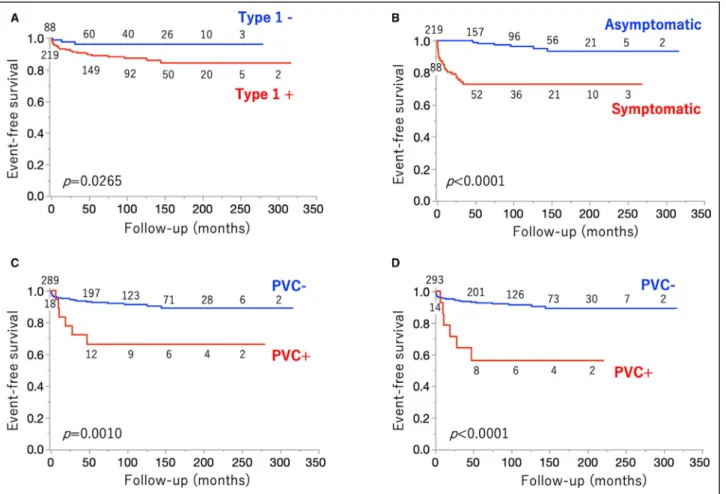
Figure 4. Event-free survival according to the clinical markers and premature ventricular contractions (PVCs) after exercise. A, Prognosis of patients with and those without spontaneous type 1 ECG. Patients with spontaneous type 1 ECG had a shorter time
to experience ventricular fibrillation (VF) events during follow-up than did patients without spontaneous type 1 ECG. B, Prognosis
according to symptoms. Symptomatic patients had a worse prognosis than did asymptomatic patients. C, Prognosis according to
occurrence of PVCs during 1.5 to 3 minutes of the recovery phase after the exercise test. Patients with PVCs more frequently had VF events than did patients without PVCs. D, Prognosis according to occurrences of PVCs during 1.5–3 minutes and after 4 minutes of
the recovery phase. Patients who had PVCs during early recovery phase (1.5–3 minutes of the recovery) but did not have PVCs during the late recovery phase (after 4 minutes of the exercise) had a significantly shorter time to experience VF events during follow-up. Numbers along with the survival curves show number of patients at risk. HR indicates hazard ratio for occurrence of VF events by univariable analysis; and PVC, premature ventricular contractions during the early recovery phase.
Table 5. Risk for VF of Baseline Characteristics and Exercise-Related Arrhythmias—Multivariable Analysis
Hazard Risk CI P Value
Symptomatic (syncope and VF) 12.24 5.46–31.18 <0.0001 Spontaneous type 1 ECG 4.69 1.62–19.89 0.0028 PVCs 1.5–3 min after exercise 4.45 1.60–10.68 0.0062
PVC indicates premature ventricular contraction; and VF, ventricular fibrillation.
The present study showed that PVCs after exer-cise were associated with VF events in patients with Brugada syndrome, but the observations of this study do not forbid participation in sports activity. All exercise tests were accomplished safely and did not induce le-thal arrhythmias. However, if patients have syncope related to exercise or monomorphic VT, sports partici-pation should be avoided.
Incidence of Exercise-Related PVCs
In the present study, we evaluated the response to ex-ercise in patients with Brugada syndrome but not in control subjects. Previous studies showed that
exer-cise-related PVCs occurred in 0% to 53%28 of subjects
who performed exercise tests, but some studies
in-cluded patients with heart diseases29,30 such as
myo-cardial infarction. Data for healthy subjects are limited, and the incidence of PVCs induced by exercise in the general population has been reported to be 14% to
21%.28 The definition of exercise-induced PVCs was
different in each study, and comparison of the data in the present study with data in previous studies is not appropriate. It has been reported that VF events were most frequent in patients with previous VF events and that the frequency of VF events was the lowest in asymptomatic patients without spontaneous type
1 ECG.31 Therefore, asymptomatic patients without
spontaneous type 1 ECG only have little arrhythmo-genic substrate and their characteristics might be close to normal subjects. When asymptomatic patients without spontaneous type 1 ECG were regarded as controls, the frequency of PVCs during the early re-covery phase was significantly higher in patients with VF events than in controls (Figure S4). Thus, PVC at the early recovery phase from the exercise would be significantly more frequent in patients with VF events than in a general population.
Limitation
This is a retrospective observational study, and we did not divide the patients to the derivation and validation co-horts. The number of events was not enough to divide the patient group. Moreover, occurrence of VF was rare in asymptomatic patients, and long follow-up periods should be required to confirm the prognosis of patients.
We measured ST-segment levels for ECG markers during exercise test. We also have reported that frag-mented QRS, that is, multiple spikes within the QRS
complex, is associated with VF events.32 The
frag-mented QRS should be evaluated with 100 to 150 Hz of low-pass ECG filter, but exercise ECGs are usually recorded with 25 Hz of low-pass filter. Lowering of the low-pass filter can mask high-frequency spikes of frag-mented QRS, and we could not evaluate fragfrag-mented QRS during exercise tests.
Nonsustained VF during sleeping will not cause any symptoms, but an ICD will detect it and cardio-version can be performed. Then, the detection of end points in patients with an ICD could be higher than that in patients without an ICD. However, some stud-ies showed that VF was not detected by an
insert-able loop recorder even in symptomatic patients,33
and nonsustained VF might be rare in patients with Brugada syndrome.
This study included 18 patients with the peak stage 1 or 2 of the Bruce protocol, and weak stress intensity could cause a false negative for occurrence of PVCs during the recovery phase. The appropriate stress in-tensity for identifying risk of patients is unclear but the interpretation of results with low stress intensity should be careful.
CONCLUSIONS
PVCs appeared as a bimodal increment during and after exercise in patients with Brugada syndrome. The occurrence of PVCs in the early recovery phase after the exercise was associated with VF events during follow-up, and it is a high-risk sign for VF. If these findings are confirmed in future studies, exercise testing should be a routine examination for risk stratification in Brugada syndrome, and patients with PVCs during the recovery phase should be referred to an electrophysiologist. ARTICLE INFORMATION
Received May 9, 2020; accepted October 16, 2020.
Affiliations
From the Department of Cardiovascular Therapeutics, Okayama University Graduate School of Medicine, Dentistry and Pharmaceutical Sciences, Okayama, Japan (H.M., N.N.); and Department of Cardiovascular Medicine, Okayama University Graduate School of Medicine and Dentistry, Okayama, Japan (S.T.A., M.M., Y.M., T.K., T.M., K.N., A.W., H.I.).
Sources of Funding
This study was supported by Grants-in-Aid for Scientific Research of the Japan Society for the Promotion of Science (JSPS KAKENHI, 15K09082 to Morita), and Tailor-made Medical Treatment Program with the BioBank Japan Project from the Japan Agency for Medical Research and Development (18ek0109275h0002 to Dr Morita).
Disclosures
Drs Morita and Nishii are affiliated with the endowed department by Japan Medtronic Inc. The remaining authors have no disclosures to report.
Supplementary Material
Tables S1–S2 Figures S1–S4
REFERENCES
1. Matsuo K, Kurita T, Inagaki M, Kakishita M, Aihara N, Shimizu W, Taguchi A, Suyama K, Kamakura S, Shimomura K. The circadian pattern of the development of ventricular fibrillation in patients with Brugada syndrome. Eur Heart J. 1999;20:465–470.
2. Takigawa M, Noda T, Shimizu W, Miyamoto K, Okamura H, Satomi K, Suyama K, Aihara N, Kamakura S, Kurita T. Seasonal and circadian dis-tributions of ventricular fibrillation in patients with Brugada syndrome.
Heart Rhythm. 2008;5:1523–1527.
3. Amin AS, de Groot EA, Ruijter JM, Wilde AA, Tan HL. Exercise-induced ECG changes in Brugada syndrome. Circ Arrhythm Electrophysiol. 2009;2:531–539.
4. Makimoto H, Nakagawa E, Takaki H, Yamada Y, Okamura H, Noda T, Satomi K, Suyama K, Aihara N, Kurita T, et al. Augmented ST-segment elevation during recovery from exercise predicts car-diac events in patients with Brugada syndrome. J Am Coll Cardiol. 2010;56:1576–1584.
5. Arai Y, Saul JP, Albrecht P, Hartley LH, Lilly LS, Cohen RJ, Colucci WS. Modulation of cardiac autonomic activity during and immediately after exercise. Am J Physiol. 1989;256:H132–H141.
6. Mizusawa Y, Morita H, Adler A, Havakuk O, Thollet A, Maury P, Wang DW, Hong K, Gandjbakhch E, Sacher F, et al. Prognostic significance of fever-induced Brugada syndrome. Heart Rhythm. 2016;13:1515–1520. 7. Papadakis M, Petzer E, Sharma S. Unmasking of the brugada
pheno-type during exercise testing and its association with ventricular arrhyth-mia on the recovery phase. Heart. 2009;95:2022.
8. Masrur S, Memon S, Thompson PD. Brugada syndrome, exercise, and exercise testing. Clin Cardiol. 2015;38:323–326.
9. Stroker E, de Asmundis C, Chierchia GB, Brugada P. Exercise-related Brugada pattern and monomorphic ventricular tachycardia in a patient with Brugada syndrome: interplay between body temperature, haemo-dynamics and vagal activity. Eur Heart J. 2016;37:655.
10. Pospiech T, Jaussaud J, Sacher F, Hooks DA, Haissaguerre M, Douard H. Characterization of repolarization in Brugada syndrome patients during exercise testing: dynamic angle evaluation. J Electrocardiol. 2015;48:879–886.
11. Ozeke O, Aras D, Celenk MK, Deveci B, Yildiz A, Topaloglu S, Maden O, Selcuk MT, Ulupinar H. Exercise-induced ventricular tachycardia as-sociated with J point ST-segment elevation in inferior leads in a patient without apparent heart disease: a variant form of Brugada syndrome? J
Electrocardiol. 2006;39:409–412.
12. Ozeke O, Cagli KE, Aras D, Ilkay E. Exercise-induced ventricular tachy-cardia associated with asymptomatic Brugada syndrome in a patient with urinary bladder stone. Turk Kardiyol Dern Ars. 2009;37:128–131. 13. Yuasa S, Sato M, Kitazawa H, Okabe M, Komatsu Y, Koshikawa T,
Miyajima S, Aizawa Y. Brugada syndrome and idiopathic left ventricular tachycardia unmasked by exercise and a class Ic drug. J Electrocardiol. 2014;47:721–724.
14. Kakishita M, Kurita T, Matsuo K, Taguchi A, Suyama K, Shimizu W, Aihara N, Kamakura S, Yamamoto F, Kobayashi J, et al. Mode of onset of ventricular fibrillation in patients with Brugada syndrome detected by implantable cardioverter defibrillator therapy. J Am Coll Cardiol. 2000;36:1646–1653.
15. Sieira J, Conte G, Ciconte G, Chierchia GB, Casado-Arroyo R, Baltogiannis G, Di Giovanni G, Saitoh Y, Julia J, Mugnai G, et al. A score model to predict risk of events in patients with Brugada syndrome. Eur
Heart J. 2017;38:1756–1763.
16. Antzelevitch C, Yan GX, Ackerman MJ, Borggrefe M, Corrado D, Guo J, Gussak I, Hasdemir C, Horie M, Huikuri H, et al. J-wave syndromes expert consensus conference report: emerging concepts and gaps in knowledge. Heart Rhythm. 2016;13:e295–e324.
17. Miyazaki T, Mitamura H, Miyoshi S, Soejima K, Aizawa Y, Ogawa S. Autonomic and antiarrhythmic drug modulation of ST segment el-evation in patients with Brugada syndrome. J Am Coll Cardiol. 1996;27:1061–1070.
18. Watanabe A, Fukushima Kusano K, Morita H, Miura D, Sumida W, Hiramatsu S, Banba K, Nishii N, Nagase S, Nakamura K, et al. Low-dose isoproterenol for repetitive ventricular arrhythmia in patients with Brugada syndrome. Eur Heart J. 2006;27:1579–1583.
19. Ackerman MJ, Zipes DP, Kovacs RJ, Maron BJ; American Heart Association Electrocardiography and Arrhythmias Committee of Council on Clinical Cardiology, Council on Cardiovascular Disease in the Young, Council on Cardiovascular and Stroke Nursing, Council on Functional Genomics and Translational Biology, American College of Cardiology. Eligibility and disqualification recommendations for com-petitive athletes with cardiovascular abnormalities: Task Force 10: the cardiac channelopathies: a scientific statement from the American Heart Association and American College of Cardiology. Circulation. 2015;132:e326–e329.
20. Papadakis M, Papatheodorou E, Mellor G, Raju H, Bastiaenen R, Wijeyeratne Y, Wasim S, Ensam B, Finocchiaro G, Gray B, et al. The diagnostic yield of Brugada syndrome after sudden death with normal autopsy. J Am Coll Cardiol. 2018;71:1204–1214.
21. Subramanian M, Prabhu MA, Harikrishnan MS, Shekhar SS, Pai PG, Natarajan K. The utility of exercise testing in risk stratification of as-ymptomatic patients with type 1 Brugada pattern. J Cardiovasc
Electrophysiol. 2017;28:677–683.
22. Mascia G, Arbelo E, Hernandez-Ojeda J, Solimene F, Brugada R, Brugada J. Brugada syndrome and exercise practice: current knowledge, shortcomings and open questions. Int J Sports Med. 2017;38:573–581.
23. Behr ER, Ensam B. Fever vs drug: battling with the Brugada syndrome substrate. Heart Rhythm. 2017;14:518–519.
24. Veltmann C, Schimpf R, Echternach C, Eckardt L, Kuschyk J, Streitner F, Spehl S, Borggrefe M, Wolpert C. A prospective study on sponta-neous fluctuations between diagnostic and non-diagnostic ECGs in Brugada syndrome: implications for correct phenotyping and risk strat-ification. Eur Heart J. 2006;27:2544–2552.
25. Take Y, Morita H, Wu J, Nagase S, Morita S, Toh N, Nishii N, Nakamura K, Kusano KF, Ohe T, et al. Spontaneous electrocardiogram alterations predict ventricular fibrillation in Brugada syndrome. Heart Rhythm. 2011;8:1014–1021.
26. Morita H, Miyamoto M, Watanabe A, Tsukuda S, Morimoto Y, Kawada S, Nakagawa K, Nishii N, Ito H. Progression of electrocardiographic ab-normalities associated with initial ventricular fibrillation in asymptomatic patients with Brugada syndrome. Heart Rhythm. 2018;15:1468–1474. 27. Nademanee K, Veerakul G, Chandanamattha P, Chaothawee L,
Ariyachaipanich A, Jirasirirojanakorn K, Likittanasombat K, Bhuripanyo K, Ngarmukos T. Prevention of ventricular fibrillation episodes in Brugada syndrome by catheter ablation over the anterior right ventricu-lar outflow tract epicardium. Circulation. 2011;123:1270–1279. 28. Barrett PA, Peter CT, Swan HJ, Singh BN, Mandel WJ. The
fre-quency and prognostic significance of electrocardiographic ab-normalities in clinically normal individuals. Prog Cardiovasc Dis. 1981;23:299–319.
29. Jouven X, Zureik M, Desnos M, Courbon D, Ducimetiere P. Long-term outcome in asymptomatic men with exercise-induced premature ven-tricular depolarizations. N Engl J Med. 2000;343:826–833.
30. Eckart RE, Field ME, Hruczkowski TW, Forman DE, Dorbala S, Di Carli MF, Albert CE, Maisel WH, Epstein LM, Stevenson WG. Association of electrocardiographic morphology of exercise-induced ventricular ar-rhythmia with mortality. Ann Intern Med. 2008;149:451–460, W482. 31. Probst V, Veltmann C, Eckardt L, Meregalli PG, Gaita F, Tan HL, Babuty
D, Sacher F, Giustetto C, Schulze-Bahr E, et al. Long-term prognosis of patients diagnosed with Brugada syndrome: results from the FINGER Brugada Syndrome Registry. Circulation. 2010;121:635–643.
32. Morita H, Watanabe A, Morimoto Y, Kawada S, Tachibana M, Nakagawa K, Nishii N, Ito H. Distribution and prognostic significance of fragmented QRS in patients with Brugada syndrome. Circ Arrhythm Electrophysiol. 2017;10:e004765. DOI: 10.1161/CIRCEP.116.004765.
33. Kubala M, Aissou L, Traulle S, Gugenheim AL, Hermida JS. Use of im-plantable loop recorders in patients with Brugada syndrome and sus-pected risk of ventricular arrhythmia. Europace. 2012;14:898–902.