Dual strands of the miR-223 duplex (miR-223-5p and miR-223-3p) inhibit
cancer cell aggressiveness: targeted genes are involved in bladder cancer
pathogenesis
(マイクロ
RNA223 の両鎖(miR-223-5p と miR-223-3p)は癌抑制的に働き、標的
遺伝子は膀胱癌の病態に関与する)
千葉大学大学院医学薬学府
先端医学薬学専攻
(主任:市川 智彦 教授)
Abstract
Analyses of microRNA (miRNA) expression signatures obtained by RNA sequencing revealed that some passenger miRNAs (miR-144-5p, miR-145-3p, miR-149-3p, miR-150-3p and miR-199a-3p) acted as anti-tumor miRNAs in several types of cancer cells. The involvement of passenger strands in the pathogenesis of human cancer is a novel concept. Based on the miRNA signature of bladder cancer (BC) obtained by RNA sequencing, we focused on both strands of the miR-223-duplex (miR-223-5p and miR-223-3p) and investigated their functional significance in BC cells. Ectopic expression of these miRNAs showed that both miR-223-3p (the guide strand) and miR-223-5p (the passenger strand) inhibited cancer cell migration and invasion of BC cells. The role of miR-223-5p (the passenger strand) has not been well studied. Combining gene expression studies and in silico database analyses, we demonstrated the presence of 20 putative target genes that could be regulated by miR-223-5p in BC cells. Among these targets, high expression of 5 genes (ANLN, INHBA, OIP5, CCNB1 and CDCA2) was significantly associated with poor prognosis of BC patients based on The Cancer Genome Atlas (TCGA) database. Moreover, we showed that a gene (ANLN) encoding a multifunctional actin-binding protein was directly regulated by miR-223-5p in BC cells. Overexpression of ANLN was observed in BC clinical specimens and high expression of ANLN was significantly associated with poor prognosis of BC patients.
We suggest that studies of regulatory cancer networks, including the passenger strands of miRNAs, may provide new insights into the pathogenic mechanisms of BC.
Keywords: miR-223-5p, miR-223-3p, microRNA, miRNA duplex, bladder cancer, ANLN, anti-tumor, passenger strand
Introduction
Bladder cancer (BC) is a commonly diagnosed urologic cancer that constitutes the ninth most common cause of cancer-related death worldwide.1 In 2015, it was estimated that
approximately 76,000 new cases of BC were diagnosed and 1,600 patients died in the United States of America.2 Approximately 70–80% of patients are categorized with non-muscle–
invasive BC (NMIBC) at the first diagnosis and high relapse rates (50%-70%) are observed in patients with NMIBC.3, 4 Unfortunately, more than 15% of recurrent BC patients develop
muscle–invasive BC (MIBC); the 5-year survival rate for patients with MIBC is approximately 60%.5 At present, there are no effective therapeutic strategies for patients with lymph node and
distant metastasis or failure of first-line treatment.6 Therefore, elucidation of the molecular
pathogenesis underlying the acquisition of MIBC phenotypes and identification of therapeutic targets are urgent issues that must be addressed to improve the poor prognosis of BC patients.
MicroRNAs (miRNAs) belong to a group of small non-coding RNA molecules that act as pivotal agents responsible for fine-tuning RNA expression in a sequence-dependent manner.7, 8
The unique nature of miRNA is that a single miRNA species might control the expression of a large number of protein-coding (or noncoding) genes in normal or diseased cells.9 Thus,
abnormal expression of miRNAs can disrupt RNA networks and can lead to human diseases.10-15
There is substantial evidence that dysregulated miRNAs are deeply involved in the development of cancer cells, as well as their metastasis and drug resistance.11-16 We have been analyzing
anti-tumor miRNAs in BC based on miRNA signatures to clarify their contribution to BC pathogenesis.17-19
The general concept of miRNA biogenesis posits that the passenger strand of miRNA (the minor strand or miRNA*) derived from duplex miRNA is degraded and does not regulate gene expression.20, 21 Contrary to this theory, construction of miRNA signatures by RNA sequencing
revealed that some miRNA passenger strands (miR-144-5p, miR-145-3p, miR-149-3p, miR-150-3p and miR-199a-miR-150-3p) were downregulated in several cancers compared with normal tissues.17, 22-24 Our previous studies of BC revealed that miR-144-5p and miR-139-3p possessed
anti-tumor roles through targeting of CCNE1/2 and MMP11, respectively.18, 25 These facts have
changed the concept of miRNA biogenesis and demonstrated the importance of analyzing miRNA passenger strands in cancer.
In this study, we focused on both strands of the miR-223 duplex, i.e., miR-223-5p, the passenger strand and miR-223-3p, the guide strand. We used miRNA signatures to investigate their anti-tumor roles and determine the relevant oncogenic networks in BC cells. We suggest that identification of novel function of passenger strands of miRNAs and the RNA networks they regulate might enhance our understanding of the molecular pathogenesis of BC.
Materials and Methods
Human tissue samples and BC cell lines
Clinical tissue samples (BC and normal tissues) were collected from patients who underwent total cystectomy or nephroureterectomy at Chiba University Hospital between 2014 and 2015. The characteristics of the patients with BC are summarized in Supplemental Table 1. Written informed consent was obtained from all patients who were told the outline of the study and the use of the samples for scientific research. The protocol of this study was approved by the Institutional Review Board of Chiba University; approval number is 484.
In this study, we used 2 human BC cell lines, BOY and T24. The BOY cell line was established at Kagoshima University and was derived from a male Asian patient. The T24 cell line was obtained from the American Type Culture Collection. Details of the 2 cell lines were described previously.26, 27
Transfection of mature miRNAs and small interfering RNAs (siRNAs)
The following molecular reagents were used for functional assays: mature miRNAs, mature miRNA precursors (hsa-miR-223-5p, assay ID: PM 12672; hsa-miR-223-3p, assay ID: PM 12301; Applied Biosystems, Foster City, CA, USA), miRNA control (assay ID: AM 17111; Applied Biosystems), and siRNA (Stealth Select RNAi siRNA; si-ANLN P/N: HSS122893 and HSS182497; Invitrogen, Carlsbad, CA, USA). The procedures for transfecting these small RNAs into cancer cell lines have been described.26, 27
Quantitative real-time reverse transcription polymerase chain reaction (qRT-PCR) To measure the expression of miR-223-5p, miR-223-3p and ANLN in normal tissues, cancer tissues and cell lines, the following procedures and reagents were used. For quantitative RT-PCR (qRT-PCR) we utilized P/N: 002098 (Applied Biosystems) for miR-223-5p and P/N: 002295 for miR-223-3p. To assess ANLN, we used an assay-on-demand gene expression product (P/N: Hs01122612_m1; Thermo Fisher Scientific). To normalize the data, we used human GUSB (P/N: Hs99999908_m1; Applied Biosystems), GAPDH (P/N: Hs02758991_m1; Applied Biosystems) and RNU48 (assay ID: 001006; Applied Biosystems). The qRT-PCR procedures were described earlier.26, 27
Functional assays for cell proliferation, migration, and invasion
The procedures for assessment of cell proliferation, migration and invasion have been described.26, 27
Preparation of the miRNA-incorporated fraction by Ago2 immunoprecipitation We investigated whether the passenger strand of miRNA (miR-223-5p) was actually incorporated into the RNA-induced silencing complex (RISC). Immunoprecipitation was carried out using a human Ago2 miRNA isolation kit (Wako, Osaka, Japan) in order to purify miRNA-incorporated fractions after miR-223-5p or miR-223-3p transfection. The procedure was carried out according to the manufacturer’s protocol.28
Western blot and Immunohistochemistry analysis
Antibodies used in this experiment were mouse monoclonal anti-ANLN antibodies (1:750 dilution; AMAb90662; Atlas Antibodies AB, Stockholm, Sweden). Anti-glyceraldehyde 3-phosphate dehydrogenase (GAPDH) antibodies (1:10000 dilution; ab8245; Abcam, Cambridge, UK) were used as an internal control. The procedures for Western blot analysis were described previously.26, 27
Tissue samples were incubated overnight at 4°C with anti-ANLN antibodies diluted 1:50 (AMAb90662; Atlas Antibodies AB) and anti-Ki67 antibodies diluted 1:50 (sc-15402; Santa Cruz Biotechnology, Santa Cruz, CA, USA). The procedure for immunohistochemistry was described previously.26, 27
Identification of the putative oncogenic targets of miR-223-5p or miR-223-3p in BC cells Oncogenic genes specifically affected by miR-223-5p or miR-223-3p in BC cells were identified by using a combination of in silico database and genome-wide gene expression analyses. TargetScan database (release 7.1) was used for prediction of putative targets of these miRNAs. Upregulated genes in BC clinical specimens were analyzed in Gene Expression Omnibus (GEO) data sets (accession numbers GSE 31684 and GSE 11783). Our original gene expression data for miR-223-5p or miR-223-3p transfected cells were deposited in GEO database (accession number GSE 107008). Strategies for identifying miRNA targeted genes were described previously.22, 29
Dual-luciferase reporter assays and vector construction
The wild-type or deletion-type sequences targeted by miR-223-5p were inserted in the psiCHECK-2 vector (C8021; Promega, Madison, WI, USA). After co-transfecting miR-223-5p and the constructed vector in T24 cells, firefly and Renilla luciferase activities were measured. Renilla luciferase intensity (Renilla/firefly) was expressed as normalized data. The procedure for dual-luciferase reporter assays was described in our previous studies.26, 27
To investigate the clinical significance of the expression status of ANLN in BC patients, we analyzed TCGA datasets (The Cancer Genome Atlas: https://tcga-data.nci.nih.gov/tcga/). A large amount of cohort data was retrieved from cBioPortal (http://www.cbioportal.org/) and OncoLnc (data downloaded on November 1, 2017). Detailed information on the method is described in a previous paper.26, 27
Statistical analysis
The relationships between 2 groups were analyzed using Mann-Whitney U tests. The relationships between three variables and numerical values were analyzed using Bonferroni-adjusted Mann-Whitney U tests. Spearman’s rank tests were used to evaluate the correlation between the expression of miR-223-5p and miR-223-3p. Data analyses were performed using Expert StatView software, version 5.0 (Cary, NC, USA). Overall survival and disease-free survival of patients were estimated using the Kaplan-Meier method and log-rank tests with JMP PRO software (version 13; SAS Institute Inc., Cary, NC, USA).
Results
Expression levels of miR-223-5p and miR-223-3p in BC clinical specimens and cell lines We evaluated the expression levels of miR-223-5p and miR-223-3p in 15 paired BC tissues and their matched adjacent non-cancer tissues. The results showed that the expression levels of miR-223-5p and miR-223-3p were significantly lower in BC tissues than in adjacent noncancerous epithelium (P < 0.0001; Fig. 1A). In BOY and T24 cells, the expression of miR-223-5p and miR-223-3p was somewhat lower than non-cancerous clinical specimens (Fig. 1A).
In clinical specimens, a positive correlation between the expression levels of miR-223-5p and miR-223-3p was found by Spearman’s rank test (P < 0.0001; Fig. 1B).
Investigation of the anti-tumor roles of miR-223-5p and miR-223-3p in BC cells
To evaluate the anti-tumor roles of both strands of the miR-223-duplex (miR-223-5p and miR-223-3p), we applied gain-of-function assays using mature miRNA restoration into two BC cell lines (BOY and T24).
Cancer cell proliferation was reduced by restoration of miR-223-5p and miR-223-3p in BC cells compared to mock or miR-control transfected cells (Fig. 1C). Likewise, cancer cell migration and invasion abilities were significantly suppressed by restoration of miR-223-5p and 223-3p in BC cells (Figs. 1D and 1E). These findings suggested that both strands of miR-223-5p and miR-223-3p acted as anti-tumor miRNAs in BC cells.
Incorporation of both strands of the miR-223 duplex into RISC in BC cell
We investigated whether both strands of the miR-223 duplex were actually functional in BC cells. The illustration of restored miRNAs incorporated into the RISC is shown in
Supplemental Fig. 1. We carried out immunoprecipitation with antibodies targeting Ago2, which plays pivotal roles in the RISC. After mature miRNA transfection, we performed qRT-PCR to assess whether transfected miRNA was actually bound to Ago2. After transfection with miR-223-5p as a passenger strand and immunoprecipitation by anti-Ago2 antibodies, miR-miR-223-5p levels were significantly higher than those of mock- or miR-control-transfected cells and those of miR-223-3p-transefected T24 cells (P < 0.0001; Supplemental Fig. 1).
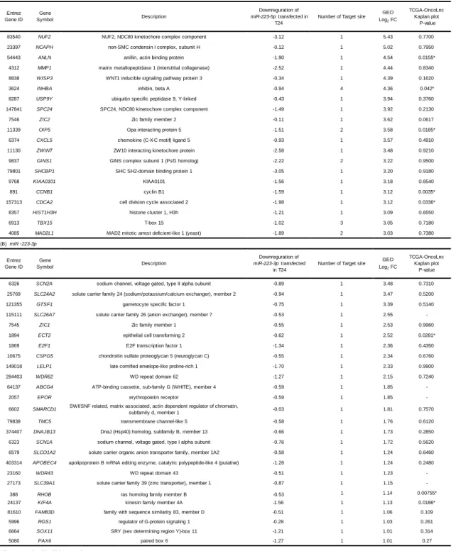
Regulation of putative oncogenic targets by miR-223-5p and miR-223-3p in BC cells Our strategy for identifying target oncogenes of miR-223-5p and miR-223-3p is shown in Supplemental Fig. 2. First, putative target genes of miR-223-5p were identified using the TargetScan database, leading us to 3,164 genes. Next, we carried out genome-wide gene expression analysis using a microarray comparing miR-223-5p- transfected and mock
transfected T24 cells (accession number: GSE 107008). As a result, a total of 2,556 genes were downregulated (log2 ratio < 0). Among these genes, we searched for genes that were upregulated
in BC cells (log2 fold-change > 3), using publicly available gene expression data sets in GEO
(accession number: GSE 31684 and GSE 11783). As a result, we identified 20 candidate target genes of miR-223-5p (Table 1A).
Using The Cancer Genome Atlas (TCGA) database, we investigated patient survival rates as a function of high or low expression levels of these genes. Among them, CCNB1 had the greatest effect on overall survival, but ANLN was higher in GEO log fold-change data (Figs. 2A and 2B). Therefore, we focused on ANLN as a candidate gene of miR-223-5p (see below). Using a similar approach, we found that 26 genes were candidate targets of miR-223-3p regulation (Table 1B).
Direct regulation of ANLN by anti-tumor miR-223-5p in BC cells
We confirmed that gene expression of ANLN was significantly decreased in BC cells transfected with miR-223-5p compared to the mock or miR-control BC cell lines (P < 0.001; Fig. 3A). Western blotting analyses revealed that ANLN protein levels of miR-223-5p-
transfected BC cells were decreased compared to mock or miR-control BC cell lines (Fig. 3B). On the other hand, the expression of ANLN was not reduced in miR-223-3p transfected BC cell lines at either the mRNA level or protein level (Figs. 3A and 3B).
Next, we performed luciferase reporter assays to determine whether miR-223-5p directly targeted the 3′-UTR region of ANLN. According to the TargetScan Human database, the binding
site for miR-223-5p in the 3′-UTR of ANLN consisted of a single region (positions 1007-1013; Fig. 3C). We showed that miR-223-5p suppressed reporter activity of ANLN wild-type vector transfectants compared to mock or miR-control transfectants, whereas transfectants of the deletion type vector were not decreased (Fig. 3D).
Effects of knockdown of ANLN in BC cells
To assess the effect of ANLN in BC cells, loss-of-function assays using siRNA were performed. We evaluated the knockdown efficiency of si-ANLN-transfected BC cell lines. Downregulation of ANLN/ANLN was detected in si-ANLN transfectants (Figs. 4A and 4B).
Cancer cell proliferation, migration and invasion abilities were significantly blocked in si-ANLN transfectants compared to those in mock or miR-control transfected BC cell lines (Figs. 4C-4E).
Expression of ANLN/ANLN in BC clinical specimens and its clinical significance The mRNA levels of ANLN were upregulated in 15 BC tissues compared to adjacent noncancerous tissues (P < 0.01; Fig. 5A). To investigate the relationship between ANLN expression and clinical features, we searched the TCGA database. Kaplan–Meier analysis showed that the high ANLN expression group had a significantly lower disease-free survival compared to the low ANLN expression group (P = 0.01; Fig. 5B). The histologically poor grade group was higher in ANLN expression (P < 0.0001; Fig. 5C).
Finally, we carried out immunostaining analyses of clinical specimens to evaluate ANLN protein expression. Immunostaining analyses revealed that ANLN was more strongly expressed in the BC clinical specimens than in the non-cancer specimens. Notably, ANLN was expressed at higher levels in the nuclei of the BC specimens. We also performed immunostaining of Ki67. The cells showing nuclear staining for ANLN were also positive for Ki67 (Fig. 5D). Therefore, it appeared that ANLN was expressed abundantly in the cancerous regions where cell
proliferation was active.
Discussion
It is generally believed that only guide strands of miRNAs are incorporated into the RISC and control target gene expression.30 Our original RNA sequencing-based miRNA expression
signatures revealed that some passenger strands of miRNAs might be involved in cancer pathogenesis. This finding expands the number of miRNAs to be studied in cancer pathology research. For example, our recent studies showed that miR-145-3p (a passenger strand of the miR-145-duplex) had anti-tumor roles, as did miR-145-5p in several cancers.19, 23, 31, 32 In
prostate cancer (PCa), expression of anti-tumor miR-145-3p was significantly reduced in castration-resistant PCa specimens, and its targeted genes (MELK, NCAPG, BUB1, and CDK1) predicted survival in such patients.23 More recently, we confirmed that miR-145-3p possessed
anti-tumor functions through its targeting of several oncogenes in head neck cancer.32
Our present data showed that both strands of the 223-duplex (223-5p and miR-223-3p) were downregulated in BC tissues and that these miRNAs had anti-tumor roles. The anti-tumor functions of miR-223-3p (guide strand) were reported in several cancers, such as breast and cervical cancers.33, 34 In contrast, oncogenic function of miR-223-3p was reported by
several cancers, T-cell acute lymphoblastic leukemia and gastric cancer.35 Our previous study of
PCa showed that miR-223-3p was significantly reduced in naïve PCa specimens and that ectopic expression of miR-223-3p significantly inhibited cancer cell migration and invasion.36
Moreover, our study found that expression of integrin α3 (ITGA3) and β1 (ITGB1) was directly regulated by miR-223-3p in PCa cells.36 Interestingly, both ITGA3 and ITGB1 were
overexpressed in PCa tissues and knockdown of these integrins inhibited cancer cell
aggressiveness through reduced downstream oncogenic signaling.36 Strategies to block
integrin-mediated oncogenic signaling might lead to new cancer treatments.
Previous studies showed that several transcription factors and oncogenes have been identified as miR-223-3p regulation in several cancer cell and disease cells.35, 37, 38 In our
present study of miR-223-3p target genes, some genes, e.g., E2F1, SMARCD1, RHOB, SOX11 and PAX6, have been reported as miR-223-3p regulation in previous reports. 35, 37-42 This
indicates that our miRNA target search strategy is reliable. Interestingly, overexpression of E2F1, a master transcriptional factor contributed to MIBC progression and its expression was significantly associated with EZH2 and SUZ12 expression.43, 44 Moreover, overexpression of
E2F1/EZH2/SUZ2 enhanced cancer cell migration, invasion and colony formation.43, 44 From
these things, screening for miR-223-3p or miR-223-5p targets could provide effective information for BC treatment.
To elucidate the involvement of both miR-223-5p and miR-223-3p in BC pathogenesis, we analyzed the molecular targets regulated by these molecules. In the present study, we identified 20 putative target genes regulated by 223-3p (the guide strand) and 26 by miR-223-5p (the passenger strand) in BC cells. Among these target genes (46 genes), high expression levels of 8 genes (ANLN, INHBA, OIP5, CCNB1, CDCA2, KIF4A, ECT2 and RHOB) were associated with poor prognosis of BC patients. Analysis of these genes is important for deeper understanding of the molecular pathogenesis of BC. We focused on ANLN because its
expression was regulated by miR-223-5p and its expression was deeply associated with poor prognosis of BC by a large number of cohort analyses.
actin-binding, myosin-binding and pleckstrin-homology domains.45 ANLN is localized
primarily in the nucleus during interphase; in telophase, it moves to the cytoplasm and forms the contractile ring and cleavage furrow.46 Overexpression of ANLN enhances the metastatic
potential of several types of cancer cells, including lung, breast and colon cancers.47-49 Recent
study of pancreatic adenocarcinoma (PDAC) showed that knockdown of ANLN significantly inhibited cancer cell migration and invasion.50 Moreover, Kaplan–Meier survival curves showed
that high expression of ANLN predicted shorter survival of patients with PDAC based on TCGA dataset analysis.50 More recently, ANLN was identified as a frequently overexpressed gene in
BC by RNA sequencing and its expression is a promising prognostic biomarker in this disease.51
These studies and our present data showed that aberrantly expressed ANLN acted as a pivotal player for BC cell aggressiveness and metastasis.51 Exploration of novel anti-tumor
miR-233-5p-mediated pathways may lead to the development of new treatment protocols for this disease. Previous studies indicated that expression of ANLN was directly regulated by 217 and miR-497 in pancreatic cancer and nasopharyngeal carcinoma, respectively.50, 52 It would be necessary
to investigate the involvement of these miRNAs in BC pathogenesis.
In conclusion, both strands of the miR-223-duplex, miR-223-5p (the passenger strand) and miR-223-3p (the guide strand), acted as anti-tumor miRNAs through their targeting of several oncogenic genes in BC cells. ANLN, a gene encoding a multifunctional actin-binding protein was directly regulated by miR-223-5p. Overexpression of ANLN was involved in the
pathogenesis of BC and acted as an oncogene. The anti-tumor function of the passenger strand of miRNA is a new concept in cancer research and searching for molecular mechanisms controlled by passenger strands of miRNA is a new challenge in studies of BC pathogenesis.
Acknowledgements
This study was supported by KAKENHI grants 17K16778(B), 17K16777(B), 16K20125(B), 17K11160(C), 16H05462(B), and 15K10801(C).
Conflict of interest
References
1. Antoni, S., Ferlay, J., Soerjomataram, I., Znaor, A., Jemal, A. & Bray, F. Bladder Cancer Incidence and Mortality: A Global Overview and Recent Trends. Eur Urol. 71, 96-108 (2017). 2. Siegel, R.L., Miller, K.D. & Jemal, A. Cancer statistics, 2015. CA Cancer J Clin. 65, 5-29
(2015).
3. Aldousari, S. & Kassouf, W. Update on the management of non-muscle invasive bladder cancer.
Can Urol Assoc J. 4, 56-64 (2010).
4. Veeratterapillay, R., Heer, R., Johnson, M.I., Persad, R. & Bach, C. High-Risk Non-Muscle-Invasive Bladder Cancer-Therapy Options During Intravesical BCG Shortage. Curr Urol Rep. 17, 68 (2016).
5. Meeks, J.J., Bellmunt, J., Bochner, B.H., Clarke, N.W., Daneshmand, S., Galsky, M.D. et al. A systematic review of neoadjuvant and adjuvant chemotherapy for muscle-invasive bladder cancer. Eur Urol. 62, 523-533 (2012).
6. DeGeorge, K.C., Holt, H.R. & Hodges, S.C. Bladder Cancer: Diagnosis and Treatment.
American family physician. 96, 507-514 (2017).
7. Bartel, D.P. MicroRNAs: genomics, biogenesis, mechanism, and function. Cell. 116, 281-297 (2004).
8. Filipowicz, W., Bhattacharyya, S.N. & Sonenberg, N. Mechanisms of post-transcriptional regulation by microRNAs: are the answers in sight? Nat Rev Genet. 9, 102-114 (2008). 9. Friedman, R.C., Farh, K.K.H., Burge, C.B. & Bartel, D.P. Most mammalian mRNAs are
conserved targets of microRNAs. Genome Research. 19, 92-105 (2009).
10. Kong, Y.W., Ferland-McCollough, D., Jackson, T.J. & Bushell, M. microRNAs in cancer management. Lancet Oncol. 13, e249-258 (2012).
11. Koshizuka, K., Hanazawa, T., Arai, T., Okato, A., Kikkawa, N. & Seki, N. Involvement of aberrantly expressed microRNAs in the pathogenesis of head and neck squamous cell carcinoma.
Cancer Metastasis Rev. 36, 525-545 (2017).
12. Yonemori, K., Kurahara, H., Maemura, K. & Natsugoe, S. MicroRNA in pancreatic cancer. J
Hum Genet. 62, 33-40 (2017).
13. Kita, Y., Yonemori, K., Osako, Y., Baba, K., Mori, S., Maemura, K. et al. Noncoding RNA and colorectal cancer: its epigenetic role. J Hum Genet. 62, 41-47 (2017).
14. Kurozumi, S., Yamaguchi, Y., Kurosumi, M., Ohira, M., Matsumoto, H. & Horiguchi, J. Recent trends in microRNA research into breast cancer with particular focus on the associations between microRNAs and intrinsic subtypes. J Hum Genet. 62, 15-24 (2017).
15. Koshizuka, K., Hanazawa, T., Fukumoto, I., Kikkawa, N., Okamoto, Y. & Seki, N. The microRNA signatures: aberrantly expressed microRNAs in head and neck squamous cell carcinoma. J Hum Genet. 62, 3-13 (2017).
16. Hobert, O. Gene regulation by transcription factors and microRNAs. Science. 319, 1785-1786 (2008).
17. Itesako, T., Seki, N., Yoshino, H., Chiyomaru, T., Yamasaki, T., Hidaka, H. et al. The microRNA expression signature of bladder cancer by deep sequencing: the functional significance of the miR-195/497 cluster. PLoS One. 9, e84311 (2014).
18. Yonemori, M., Seki, N., Yoshino, H., Matsushita, R., Miyamoto, K., Nakagawa, M. et al. Dual tumor-suppressors miR-139-5p and miR-139-3p targeting matrix metalloprotease 11 in bladder cancer. Cancer Sci. 107, 1233-1242 (2016).
19. Matsushita, R., Yoshino, H., Enokida, H., Goto, Y., Miyamoto, K., Yonemori, M. et al.
Regulation of UHRF1 by dual-strand tumor-suppressor microRNA-145 (145-5p and miR-145-3p): Inhibition of bladder cancer cell aggressiveness. Oncotarget. 7, 28460-28487 (2016). 20. Chendrimada, T.P., Gregory, R.I., Kumaraswamy, E., Norman, J., Cooch, N., Nishikura, K. et al.
TRBP recruits the Dicer complex to Ago2 for microRNA processing and gene silencing. Nature. 436, 740-744 (2005).
21. Bartel, D.P. MicroRNAs: target recognition and regulatory functions. Cell. 136, 215-233 (2009). 22. Koshizuka, K., Nohata, N., Hanazawa, T., Kikkawa, N., Arai, T., Okato, A. et al. Deep
sequencing-based microRNA expression signatures in head and neck squamous cell carcinoma: dual strands of pre-miR-150 as antitumor miRNAs. Oncotarget. 8, 30288-30304 (2017). 23. Goto, Y., Kurozumi, A., Arai, T., Nohata, N., Kojima, S., Okato, A. et al. Impact of novel
miR-145-3p regulatory networks on survival in patients with castration-resistant prostate cancer. Br J
Cancer. 117, 409-420 (2017).
24. Yonemori, K., Seki, N., Idichi, T., Kurahara, H., Osako, Y., Koshizuka, K. et al. The microRNA expression signature of pancreatic ductal adenocarcinoma by RNA sequencing: anti-tumour functions of the microRNA-216 cluster. Oncotarget. 8, 70097-70115 (2017).
25. Matsushita, R., Seki, N., Chiyomaru, T., Inoguchi, S., Ishihara, T., Goto, Y. et al. Tumour-suppressive microRNA-144-5p directly targets CCNE1/2 as potential prognostic markers in bladder cancer. Br J Cancer. 113, 282-289 (2015).
26. Arai, T., Okato, A., Kojima, S., Idichi, T., Koshizuka, K., Kurozumi, A. et al. Regulation of spindle and kinetochore-associated protein 1 by antitumor miR-10a-5p in renal cell carcinoma.
Cancer Sci. 108, 2088-2101 (2017).
27. Yamada, Y., Nishikawa, R., Kato, M., Okato, A., Arai, T., Kojima, S. et al. Regulation of
HMGB3 by antitumor miR-205-5p inhibits cancer cell aggressiveness and is involved in prostate cancer pathogenesis. J Hum Genet. 63, 195-205 (2018).
28. Okato, A., Arai, T., Yamada, Y., Sugawara, S., Koshizuka, K., Fujimura, L. et al. Dual Strands of Pre-miR-149 Inhibit Cancer Cell Migration and Invasion through Targeting FOXM1 in Renal Cell Carcinoma. Int J Mol Sci. 18 (2017).
29. Fukumoto, I., Hanazawa, T., Kinoshita, T., Kikkawa, N., Koshizuka, K., Goto, Y. et al.
MicroRNA expression signature of oral squamous cell carcinoma: functional role of microRNA-26a/b in the modulation of novel cancer pathways. Br J Cancer. 112, 891-900 (2015).
30. Mah, S.M., Buske, C., Humphries, R.K. & Kuchenbauer, F. miRNA*: a passenger stranded in RNA-induced silencing complex? Critical reviews in eukaryotic gene expression. 20, 141-148 (2010).
31. Mataki, H., Seki, N., Mizuno, K., Nohata, N., Kamikawaji, K., Kumamoto, T. et al. Dual-strand tumor-suppressor microRNA-145 (miR-145-5p and miR-145-3p) coordinately targeted MTDH in lung squamous cell carcinoma. Oncotarget. 7, 72084-72098 (2016).
32. Yamada, Y., Koshizuka, K., Hanazawa, T., Kikkawa, N., Okato, A., Idichi, T. et al. Passenger strand of miR-145-3p acts as a tumor-suppressor by targeting MYO1B in head and neck squamous cell carcinoma. Int J Oncol. 52, 166-178 (2018).
33. Pinatel, E.M., Orso, F., Penna, E., Cimino, D., Elia, A.R., Circosta, P. et al. miR-223 is a
coordinator of breast cancer progression as revealed by bioinformatics predictions. PLoS One. 9, e84859 (2014).
34. Tang, Y., Wang, Y., Chen, Q., Qiu, N., Zhao, Y. & You, X. MiR-223 inhibited cell metastasis of human cervical cancer by modulating epithelial-mesenchymal transition. Int J Clin Exp Pathol. 8, 11224-11229 (2015).
35. Gao, Y., Lin, L., Li, T., Yang, J. & Wei, Y. The role of miRNA-223 in cancer: Function, diagnosis and therapy. Gene. 616, 1-7 (2017).
36. Kurozumi, A., Goto, Y., Matsushita, R., Fukumoto, I., Kato, M., Nishikawa, R. et al. Tumor-suppressive microRNA-223 inhibits cancer cell migration and invasion by targeting ITGA3/ITGB1 signaling in prostate cancer. Cancer Sci. 107, 84-94 (2016).
37. Aziz, F. The emerging role of miR-223 as novel potential diagnostic and therapeutic target for inflammatory disorders. Cell Immunol. 303, 1-6 (2016).
38. Haneklaus, M., Gerlic, M., O'Neill, L.A. & Masters, S.L. miR-223: infection, inflammation and cancer. Journal of internal medicine. 274, 215-226 (2013).
39. Zeng, Y., Zhang, X., Kang, K., Chen, J., Wu, Z., Huang, J. et al. MicroRNA-223 Attenuates Hypoxia-induced Vascular Remodeling by Targeting RhoB/MLC2 in Pulmonary Arterial Smooth Muscle Cells. Sci Rep. 6, 24900 (2016).
40. Huang, B.S., Luo, Q.Z., Han, Y., Huang, D., Tang, Q.P. & Wu, L.X. MiR-223/PAX6 Axis Regulates Glioblastoma Stem Cell Proliferation and the Chemo Resistance to TMZ via Regulating PI3K/Akt Pathway. J Cell Biochem. 118, 3452-3461 (2017).
41. Arts, F.A., Keogh, L., Smyth, P., O'Toole, S., Ta, R., Gleeson, N. et al. miR-223 potentially targets SWI/SNF complex protein SMARCD1 in atypical proliferative serous tumor and high-grade ovarian serous carcinoma. Hum Pathol. 70, 98-104 (2017).
42. Zhou, K., Feng, X., Wang, Y., Liu, Y., Tian, L., Zuo, W. et al. miR-223 is repressed and
correlates with inferior clinical features in mantle cell lymphoma through targeting SOX11. Exp
Hematol. 58, 27-34.e21 (2018).
43. Lee, J.S., Leem, S.H., Lee, S.Y., Kim, S.C., Park, E.S., Kim, S.B. et al. Expression signature of E2F1 and its associated genes predict superficial to invasive progression of bladder tumors. J
Clin Oncol. 28, 2660-2667 (2010).
44. Lee, S.R., Roh, Y.G., Kim, S.K., Lee, J.S., Seol, S.Y., Lee, H.H. et al. Activation of EZH2 and SUZ12 Regulated by E2F1 Predicts the Disease Progression and Aggressive Characteristics of Bladder Cancer. Clin Cancer Res. 21, 5391-5403 (2015).
45. Oegema, K., Savoian, M.S., Mitchison, T.J. & Field, C.M. Functional analysis of a human homologue of the Drosophila actin binding protein anillin suggests a role in cytokinesis. The
Journal of cell biology. 150, 539-552 (2000).
46. Giansanti, M.G., Bonaccorsi, S. & Gatti, M. The role of anillin in meiotic cytokinesis of Drosophila males. Journal of cell science. 112 ( Pt 14), 2323-2334 (1999).
47. Suzuki, C., Daigo, Y., Ishikawa, N., Kato, T., Hayama, S., Ito, T. et al. ANLN plays a critical role in human lung carcinogenesis through the activation of RHOA and by involvement in the phosphoinositide 3-kinase/AKT pathway. Cancer research. 65, 11314-11325 (2005).
48. Wang, Z., Chen, J., Zhong, M.Z., Huang, J., Hu, Y.P., Feng, D.Y. et al. Overexpression of ANLN contributed to poor prognosis of anthracycline-based chemotherapy in breast cancer patients.
Cancer Chemother Pharmacol. 79, 535-543 (2017).
49. Wang, G., Shen, W., Cui, L., Chen, W., Hu, X. & Fu, J. Overexpression of Anillin (ANLN) is correlated with colorectal cancer progression and poor prognosis. Cancer biomarkers : section A
of Disease markers. 16, 459-465 (2016).
50. Idichi, T., Seki, N., Kurahara, H., Yonemori, K., Osako, Y., Arai, T. et al. Regulation of actin-binding protein ANLN by antitumor miR-217 inhibits cancer cell aggressiveness in pancreatic ductal adenocarcinoma. Oncotarget. 8, 53180-53193 (2017).
51. Zeng, S., Yu, X., Ma, C., Song, R., Zhang, Z., Zi, X. et al. Transcriptome sequencing identifies ANLN as a promising prognostic biomarker in bladder urothelial carcinoma. Sci Rep. 7, 3151 (2017).
52. Wang, S., Mo, Y., Midorikawa, K., Zhang, Z., Huang, G., Ma, N. et al. The potent tumor suppressor miR-497 inhibits cancer phenotypes in nasopharyngeal carcinoma by targeting ANLN and HSPA4L. Oncotarget. 6, 35893-35907 (2015).
Figure Legends
Figure 1. Expression levels of miR-223-5p and miR-223-3p in BC clinical specimens and cell lines: functional analyses of those miRNAs in BC cells.
(A) Quantitative real-time RT-PCR showed that the expression levels of 223-5p and miR-223-3p were significantly lower in BC tissues than in normal bladder tissues. Expression in 2 BC cell lines, BOY and T24, was lower than in normal bladder tissues. RNU48 was used as an internal control. (B) Correlations among the relative expression levels of 223-5p and miR-223-3p. (C-E) Effects of restoration of miR-223-5p and miR-223-3p in BC cells. Cell
proliferation (72 h after transfection with 10 nM miRNA) and migration and invasion (48 h after transfection with 10 nM miRNA) were characterized after transfection with 223-5p of miR-223-3p. *,P < 0.0001; **, P = 0.001
Figure 2. Kaplan–Meier plots of overall survival versus expression of the candidate genes regulated by miR-223-5p and miR-223-3p in BC cells.
The Cancer Genome Atlas (TCGA) database was used to analyze putative targets of miR-223-5p (A) and miR-223-3p (B) in BC. Kaplan-Meier plots of overall survival with log-rank tests for 8 genes with high and low expression in the BC TCGA database.
Figure 3. Direct regulation of ANLN by miR-223-5p in BC cell lines.
(A) ANLN mRNA expression in BC cell lines was evaluated by qRT-PCR 72 h after transfection with miR-223-5p. GUSB was used as an internal control. *, P < 0.0001. (B) ANLN protein expression in BC cell lines was evaluated by Western blot analyses 72 h after transfection with miR-223-5p. GAPDH was used as a loading control. (C) miR-223-5p binding sites in the 3′-UTR of ANLN mRNA. Dual luciferase reporter assays using vectors encoding putative miR-223-5p target sites (positions 1007-1013) of the ANLN 3′-UTR for both wild-type and deleted regions. Normalized data were calculated as ratios of Renilla/firefly luciferase activities. *, P < 0.005.
Figure 4. Effects of ANLN silencing in BC cell lines.
(A) ANLN mRNA expression in BC cell lines was evaluated by qRT-PCR 72 h after transfection with si-ANLN-1 or si-ANLN-2. GUSB was used as an internal control. (B) ANLN protein expression in BC cell lines was evaluated by Western blot analysis 72 h after transfection with si-ANLN-1 or si-ANLN-2. GAPDH was used as a loading control. (C) Cell proliferation was determined with the XTT assays 72 h after transfection with 10 nM si-ANLN-1 and si-ANLN-2. *,P < 0.0001. (D) Cell migration activity was determined by migration assays. *,P < 0.0001. (E) Cell invasion activity was determined using Matrigel invasion assays. *, P < 0.0001.
Figure 5. Expression levels of ANLN mRNA and immunohistochemical staining of ANLN protein in BC specimens.
(A) Expression levels of ANLN mRNA in BC or normal bladder tissues and BC cell lines. (B) The high ANLN expression group had significantly lower disease-free survival. p = 0.01 (C) The relation between the pathological grade of BC and ANLN expression. The high grade BC group shows higher ANLN expression. *, P < 0.0001. (D) Immunohistochemical staining of ANLN and Ki67 in BC specimens.
Supplemental Figure 1. Incorporation of miR-223-5p and miR-223-3p into the RNA-induced silencing complex (RISC) in T24 cells.
(A) Mature microRNA was transfected into T24 cells where it was incorporated into the RISC. The incorporated microRNA was prepared to high purity using immunoprecipitation with anti-human Ago2 monoclonal antibody. Expression of the microRNA was quantified with RT-PCR. (B) Expression of miR-223-5p was significantly higher than that in cells transfected with mock, miR control or miR-223-3p. P < 0.0001. (C) Expression of miR-223-3p was significantly higher than that in cells transfected with mock, miR control or miR-223-5p. *, P < 0.0001.
Supplemental Figure 2. Flow chart illustrates the strategy for analysis of miR-223-5p and
83540 NUF2 NUF2, NDC80 kinetochore complex component -3.12 1 5.43 0.7700
23397 NCAPH non-SMC condensin I complex, subunit H -0.12 1 5.02 0.7950
54443 ANLN anillin, actin binding protein -1.90 1 4.54 0.0155*
4312 MMP1 matrix metallopeptidase 1 (interstitial collagenase) -2.52 1 4.44 0.8340
8838 WISP3 WNT1 inducible signaling pathway protein 3 -0.34 1 4.39 0.1620
3624 INHBA inhibin, beta A -0.94 4 4.36 0.042*
8287 USP9Y ubiquitin specific peptidase 9, Y-linked -0.43 1 3.94 0.3760
147841 SPC24 SPC24, NDC80 kinetochore complex component -1.49 1 3.92 0.2130
7546 ZIC2 Zic family member 2 -0.11 1 3.62 0.0617
11339 OIP5 Opa interacting protein 5 -1.51 2 3.58 0.0185*
6374 CXCL5 chemokine (C-X-C motif) ligand 5 -0.93 1 3.57 0.4910
11130 ZWINT ZW10 interacting kinetochore protein -2.58 1 3.48 0.9210
9837 GINS1 GINS complex subunit 1 (Psf1 homolog) -2.22 2 3.22 0.9500
79801 SHCBP1 SHC SH2-domain binding protein 1 -3.05 1 3.20 0.9180
9768 KIAA0101 KIAA0101 -1.56 1 3.18 0.6540
891 CCNB1 cyclin B1 -1.59 1 3.12 0.0035*
157313 CDCA2 cell division cycle associated 2 -1.98 1 3.12 0.0336*
8357 HIST1H3H histone cluster 1, H3h -1.21 1 3.09 0.6550
6913 TBX15 T-box 15 -1.02 3 3.05 0.7180
4085 MAD2L1 MAD2 mitotic arrest deficient-like 1 (yeast) -1.89 2 3.03 0.7380
6326 SCN2A sodium channel, voltage gated, type II alpha subunit -0.89 1 3.48 0.7310 25769 SLC24A2 solute carrier family 24 (sodium/potassium/calcium exchanger), member 2 -0.94 1 3.47 0.5200
121355 GTSF1 gametocyte specific factor 1 -0.75 1 3.39 0.5140
115111 SLC26A7 solute carrier family 26 (anion exchanger), member 7 -0.53 1 2.55
-7545 ZIC1 Zic family member 1 -0.55 1 2.53 0.9960
1894 ECT2 epithelial cell transforming 2 -0.62 1 2.52 0.0281*
1869 E2F1 E2F transcription factor 1 -1.34 1 2.36 0.4350
10675 CSPG5 chondroitin sulfate proteoglycan 5 (neuroglycan C) -0.55 1 2.34 0.6760
149018 LELP1 late cornified envelope-like proline-rich 1 -1.70 1 2.33 0.9900
284403 WDR62 WD repeat domain 62 -1.27 1 2.15 0.7240
64137 ABCG4 ATP-binding cassette, sub-family G (WHITE), member 4 -0.59 1 1.85
-2057 EPOR erythropoietin receptor -0.59 1 1.85
-6602 SMARCD1 SWI/SNF related, matrix associated, actin dependent regulator of chromatin,
subfamily d, member 1 -0.03 1 1.81 0.7570
79838 TMC5 transmembrane channel-like 5 -0.58 1 1.76 0.6120
374407 DNAJB13 DnaJ (Hsp40) homolog, subfamily B, member 13 -0.66 1 1.73 0.2850
6323 SCN1A sodium channel, voltage gated, type I alpha subunit -0.76 1 1.72 0.5620 6579 SLCO1A2 solute carrier organic anion transporter family, member 1A2 -0.58 1 1.24 0.6460 403314 APOBEC4 apolipoprotein B mRNA editing enzyme, catalytic polypeptide-like 4 (putative) -1.28 1 1.24 0.2480
23160 WDR43 WD repeat domain 43 -0.51 1 1.23
-27173 SLC39A1 solute carrier family 39 (zinc transporter), member 1 -0.87 1 1.15
-388 RHOB ras homolog family member B -0.53 1 1.14 0.00755*
24137 KIF4A kinesin family member 4A -1.56 1 1.13 0.0186*
81610 FAM83D family with sequence similarity 83, member D -0.51 1 1.06 0.109
5996 RGS1 regulator of G-protein signaling 1 -0.28 1 1.03 0.261
6664 SOX11 SRY (sex determining region Y)-box 11 -1.21 1 1.01 0.314
5080 PAX6 paired box 6 -1.27 1 1.01 0.27
GEO Log2 FC (A) miR ‑ 223-5p
* Poor prognosis with a high expression
Table 1 Candidate target genes regulated by miR ‑ 223-5p and miR-223-3p in BC.
(B) miR ‑ 223-3p Entrez Gene ID Gene Symbol Description Downreguration of miR-223-3p transfected in T24
Number of Target site GEO Log2 FC TCGA-OncoLnc Kaplan plot P-value TCGA-OncoLnc Kaplan plot P-value Entrez Gene ID Gene Symbol Description Downreguration of miR-223-5p transfected in T24
No. Age Sex T N M operation method neoadjuvant therapy pathology grade remarks
1 51 M a 0 0 nephroureterectomy - Urothelial Carcinoma 3 RT-PCR
2 61 M a 0 0 cystectomy - Urothelial Carcinoma 2 RT-PCR
3 65 M 1 0 0 nephroureterectomy - Urothelial Carcinoma 3 RT-PCR
4 67 M 2 0 0 nephroureterectomy - Urothelial Carcinoma 3 RT-PCR
5 67 M 3b 0 0 cystectomy GCBDCA 4 course Urothelial Carcinoma 3 RT-PCR
6 70 M 2b 0 0 cystectomy GC 2 course Urothelial Carcinoma 3 RT-PCR / IHC
7 72 M 2 0 0 cystectomy MVAC 2 course Urothelial Carcinoma 3 RT-PCR
8 72 F 3 0 0 nephroureterectomy - Urothelial Carcinoma 2 RT-PCR
9 73 F 3 0 0 nephroureterectomy - Urothelial Carcinoma 3 RT-PCR
10 74 F 3 0 0 nephroureterectomy - Urothelial Carcinoma 3 RT-PCR
11 78 F 2 0 0 nephroureterectomy - Urothelial Carcinoma 3 RT-PCR
12 80 F 1 0 0 nephroureterectomy - Urothelial Carcinoma 3 RT-PCR
13 81 M 2 0 0 nephroureterectomy - Urothelial Carcinoma 3 RT-PCR
14 81 M 3 0 0 nephroureterectomy - Urothelial Carcinoma 2 RT-PCR
15 81 M 3 0 0 nephroureterectomy - Urothelial Carcinoma 3 RT-PCR
16 68 F 1 0 0 cystectomy GC 2 course Urothelial Carcinoma 2 IHC
GCBDCA: Gemcitabine Carboplatin; GC: Gemcitabine Cisplatin; MVAC: Methotrexate Vinblastine Doxorubicin Cisplatin Supplemental Table 1. Clinical features of patients