AHA/ASA Guideline
2015 AHA/ASA Focused Update of the 2013 Guidelines for the Early Management of Patients With Acute Ischemic Stroke Regarding Endovascular Treatment
A Guideline for Healthcare Professionals From the American Heart Association/American Stroke Association
The American Academy of Neurology affirms the value of this guideline as an educational tool for neurologists.
Endorsed by the American Association of Neurological Surgeons (AANS); Congress of Neurological Surgeons (CNS); AANS/CNS Cerebrovascular Section; American Society of
Neuroradiology; and Society of Vascular and Interventional Neurology
William J. Powers, MD, FAHA, Chair; Colin P. Derdeyn, MD, FAHA, Vice Chair; José Biller, MD, FAHA; Christopher S. Coffey, PhD; Brian L. Hoh, MD, FAHA;
Edward C. Jauch, MD, MS, FAHA; Karen C. Johnston, MD, MSc;
S. Claiborne Johnston, MD, PhD, FAHA; Alexander A. Khalessi, MD, MS, FAHA; Chelsea S. Kidwell, MD, FAHA; James F. Meschia, MD, FAHA;
Bruce Ovbiagele, MD, MSc, MAS, FAHA; Dileep R. Yavagal, MD, MBBS; on behalf of the American Heart Association Stroke Council
by guest on September 4, 2017 http://stroke.ahajournals.org/ Downloaded from by guest on September 4, 2017 http://stroke.ahajournals.org/ Downloaded from by guest on September 4, 2017 http://stroke.ahajournals.org/ Downloaded from by guest on September 4, 2017 http://stroke.ahajournals.org/ Downloaded from by guest on September 4, 2017 http://stroke.ahajournals.org/ Downloaded from by guest on September 4, 2017 http://stroke.ahajournals.org/ Downloaded from
The American Heart Association makes every effort to avoid any actual or potential conflicts of interest that may arise as a result of an outside relationship or a personal, professional, or business interest of a member of the writing panel. Specifically, all members of the writing group are required to complete and submit a Disclosure Questionnaire showing all such relationships that might be perceived as real or potential conflicts of interest.
This statement was approved by the American Heart Association Science Advisory and Coordinating Committee on June 5, 2015, and the American Heart Association Executive Committee on June 12, 2015. A copy of the document is available at http://my.americanheart.org/statements by selecting either the “By Topic” link or the “By Publication Date” link. To purchase additional reprints, call 843-216-2533 or e-mail kelle.ramsay@wolterskluwer.com.
The American Heart Association requests that this document be cited as follows: Powers WJ, Derdeyn CP, Biller J, Coffey CS, Hoh BL, Jauch EC, Johnston KC, Johnston SC, Khalessi AA, Kidwell CS, Meschia JF, Ovbiagele B; Yavagal DR; on behalf of the American Heart Association Stroke Council. 2015 AHA/ASA focused update of the 2013 guidelines for the early management of patients with acute ischemic stroke regarding endovascular treatment: a guideline for healthcare professionals from the American Heart Association/American Stroke Association. Stroke. 2015;46:•••–•••.
Expert peer review of AHA Scientific Statements is conducted by the AHA Office of Science Operations. For more on AHA statements and guidelines development, visit http://my.americanheart.org/statements and select the “Policies and Development” link.
Permissions: Multiple copies, modification, alteration, enhancement, and/or distribution of this document are not permitted without the express permission of the American Heart Association. Instructions for obtaining permission are located at http://www.heart.org/HEARTORG/General/Copyright-Permission-Guidelines_UCM_300404_Article.jsp. A link to the “Copyright Permissions Request Form” appears on the right side of the page.
(Stroke. 2015;46:000-000.)
© 2015 American Heart Association, Inc.
Stroke is available at http://stroke.ahajournals.org.
DOI: 10.1161/STR.0000000000000074
by guest on September 4, 2017
http://stroke.ahajournals.org/
Abstract
Purpose—The aim of this guideline is to provide a focused update of the current recommendations for the endovascular treatment of acute ischemic stroke. Where there is overlap, the recommendations made here supersede those of previous guidelines.
Methods—This focused update analyzes results from 8 randomized clinical trials of endovascular treatment and other relevant data published since 2013. It is not intended to be a complete literature review from the date of the previous guideline publication but rather to include pivotal new evidence that justifies changes in current recommendations. Members of the writing committee were appointed by the American Heart Association/American Stroke Association Stroke Council’s Scientific Statement Oversight Committee and the American Heart Association/American Stroke Association Manuscript Oversight Committee (MOC). Strict adherence to the American Heart Association conflict of interest policy was maintained throughout the consensus process. Recommendations follow the American Heart Association/American Stroke Association methods of classifying the level of certainty of the treatment effect and the class of evidence. Prerelease review of the draft guideline was performed by 6 expert peer reviewers and by the members of the Stroke Council Scientific Statement Oversight Committee and Stroke Council Leadership Committee.
Results—Evidence-based guidelines are presented for the selection of patients with acute ischemic stroke for endovascular treatment, the endovascular procedure and for systems of care to facilitate endovascular treatment.
by guest on September 4, 2017
http://stroke.ahajournals.org/
Conclusions—Certain endovascular procedures have been demonstrated to provide clinical benefit in selected patients with acute ischemic stroke. Systems of care should be organized to facilitate the delivery of this care.
Key Words: AHA Scientific Statements; stroke treatment; endovascular stroke treatment; intra-arterial stroke treatment; neurointerventional stroke treatment; stent retriever; ischemic stroke
by guest on September 4, 2017
http://stroke.ahajournals.org/
INTRODUCTION
Since the publication of the most recent “Guidelines for the Early Management of Patients With Acute Ischemic Stroke” in 2013,1 substantial new high-quality evidence regarding the clinical efficacy of endovascular treatments of acute ischemic stroke has become available. This focused update on endovascular treatment of acute ischemic stroke analyzes results from 8 randomized clinical trials of endovascular treatment and other relevant data published since 2013, while taking into account the previous evidence summarized in the 2013 guidelines. This focused update is not intended to be based on a complete literature review from the date of the previous guideline publication but rather to include pivotal new evidence that justifies changes in current recommendations. Where there is overlap, the recommendations made here supersede those of previous guidelines.
Members of the writing committee were appointed by the American Heart Association/American Stroke Association (AHA/ASA) Stroke Council’s Scientific Statement Oversight Committee and the AHA/ASA Manuscript Oversight Committee, representing various areas of medical expertise. Strict adherence to the AHA conflict of interest policy was maintained throughout the consensus process. Panel members were assigned topics relevant to their areas of expertise, reviewed the stroke literature with emphasis on publications since the prior guidelines, and drafted recommendations in accordance with the American College of Cardiology/AHA’s Level of Evidence grading algorithm (Table 1). All recommendations were unanimously approved by the members of the writing group.
by guest on September 4, 2017
http://stroke.ahajournals.org/
TREATMENT WITH INTRAVENOUS RECOMBINANT TISSUE-TYPE PLASMINOGEN ACTIVATOR
Rapid administration of intravenous recombinant tissue-type plasminogen activator (r-tPA) to appropriate patients remains the mainstay of early treatment of acute ischemic stroke.1 Timely restoration of blood flow in ischemic stroke patients is effective in reducing long term morbidity. For patients who meet national and international eligibility guidelines, intravenous r-tPA administration improves functional outcomes at 3 to 6 months when given within 4.5 hours of ischemic stroke onset and should be administered. Every effort should be made to shorten any delays in initiation of treatment as earlier treatments are associated with increased benefits. If patients who are eligible for intravenous r-tPA do not have intracranial vascular imaging as part of their initial evaluation, they should begin receiving intravenous r-tPA before being transported for additional imaging and before being transferred for endovascular treatment. This approach will help minimize onset-to-treatment times, a key driver of efficacy for r-tPA.1-6
NEW RANDOMIZED CLINICAL TRIALS OF ENDOVASCULAR STROKE TREATMENT
Studies With Primarily Intra-Arterial Fibrinolysis and/or First-Generation Mechanical Embolectomy Devices (Tables 2-4)
SYNTHESIS Expansion was a prospective, randomized, open-label, blinded-end point (PROBE) 2-arm superiority trial that enrolled 362 patients with ischemic stroke eligible for intravenous r-tPA within 4.5 hours of onset and for whom endovascular treatment was possible within 6 hours. No imaging other than nonenhanced computed tomography (CT) was required. The patients were randomized 1:1 to standard dose intravenous r-tPA 0.9 mg/kg or endovascular therapy
by guest on September 4, 2017
http://stroke.ahajournals.org/
arterial r-tPA, mechanical clot disruption or retrieval, or combination of these approaches).Only 8% had posterior circulation strokes. Median onset to treatment time interval was 2.75 hours in the intravenous r-tPA group and 3.75 hours in the endovascular group. Among the patients who received endovascular treatment, 66% underwent infusion of intra-arterial r-tPA and thrombus fragmentation with a guidewire only; in 34% a device was also deployed. Stent retrievers were used in 14%. Data on rates and efficacy of recanalization were not published. There was no difference in the primary end point of the percentage with good outcome defined as modified Rankin scale (mRS)7,8 score of 0 or 1 or in death at 3 months or in symptomatic intracerebral hemorrhage (sICH) at 7 days. There were no significant differences in outcome in subgroups including time to treatment (0-3 or 3-4.5 hours), baseline National Institutes of Health Stroke Scale (NIHSS)9 score (<11, ≥11), and age (≤67 years, >67 years).10
The Interventional Management of Stroke Trial III (IMS III) was a PROBE, 2-arm, superiority trial that enrolled patients with a major ischemic stroke defined by NIHSS score ≥10 who received intravenousr-tPA within 3 hours and were likely to or known to have occlusion of a major cerebral artery. Those who showed clear hypodensity in greater than one third of the middle cerebral artery (MCA) territory on nonenhanced CT were excluded. No other imaging was required. An amendment midway through the trial allowed screening with computed tomographic angiography (CTA) for patients with NIHSS score of >8. Over 95% received a clinical diagnosis of anterior circulation stroke. Patients were randomly allocated 1:2 to standard dose intravenous r-tPA (0.9 mg/kg) or to intravenous r-r-tPA 0.6 mg/kg followed by endovascular therapy with a device and/or intra-arterial r-tPA, if occlusion persisted and if the endovascular intervention could be begun within 5 hours and completed within 7 hours of onset. In the endovascular group, groin puncture occurred at a mean of 208±47 (SD) minutes after stroke onset. Endovascular therapy was
by guest on September 4, 2017
http://stroke.ahajournals.org/
administered in 77% randomized to this treatment group. Intra-arterial r-tPA alone was used in 41% and a device with or without intra-arterial r-tPA in 59%, in only 1.5% were stent retrievers used. Recanalization occurred 325±52 (SD) minutes after stroke onset achieving Thrombolysis In Cerebral Infarction (TICI) grade11 of 2b/3 in 41%. The trial was stopped early for futility after 656 of projected 900 subjects were enrolled. There was no significant difference in outcome between the intravenous r-tPA only group and the endovascular group for the primary end point of the percentage of patients with a good outcome as measured by mRS score of 0 to 2 or for death at 90 days. In the endovascular group, there was no difference in outcome between those treated <90 minutes versus >90 minutes from intravenousr-tPA to groin puncture. The proportion of patients with mRS score of 0 to 2 at 90 days increased with increasing recanalization.12
MR and Recanalization of Stroke Clots Using Embolectomy (MR RESCUE) was a PROBE, 2-arm, superiority trial that enrolled 118 patients with large artery occlusion and anterior circulation ischemic stroke within 8 hours who were ineligible for intravenous r-tPA or had persistent vessel occlusion after intravenous r-tPA. Patients were divided into 2 subgroups by pretreatment CT or MRI into those with a favorable or an unfavorable penumbral pattern using imaging criteria based on a previous study.13 Patients were randomly allocated 1:1 to standard medical care or endovascular therapy (MERCI or Penumbra device with optional intra-arterial r-tPA). Onset to groin puncture in endovascular group was 6.35±1.2 (SD) hours. TICI 2b/3 recanalization was achieved in 25% of the endovascular group. Among all patients, mean scores on the mRS at 90 days did not differ between endovascular and standard medical care, nor was endovascular therapy superior to standard medical care in patients with a favorable penumbral pattern (mean score, 3.9 vs 3.4; P=0.23) or in patients with an unfavorable penumbral pattern, (mean score, 4.0 vs 4.4; P=0.32).14
by guest on September 4, 2017
http://stroke.ahajournals.org/
Studies With Primarily Stent Retrievers (Tables 2-4)
The Multicenter Randomized Clinical Trial of Endovascular Treatment for Acute Ischemic Stroke (MR CLEAN) was a PROBE, 2-arm, superiority trial that studied 500 patients with acute ischemic stroke caused by an proximal intracranial occlusion in the anterior circulation (distal intracranial carotid artery, MCA [M1 or M2], or anterior cerebral artery [A1 or A2]) established with CTA, magnetic resonance angiography (MRA), or digital-subtraction angiography (DSA), and a score of ≥2 on the NIHSS. The steering committee recommended that neuroimaging studies to assess vessel patency should preferably be done before or simultaneously with treatment with intravenous r-tPA. Initiation of endovascular treatment within 6 hours of stroke onset had to be possible. There were different specific exclusion criteria for patients with coagulation abnormalities, previous ischemic stroke, ICH, or severe head trauma depending on whether intra-arterial fibrinolysis was contemplated. Patients who were eligible in agreement with national guidelines received intravenous r-tPA. Those with a nonfavorable response were eligible for inclusion. There was no specified time for observation to determine the response to intravenous r-tPA nor was there an exact definition of what constituted a nonfavorable response, although recovery to level that would not result in administration of intravenous r-tPA was suggested. Patients were randomly allocated 1:1 to either usual care alone or intra-arterial treatment plus usual care. Intra-arterial treatment consisted of arterial catheterization with a microcatheter to the level of occlusion and delivery of a fibrinolytic agent, mechanical thrombectomy, or both. The method of intra-arterial treatment was left to the discretion of the local interventionist. Sixty-four percent of participants had M1 occlusion alone and an additional 27% had occlusion of M1 and the internal carotid artery (ICA). Of the 195 patients in the endovascular group of 233 who received endovascular treatment, onset
by guest on September 4, 2017
http://stroke.ahajournals.org/
to groin puncture was 260 minutes (interquartile range [IQR], 210–313), a stent retriever was used in 81.5% and TICI 2b/3 recanalization was achieved in 59%. The treatment effect was estimated as an odds ratio (OR), adjusted for prespecified prognostic factors that intra-arterial treatment would lead to lower mRS score at 90 days, compared with usual care alone (shift analysis). The adjusted OR was 1.67 (95% confidence interval [CI], 1.21–2.30) in favor of intervention. There was an absolute difference of 13.5% (95% CI, 5.9–21.2) in the rate of functional independence (mRS score, 0-2) in favor of the intervention (32.6% vs 19.1%). There were no significant differences in mortality or the occurrence of sICH. Most patients received intravenous r-tPA (445/500) and showed benefit in subgroup analysis. There were too few patients who did not receive intravenous r-tPA to draw any conclusions.15 In a subsequent presentation at the 2015 International Stroke Conference, the MR CLEAN investigators reported a stroke onset to reperfusion time of 332 minutes (IQR, 279–394) and demonstrated a marked decline in clinical benefit with time such that the benefit was no longer statistically significant if reperfusion occurred after 6 hours and 19 minutes.16
The Endovascular Treatment for Small Core and Anterior Circulation Proximal Occlusion with Emphasis on Minimizing CT to Recanalization Times (ESCAPE) was a PROBE, 2-arm superiority trial of 316 patients with disabling acute ischemic stroke (NIHSS score >5) who could be randomized up 12 hours after the onset. Groin puncture had to be possible within 60 minutes of CT/CTA. Nonenhanced CT and CTA (preferably multiphase) were performed rapidly with a target door-to-imaging time of 25 minutes to identify participants with a small infarct core (by Alberta Stroke Program Early CT Score [ASPECTS]17 6-10 or CT perfusion), an occluded proximal intracranial artery in the anterior circulation (internal carotid, M1 MCA, or ≥2 M2s), and moderate-to-good collateral circulation defined as “the filling of 50% or more of the
by guest on September 4, 2017
http://stroke.ahajournals.org/
cerebral artery pial arterial circulation on CTA (preferably on multiphase CTA).” There were no exclusions for coagulopathy, prior stroke or head trauma. Fifty-eight patients received intravenous r-tPA at a community hospital and then were transferred to an ESCAPE endovascular center.
Participants were randomly assigned 1:1 to receive guideline-based care alone or guideline-based care plus endovascular treatment with the use of available thrombectomy devices. The use of retrievable stents and suction through a balloon guide catheter during thrombus retrieval was also recommended. Participants in both groups received intravenous r-tPA within 4.5 hours after onset if they met accepted local guidelines. The primary outcome was the OR that the intervention would lead to lower scores on the mRS at 90 days (shift analysis). After the release of the MR CLEAN results, an interim analysis was conducted earlier than planned that showed that a stopping criterion based on the prespecified O’Brien-Fleming stopping boundary had been crossed and the trial was stopped. For the primary end point, the adjusted OR (indicating the odds of improvement of 1 point on the mRS) was 3.1 (95% CI, 2.0–4.7) favoring endovascular intervention. The proportion of patients with an mRS score of 0 to 2 at 90 days was 53.0% in the intervention group and 29.3% in the control group (P<0.001). Mortality at 90 days was 10.4% in the intervention group and 19.0% in the control group (adjusted rate ratio, 0.5; 95% CI, 0.3–0.8). The rate of sICH clinically determined at the study sites was 3.6% in the endovascular intervention group and 2.7% in the control group (adjusted rate ratio, 1.2; 95% CI, 0.3–4.6). Retrievable stents were used in 130 of the 151 participants (86.1%) who underwent an endovascular procedure. TICI 2b/3 recanalization was observed in 72.4% in the endovascular group. In subgroup analysis, similar benefit was observed in the 235 patients who received intravenous r-tPA (OR, 2.5 [1.6–4.0]) and the 76 who did not (OR, 2.6 [1.1– 5.9]). Only 49 participants (15.5%) underwent randomization ≥6 hours after symptom onset; too few to assess efficacy in the 6- to 12-hour time window.18
by guest on September 4, 2017
http://stroke.ahajournals.org/
Solitaire FR with the Intention for Thrombectomy as Primary Endovascular Treatment of Acute Ischemic Stroke (SWIFT PRIME) was a PROBE design trial randomizing 196 patients with acute ischemic stroke and NIHSS scores 8 to 29 who received intravenous r-tPA within 4.5 hours of onset and had CTA or MRA confirmation of intracranial ICA, M1 or carotid terminus occlusion. If CTA or MRA was part of local standard of care, it was performed at initial evaluation prior to commencing intravenous r-tPA; if not, it was performed after review of the initial imaging and signing of informed consent. Groin puncture had to be possible within 6 hours of stroke onset. There were exclusion criteria for coagulopathies. Initially, CT perfusion or multimodal MRI was required and enrollment was restricted to patients with the target mismatch profile (as assessed by specialized software19) and defined as: the ischemic core lesion measured ≤50 mL, the volume of tissue with a time to maximum delay of >10 seconds was ≤100 mL, and the mismatch volume was at least 15 mL and the mismatch ratio was >1.8. Midway through the trial, the inclusion criteria there were modified to accommodate sites with limited perfusion imaging capability. Sites with perfusion imaging were encouraged to continue to use the target mismatch criteria. Sites without perfusion imaging used ASPECTS (ASPECTS >6 was required). A total of 71 patients were enrolled under the initial imaging entry criteria and 125 patients under the revised imaging entry criteria. Perfusion imaging was performed and used for selection in 82.6%. Seventy-three percent of participants had M1 occlusion and 17% had internal carotid artery occlusion. Intravenous r-tPA was administered at an outside hospital in 35%. Participants were randomized 1:1 to treatment with intravenous r-tPA alone or treatment with intravenous r-tPA followed by neurovascular thrombectomy with the use of a stent retriever. After the results of the MR CLEAN trial and the passing of stopping boundaries in the ESCAPE trial were announced, a decision was made to conduct the first interim efficacy analysis a little earlier than originally planned. The results of this
by guest on September 4, 2017
http://stroke.ahajournals.org/
interim efficacy analysis demonstrated that the prespecified criteria for stopping the trial at the first interim analysis were met. The 2 simultaneous success criteria used for the primary end point were both in favor of endovascular intervention: improved distribution (shift analysis) of mRS score at 90 days (P<0.001) and increased proportion with mRS score of 0 to 2 at 90 days (60% in the endovascular group and 35% in the nonendovascular group; risk ratio 1.70; 95% CI, 1.23– 2.33). There were no significant differences in death or sICH. TICI 2b/3 recanalization was observed in 88% of the endovascular group.20
The Extending the Time for Thrombolysis in Emergency Neurological Deficits-Intra-Arterial (EXTEND-IA) was similar in design to SWIFT PRIME. Seventy participants who were
eligible using “standard criteria” to receive intravenous r-tPA within 4.5 hours of stroke onsetwere randomized in a PROBE design either to receive either intravenous tPA only or intravenous r-tPA plus endovascular therapy with a stent retriever. Groin puncture had to be within 6 hours and endovascular treatment had to be completed within 8 hours after stroke onset. CT or MRI had to be performed before commencing intravenous r-tPA. Occlusion of the ICA or of M1 or M2 on CTA was required. In addition, CT or MRI perfusion imaging had to show (1) mismatch ratio of >1.2, (2) absolute mismatch volume of >10 mL, and (3) infarct core lesion volume of <70 mL based on specialized software.19 There were specified exclusion criteria for coagulopathies.
Occlusion of the ICA was present in 31% and of M1 in 54%. The coprimary outcomes were reperfusion at 24 hours and early neurologic improvement (≥8-point reduction on the NIHSS or a score of 0 or 1 at day 3). The mRS score at 90 days was a secondary outcome. After the release of the MR CLEAN results, an unplanned interim efficacy analysis was implemented based on a Haybittle-Peto stopping rule. The results of the interim analysis showed that the stopping criteria for efficacy were met and the trial was halted. The percentage of ischemic territory that had
by guest on September 4, 2017
http://stroke.ahajournals.org/
undergone reperfusion at 24 hours was greater in the endovascular therapy group than in the intravenous r-tPA–only group (median, 100% vs 37%; P<0.001). Endovascular therapy, initiated at a median of 210 minutes (IQR, 166–251) after the onset of stroke, increased early neurologic improvement at 3 days (80% vs 37%; P=0.002) More patients achieved functional independence in the endovascular group (score of 0 to 2 on the mRS, 71% vs 40%; P=0.01). There were no significant differences in rates of death or sICH. Recanalization to TICI 2b/3 was achieved in 86% of patients in the endovascular group at a median of 248 minutes (IQR, 204–277) after stroke onset.21
Randomized Trial of Revascularization With Solitaire FR Device Versus Best Medical Therapy
REVASCAT (Endovascular Revascularization With Solitaire Device Versus Best Medical Therapy in Anterior Circulation Stroke Within 8 Hours) was a PROBE design trial randomizing 206 patients with acute ischemic stroke and NIHSS score of ≥6 who had intracranial ICA or M1 occlusion by CTA, MRA, or DSA. Patients who had received intravenous r-tPA were eligible, if there was if there was no significant neurological improvement (criteria specified in the protocol) at 30 minutes postinitiation of the infusion and vascular imaging at this time confirmed an eligible occlusion. Groin puncture had to be possible within 8 hours of stroke onset. There were exclusion criteria for coagulopathies. The main exclusion criteria on imaging were ASPECTS of <7 on nonenhanced CT or <6 on DWI-MRI. After the enrollment of 160 patients, the inclusion criteria were modified to include patients up to the age of 85 years (initially 80 years was maximum allowed) with an ASPECTS of >8. Twenty-six percent had ICA occlusion and 65% had M1 occlusion. Participants were randomized 1:1 to receive either medical therapy alone or
by guest on September 4, 2017
http://stroke.ahajournals.org/
thrombectomy with a stent retriever. Intravenous r-tPA was administered to 73%. When results of other similar trials became known, the Data Safety Monitoring Board recommended to stop recruitment because the emerging results showed that equipoise was lost, although the interim results did not reach the prespecified stopping boundaries. The masked steering committee agreed. Because just 1 analysis was performed, adjustment for multiple comparisons was no longer performed, and 95% CIs were reported. The primary outcome analysis showed a common OR of improvement in the distribution of the mRS score (shift analysis) favoring endovascular treatment (adjusted OR, 1.7; 95% CI, 1.05–2.8). The proportion of patients with a mRS score of 0 to 2 at 90 days was 43.7% in the intervention group and 28.2% in the control group (adjusted OR, 2.1; 95% CI, 1.1–4.0). There were no significant differences in death or sICH. Ninety-five percent of those in the endovascular group underwent thrombectomy. TICI 2b/3 recanalization was observed in 66% of the endovascular group. Across the prespecified subgroups, there were no significant interactions according to NIHSS score, vessel-occlusion site, baseline ASPECTS, administration of intravenous r-tPA, age or time of randomization, although for the latter dichotomized at 4.5 hours the P value for interaction was 0.9 with the latter group doing worse. No data are given for those who underwent groin puncture after 6 hours.22
ANALYSIS AND CONCLUSIONS
None of the 3 earlier studies carried out with primarily intra-arterial fibrinolysis and/or first-generation mechanical embolectomy devices showed a benefit of endovascular treatment over intravenous r-tPA in intravenous r-tPA–eligible patients either as a substitute for initial treatment (SYNTHESIS Expansion [Intra-arterial Versus Systemic Thrombolysis for Acute Ischemic Stroke]) or as subsequent intervention in those with persistent large artery occlusion after
by guest on September 4, 2017
http://stroke.ahajournals.org/
intravenous r-tPA (IMS III and MR RESCUE). MR RESCUE also showed no benefit for other patients treated within 8 hours even if selected by multimodal neuroimaging criteria. These studies, using almost exclusively intra-arterial r-tPA and first-generation endovascular devices alone or in combination, achieved recanalization rates of 25% to 41%. The subsequent trials using almost exclusively stent retrievers demonstrated improved results for both recanalization rates and outcome. Studies have shown that clinical outcome improved with increasing effectiveness of recanalization. Those with partial recanalization (TICI 2a) did not do as well as those with near complete/complete recanalization. TICI 2b/3 reflected as differences in discharge disposition (41.0% of TICI 2b/3 group discharged home vs 17.4% of TICI 2a) and functional outcome (34% with a TICI grade of 2a had an mRS score of 0 to 2 at 90 days vs in 49% a TICI grade of 2b/3).12,23 TICI 2b/3 recanalization was achieved in 59% to 88% of endovascularly treated subjects in the 5 stent retriever trials, whereas in the previous 3 studies the rate had been 25% to 41%, as mentioned above. All 5 stent retriever studies showed clinical benefit in the endovascular group.
Of the 5 stent retriever trials, MR CLEAN, ESCAPE, and SWIFT PRIME permitted use of salvage intra-arterial fibrinolytic drugs whereas EXTEND-IA and REVASCAT did not. These data do not establish the benefit of intra-arterial fibrinolytic salvage nor can they establish lack of benefit. Such salvage techniques may be reasonable to employ in some clinical circumstances.
The MR RESCUE trial enrolled patients up to 8 hours from symptom onset and showed no benefit from endovascular therapy with first-generation devices regardless of penumbral imaging pattern. Three of the 5 stent retriever studies specified a 6-hour window after stroke onset (2 specified 6 hours to groin puncture; the third specified 6 hours to start treatment). Aggregate data from REVASCAT and ESCAPE with treatment permitted out to 8 and 12 hours show a benefit, but ESCAPE enrolled too few patients after 6 hours to provide useful data and
by guest on September 4, 2017
http://stroke.ahajournals.org/
REVASCAT provides no data about patients who underwent groin puncture between 6 and 8 hours. How much the overall positivity in these 2 trials was completely driven by those treated at shorter times is unknown at this time. The only time dependent data are from the MR CLEAN presentation, which are not consistent with a benefit of treatment beginning after 6 hours. It will take patient level meta-analyses to sort this out.
Every, or nearly every, patient in the 5 stent retriever studies first received intravenous tPA. Only REVASCAT stipulated specific the guidelines to be used to determine intravenous r-tPA eligibility (“guidelines provided by the European Stroke Organization [ESO]”). EXTEND-IA refers to “standard criteria” and the 3 other trials used “national guidelines”. Because it is not the purpose of this update is to address eligibility criteria for intravenous r-tPA, we have used the phrase “guidelines from professional medical societies” to address this issue in our recommendations. Too few data are available from the small number of those who did not receive intravenous r-tPA, either for time-based or nontime-based exclusion criteria, to determine with certainty if there are characteristics that identify those who benefited from endovascular treatment. Two trials (MR CLEAN and REVASCAT) stipulated waiting for a period of time after beginning administration of intravenous r-tPA before proceeding to endovascular therapy, whereas 3 (ESCAPE, SWIFT PRIME, and EXTEND-IA) did not. Based on these data, a waiting period is not necessary to achieve beneficial outcome in these patients.
All of these studies enrolled participants ≥18 years of age. There are no randomized trials of endovascular therapy in patients <18 years of age. Ischemic stroke due to large vessel occlusion is rare in children and young adults relative to older individuals, posing challenges to rigorous study of this clinical scenario. Case reports and case series have documented that high rates of recanalization and favorable outcomes in young patients can be achieved with endovascular
by guest on September 4, 2017
http://stroke.ahajournals.org/
therapy.24-26 Ideally, appropriate trials would be done to test the efficacy of endovascular therapy in young patients. Studies in the United States, United Kingdom, Australia, and Canada have shown median times from onset of symptoms to initial brain imaging for pediatric stroke of 8.8 to 16 hours.27 This problem of diagnostic delay will need to be addressed if acute trials are to be conducted successfully in this population.
Four stent retriever trials used NIHSS scores as eligibility criteria (>2, >5, 8–29, and >5) and the fifth enrolled patients with a similar distribution of NIHSS scores. Based on these trials, there are insufficient data in patients with NIHSS scores <6 to determine if there is an overall net benefit from endovascular therapy in this population. Further randomized trials in patients with low NIHSS scores may be warranted. A NIHSS score of ≥6 was the minimum score used in 2 trials thus fulfilling the AHA’s Level of Evidence grading algorithm for Level A evidence.
Four of the 5 stent retriever trials used a prestroke function eligibility criterion. REVASCAT and SWIFT PRIME used a prestroke mRS score of 0 to 1, EXTEND-IA used mRS scores of 0 to 2, and ESCAPE used Barthel scores of ≥90 to 100. MR CLEAN did not set a threshold and did not provide data on prestroke function. Thus, there are good data from 4 trials for patients with good baseline function (including 2 that required mRS score of 0 to 1) and very little data for those without.
All 5 stent retriever studies required baseline nonenhanced CT or MRI. MR CLEAN did not use a specific ASPECTS criterion for eligibility; it was the only positive trial that permitted enrollment in patients with ASPECTS <6. Although the treatment effect in that trial favored intervention in all 3 ASPECTS subgroups of 0 to 4 (28 patients), 5 to 7 (92 patients), and 8 to 10 (376 patients), the point estimate in the subgroup with an ASPECTS of 0 to 4 was close to unity with wide CIs (adjusted common OR, 1.09; 95% CI, 0.14–8.46). In the ESCAPE trial secondary
by guest on September 4, 2017
http://stroke.ahajournals.org/
analyses based on ASPECTS, the risk ratio favoring intervention was 1.78 (95% CI, 1.31–2.42) for patients with an ASPECTS of 8 to 10, and 2.07 (95% CI, 0.8–5.07) for those with a score of 6 to 8. EXTEND-IA did not reported secondary analyses based on ASPECTS. SWIFT PRIME reported similar benefit for those with ASPECTS 8 to 10 (OR, 2.78; 95% CI, 1.4–5.5) and 6 to 7 (OR, 2.68; 95% CI, 0.6–10.53), although the small number of 43 patients in the latter group produced wide confidence bounds. REVASCAT reported greater benefit those with ASPECTS ≥8 (OR, 2.2; 95% CI, 1.1–4.4) than for those with ASPECTS <8 (OR, 1.4; 95% CI, 0.7–2.7) Based on these data, the benefit from endovascular therapy in patients with ASPECTS <6 is uncertain and further randomized, controlled trials are warranted. An ASPECTS of ≥6 was the minimum score used in 2 trials thus fulfilling the AHA’s Level of Evidence grading algorithm for Level A evidence.
Each of the 5 stent retriever trials used different strategies of imaging-based selection criterion in addition to nonenhanced CT or MRI. Common to all was required demonstration, usually with a noninvasive vessel imaging study (CTA or MRA), of a large vessel occlusion prior to randomization. MR CLEAN and REVASCAT also allowed DSA screening to identify a target occlusion. Two trials required noninvasive imaging to be performed at initial evaluation prior to commencing intravenous r-tPA (combined occurrence of no clot at endovascular intervention in 12/200 [6.0%]), a third recommended the same (no clot at endovascular intervention in 8/233 [3.4%]), a fourth stipulated that it be done at all centers for which this was part of local standard of care but otherwise after consent was obtained (no clot at endovascular intervention in 7/98 [7.1%]). REVASCAT stipulated that the imaging study must be completed no more than 90 minutes but ideally within 60 minutes prior to groin puncture and, for patients who had received intravenous tPA, an imaging study assessing vessel patency must be obtained at a minimum of 30
by guest on September 4, 2017
http://stroke.ahajournals.org/
minutes after intravenous r-tPA infusion start (no clot at endovascular intervention in 5/103 [4.9%]). The REVASCAT strategy did not result in a decrease in the number who failed to have a clot present at the time of endovascular intervention compared with the other studies. The goal of intravenous r-tPA and of endovascular therapy is to recanalize the occluded vessel as soon as possible. After initiating intravenous r-tPA, some patients will experience successful recanalization, obviating the need to pursue follow-on endovascular therapy.28 However, because recanalization occurs in only a minority of patients with large vessel occlusion receiving intravenous r-tPA alone (eg, 37.3% in the ESCAPE trial), noninvasive intracranial vascular imaging should proceed without delay before or immediately after initiation of r-tPA to identify the majority of patients who will benefit from follow-on endovascular therapy and expedite its performance. This approach was explicitly taken by investigators in the ESCAPE trial, helping them achieve a median CT to groin puncture time of only 51 minutes.
The ESCAPE, EXTEND-IA, and SWIFT PRIME trials were all initially designed with the intent to select and enroll only patients with small regions of ischemic cores as well as the presence of salvageable brain tissue (SWIFT PRIME and EXTEND-IA) and/or adequate collateral flow (ESCAPE). In ESCAPE, nonenhanced CT and CTA (preferably multiphase) were used to select patients with a target occlusion, small infarct core (ASPECTS 6-10), and moderate to good collateral circulation (filling of ≥50% pial arterial circulation visualized on CTA). EXTEND-IA required demonstration of potentially salvageable brain tissue on perfusion CT (mismatch ratio of >1.2, absolute mismatch volume of >10 mL), as well as ischemic core <70 mL (relative cerebral blood flow <30% of normal). All images were processed on site with a specialized software package.29 Penumbral tissue was defined as regions with Tmax perfusion values >6 seconds that
were not included in the ischemic core. SWIFT PRIME excluded patients with evidence of frank
by guest on September 4, 2017
http://stroke.ahajournals.org/
ischemia in greater than one third of the MCA territory or involving >100 mL of tissue. For the first 71 patients enrolled, an additional inclusion criterion was presence of target mismatch defined as: infarct core ≤50 mL (as assessed by specialized software19) and ischemic penumbra ≥15 mL with a mismatch ratio >1.8. After enrollment of the first 71 patients, the investigators switched to the criterion to ASPECTS of ≥6 for sites that did not have CT perfusion capability. To date, subgroup analysis using the various imaging criteria have not been published. In these trials, use of advanced imaging selection criteria had the potential advantage of increasing the likelihood of showing treatment benefit by enhancing the study population with patients most likely to respond to therapy. However, the inherent disadvantage of this study design is the possibility that patients who may have responded to therapy were excluded. In contrast, the MR RESCUE trial was designed specifically to validate imaging biomarkers as a selection tool for endovascular therapy. However, the trial was unable to demonstrate an overall benefit from endovascular therapy with first-generation devices nor in the subgroup with a favorable penumbral pattern. None of the 5 stent retriever studies was designed to validate the utility of the advanced imaging selection criteria themselves in either the early or late time windows. As such, the role of these techniques for patient selection requires further study.
The overwhelming majority of patients in the stent retriever trials had internal carotid artery or proximal MCA (M1) occlusion. The number of patients with isolated M2 lesions was small:
ESCAPE, REVASCAT, and SWIFT PRIME excluded patients with isolated M2 occlusions, although small numbers of these patients were enrolled in these trials. The distinction of M1 from M2 can be difficult in some patients owing to early branches of the M1 such as the anterior temporal branch. Inadequate numbers of patients with occlusion of other vessels, including M3,
by guest on September 4, 2017
http://stroke.ahajournals.org/
anterior cerebral arteries and those in the vertebrobasilar circulation, were enrolled to allow assessment of clinical efficacy in these territories as well.
The usefulness of mechanical thrombectomy devices other than stent retrievers is not well established, either for technical efficacy or clinical benefit. Most of the patients in MR CLEAN and ESCAPE, and all of the patients in EXTEND-IA, SWIFT-PRIME, and REVASCAT who underwent an endovascular procedure were treated with a stent retriever (81.5% in MR CLEAN, 86.1% in ESCAPE). These trials were not designed to demonstrate the superiority of stent retrievers over other devices, such as snares or suction aspiration systems. Therefore, the recommendation that stent retrievers are preferred over MERCI (Mechanical Embolus Removal in Cerebral Ischemia) is unchanged from the previous guidelines based on the SWIFT and TREVO 2 [Trevo versus Merci retrievers for thrombectomy revascularisation of large vessel occlusions in acute ischaemic stroke] studies.30,31 At the time these guidelines are written, there are no published randomized clinical trials demonstrating clinical benefit nor comparing its relative effectiveness of other devices versus stent retrievers.
None of these studies specified requirements for use of a proximal balloon guide catheter, large bore distal access catheter or cervical guide catheter alone in conjunction with stent retrievers. The concomitant use of distal access suction catheters during stent retriever mechanical thrombectomy has been described in retrospective case series.32-34 The advantages of the combined stent-aspiration technique include: a flexible large bore catheter in a tri-axial technique which provides stability for the retriever, flow reversal to prevent distal embolization during stent-retrieval of the thrombus, and the potential synergistic effect of both techniques of suction aspiration and stent retrieval used simultaneously.32,34 Clinical experience has shown the
by guest on September 4, 2017
http://stroke.ahajournals.org/
combination of balloon guide catheters or distal access/aspiration catheters with stent retrievers to provide rapid, effective and safe recanalization.35,36
All the stent retriever trials allowed inclusion of patients with proximal cervical carotid stenosis and all but one allowed inclusion of patients with complete atherosclerotic cervical carotid occlusion (SWIFT PRIME). One difficulty with this exclusion is that differentiating complete cervical carotid occlusion from a distal ICA occlusion is often not possible on CTA or MRA.37 The number of patients with cervical carotid occlusion or stenosis was not consistently reported but substantial, ranging from 18.6% (REVASCAT) to 32.2% (MR CLEAN). Stenting of the underlying stenosis or occlusion was discouraged in the ESCAPE protocol. Thirty of the 75 patients with carotid stenosis or occlusion in the intervention arm were stenting during the thrombectomy procedure in MR CLEAN. Nine of the 19 patients with carotid occlusion in REVASCATS were stented at the time of thrombectomy. The management of the underlying lesion was not reported in the other trials. Outcomes for the subgroup of patients with carotid occlusion were reported in ESCAPE (adjusted OR, 9.6; 95% CI, 2.6–35.5) and MR CLEAN (adjusted OR, 1.43; 95% CI, 0.78–2.64). Although thrombectomy for patients with cervical ICA occlusion is clearly indicated by these data, the optimal management of the underlying stenosis is not clear. There are several potential advantages and disadvantages for angioplasty and stenting at the time of thrombectomy.Although immediate revascularization may reduce the risk of recurrent stroke, urgent stenting generally requires antiplatelet prophylaxis which has been associated with intracranial hemorrhage in this setting. Carotid stenting and intracranial thrombectomy for treatment of acute stroke due to tandem occlusions with aggressive antiplatelet therapy may be associated with a high incidence of intracranial hemorrhage.38,39 In addition, there is some risk for
thromboembolic stroke at the time of stenting. Further studies are indicated.
by guest on September 4, 2017
http://stroke.ahajournals.org/
General anesthesia with intubation and conscious sedation are the 2 most frequently used anesthetic approaches for patients with an acute ischemic stroke receiving endovascular therapy.40
There are no dedicated randomized controlled clinical trials addressing this issue. The MR CLEAN investigators have reported that the outcomes of the 79 patients in the endovascular group who received general anesthesia were not different from the 267 nonendovascular control patients (adjusted OR, 1.09; 95% CI, 1.69–1.71.), whereas for the 137 endovascular patients who did not receive general anesthesia the outcomes were better than for the 267 control patients (adjusted OR, 2.13; 95% CI, 1.46–3.11).41 Similar data showing worse outcomes in those undergoing general anesthesia as compared to conscious sedation for endovascular were reported in a recent meta-analysis of 9 nonrandomized studies comprising 1956 patients (814 received general anesthesia and 1142 received conscious sedation) with the largest study having 1079 patients and the smallest study having 66 patients.42 In this meta-analysis, compared with conscious sedation, general
anesthesia was linked to lower odds of a favorable functional outcome (OR, 0.43; 95% CI, 0.35– 0.80; P<0.01), higher odds of mortality (OR, 2.59; 95% CI, 1.87–3.58; P<0.01), and fewer adverse respiratory events (OR, 2.09; 95% CI, 1.36–3.23; P<0.01). No significant differences in the rates of asymptomatic ICH, sICH, or other vascular complications were seen between the groups. Furthermore, mean time to groin puncture, mean procedure time, and mean time from symptom onset to revascularization were not significantly different between the 2 techniques. There was substantial heterogeneity (I2>50%) across the included studies for the outcomes of functional status (I2=55%), time to revascularization (I2=60%), time to groin puncture (I2=83%), and procedure time (I2=91%). In most of the included studies, patients who received general anesthesia were typically in worse clinical condition at baseline as reflected by their comparatively higher NIHSS scores. Only 6 of the 9 studies included information on baseline NIHSS score. Adjusting
by guest on September 4, 2017
http://stroke.ahajournals.org/
for NIHSS score by using metaregression for the odds of having good functional outcomes yielded an OR of 0.38; which was similar to the unadjusted estimate of 0.43; however, the 95% CI became statistically insignificant (0.12–1.22). As such, even after adjusting for initial stroke severity, the possibility of selection bias cannot be completely excluded. Patients with more severe strokes or poorer baseline conditioning may have received general anesthesia or may have been intubated before the procedure due to an actual or expected inability to maintain airway patency. Moreover, it is also possible that lower recanalization rates observed with general anesthesia in some studies were due to greater numbers of more technically difficult vascular occlusions in those who received general anesthesia. On balance, data from published data broadly indicate that conscious sedation might be safer and more effective than general anesthesia in the setting of endovascular therapy for acute ischemic stroke. However, specific randomized controlled trial data are warranted to definitively establish conscious sedation as the preferred anesthetic technique in patients receiving endovascular treatment for acute ischemic stroke. Clinical trials are ongoing (NCT01872884, NCT02317237).
The AHA’s Level of Evidence grading algorithm requires high-quality evidence from >1 randomized controlled trial for Level of Evidence A. In accordance with this algorithm and the results from the 5 recent studies with stent retrievers summarized above, we concluded that the data supported Class I, Level of Evidence A recommendations but only for a carefully defined group of patients (see recommendation 2). Subsequent meta-analysis of patient level data may allow these recommendations to be expanded.
by guest on September 4, 2017
http://stroke.ahajournals.org/
RECOMMENDATIONS Endovascular Interventions
1. Patients eligible for intravenous r-tPA should receive intravenous r-tPA even if endovascular treatments are being considered (Class I; Level of Evidence A). (Unchanged from the 2013 guideline)
2. Patients should receive endovascular therapy with a stent retriever if they meet all the following criteria (Class I; Level of Evidence A). (New recommendation):
(a) prestroke mRS score 0 to 1,
(b) acute ischemic stroke receiving intravenous r-tPA within 4.5 hours of onset according to guidelines from professional medical societies,
(c) causative occlusion of the internal carotid artery or proximal MCA (M1), (d) age ≥18 years,
(e) NIHSS score of ≥6, (f) ASPECTS of ≥6, and
(g) treatment can be initiated (groin puncture) within 6 hours of symptom onset 3. As with intravenous r-tPA, reduced time from symptom onset to reperfusion with
endovascular therapies is highly associated with better clinical outcomes. To ensure benefit, reperfusion to TICI grade 2b/3 should be achieved as early as possible and within 6 hours of stroke onset (Class I; Level of Evidence B-R). (Revised from the 2013 guideline) 4. When treatment is initiated beyond 6 hours from symptom onset, the effectiveness of endovascular therapy is uncertain for patients with acute ischemic stroke who have causative occlusion of the internal carotid artery or proximal MCA (M1) (Class IIb; Level of Evidence C). Additional randomized trial data are needed. (New recommendation)
by guest on September 4, 2017
http://stroke.ahajournals.org/
5. In carefully selected patients with anterior circulation occlusion who have contraindications to intravenous r-tPA, endovascular therapy with stent retrievers completed within 6 hours of stroke onset is reasonable (Class IIa; Level of Evidence C). There are inadequate data available at this time to determine the clinical efficacy of endovascular therapy with stent retrievers for those patients whose contraindications are time-based or nontime based (eg, prior stroke, serious head trauma, hemorrhagic coagulopathy, or receiving anticoagulant medications). (New recommendation)
6. Although the benefits are uncertain, use of endovascular therapy with stent retrievers may be reasonable for carefully selected patients with acute ischemic stroke in whom treatment can be initiated (groin puncture) within 6 hours of symptom onset and who have causative occlusion of the M2 or M3 portion of the MCAs, anterior cerebral arteries, vertebral arteries, basilar artery, or posterior cerebral arteries (Class IIb; Level of Evidence C). (New recommendation)
7. Endovascular therapy with stent retrievers may be reasonable for some patients <18 years of age with acute ischemic stroke who have demonstrated large vessel occlusion in whom treatment can be initiated (groin puncture) within 6 hours of symptom onset, but the benefits are not established in this age group (Class IIb; Level of Evidence C). (New recommendation)
8. Although the benefits are uncertain, use of endovascular therapy with stent retrievers may be reasonable for patients with acute ischemic stroke in whom treatment can be initiated (groin puncture) within 6 hours of symptom onset and who have prestroke mRS score of >1, ASPECTS <6, or NIHSS score <6 and causative occlusion of the internal carotid artery
by guest on September 4, 2017
http://stroke.ahajournals.org/
or proximal MCA (M1) (Class IIb; Level of Evidence B-R). Additional randomized trial data are needed. (New recommendation)
9. Observing patients after intravenous r-tPA to assess for clinical response before pursuing endovascular therapy is not required to achieve beneficial outcomes and is not recommended. (Class III; Level of Evidence B-R). (New recommendation)
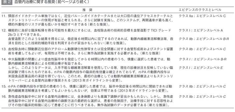
10. Use of stent retrievers is indicated in preference to the MERCI device. (Class I; Level of Evidence A). The use of mechanical thrombectomy devices other than stent retrievers may be reasonable in some circumstances (Class IIb, Level B-NR). (New recommendation) 11. The use of proximal balloon guide catheter or a large bore distal access catheter rather than
a cervical guide catheter alone in conjunction with stent retrievers may be beneficial (Class IIa; Level of Evidence C). Future studies should examine which systems provide the highest recanalization rates with the lowest risk for nontarget embolization. (New recommendation)
12. The technical goal of the thrombectomy procedure should be a TICI 2b/3 angiographic result to maximize the probability of a good functional clinical outcome (Class I; Level of Evidence A). Use of salvage technical adjuncts including intra-arterial fibrinolysis may be reasonable to achieve these angiographic results, if completed within 6 hours of symptom onset (Class IIb; Level of Evidence B-R).(New recommendation)
13. Angioplasty and stenting of proximal cervical atherosclerotic stenosis or complete occlusion at the time of thrombectomy may be considered but the usefulness is unknown (Class IIb; Level of Evidence C). Future randomized studies are needed.
14. Initial treatment with intra-arterial fibrinolysis is beneficial for carefully selected patients with major ischemic strokes of <6 hours’ duration caused by occlusions of the MCA (Class
by guest on September 4, 2017
http://stroke.ahajournals.org/
I; Level of Evidence B-R). However, these data derive from clinical trials that no longer reflect current practice, including use of fibrinolytic drugs that are not available. A clinically beneficial dose of intra-arterial r-tPA is not established, and r-tPA does not have FDA approval for intra-arterial use. As a consequence, endovascular therapy with stent retrievers is recommended over intra-arterial fibrinolysis as first-line therapy (Class I; Level of Evidence E). (Revised from the 2013 guideline)
15. Intra-arterial fibrinolysis initiated within 6 hours of stroke onset in carefully selected patients who have contraindications to the use of intravenous r-tPA might be considered, but the consequences are unknown (Class IIb; Level of Evidence C). (Revised from 2013 guideline)
16. It might be reasonable to favor conscious sedation over general anesthesia during endovascular therapy for acute ischemic stroke. However, the ultimate selection of anesthetic technique during endovascular therapy for acute ischemic stroke should be individualized based on patient risk factors, tolerance of the procedure, and other clinical characteristics. Randomized trial data are needed (Class IIb; Level of Evidence C). (New recommendation)
Imaging
1. Emergency imaging of the brain is recommended before initiating any specific treatment for acute stroke (Class I; Level of Evidence A). In most instances, nonenhanced CT will provide the necessary information to make decisions about emergency management. (Unchanged from the 2013 guideline)
by guest on September 4, 2017
http://stroke.ahajournals.org/
2. If endovascular therapy is contemplated, a noninvasive intracranial vascular study is strongly recommended during the initial imaging evaluation of the acute stroke patient but should not delay intravenous r-tPA if indicated. For patients who qualify for intravenous tPA according to guidelines from professional medical societies, initiating intravenous r-tPA before noninvasive vascular imaging is recommended for patients who have not had noninvasive vascular imaging as part of their initial imaging assessment for stroke. Noninvasive intracranial vascular imaging should then be obtained as quickly as possible (Class I; Level of Evidence A). (New recommendation)
3. The benefits of additional imaging beyond CT and CTA or MR and MRA, such as CT perfusion or diffusion- and perfusion-weighted imaging, for selecting patients for endovascular therapy are unknown (Class IIb; Level of Evidence C). Further randomized, controlled trials may be helpful to determine whether advanced imaging paradigms employing CT perfusion, CTA, and MRI perfusion and diffusion imaging, including measures of infarct core, collateral flow status, and penumbra, are beneficial for selecting patients for acute reperfusion therapy who are within 6 hours of symptom onset and have an ASPECTS <6. Further randomized, controlled trials should be done to determine whether advanced imaging paradigms using CT perfusion and MRI perfusion, CTA, and diffusion imaging, including measures of infarct core, collateral flow status, and penumbra, are beneficial for selecting patients for acute reperfusion therapy who are beyond 6 hours from symptom onset. (New recommendation)
by guest on September 4, 2017
http://stroke.ahajournals.org/
Systems of Stroke Care
1. Patients should be transported rapidly to the closest available certified primary stroke center or comprehensive stroke center or, if no such centers exist, the most appropriate institution that provides emergency stroke care as described in the 2013 guidelines (Class
I; Level of Evidence A). In some instances, this may involve air medical transport and
hospital bypass. (Unchanged from the 2013 guideline)
2. Regional systems of stroke care should be developed. These should consist of consisting of:
(a) Healthcare facilities that provide initial emergency care including administration of intravenous r-tPA, including primary stroke centers, comprehensive stroke centers, and other facilities.
(b) Centers capable of performing endovascular stroke treatment with comprehensive periprocedural care, including comprehensive stroke centers and other healthcare facilities, to which rapid transport can be arranged when appropriate (Class I; Level of Evidence A). (Revised from the 2013 guideline)
3. It may be useful for primary stroke centers and other healthcare facilities that provide initial emergency care including administration of intravenous r-tPA to develop the capability of performing emergency noninvasive intracranial vascular imaging to most appropriately select patients for transfer for endovascular intervention and reduce time to endovascular treatment (Class IIb; Level of Evidence C). (Revised from the 2013 guideline)
4. Endovascular therapy requires the patient to be at an experienced stroke center with rapid access to cerebral angiography and qualified neurointerventionalists. Systems should be designed, executed and monitored to emphasize expeditious assessment and treatment.
by guest on September 4, 2017
http://stroke.ahajournals.org/
Outcomes on all patients should be tracked. Facilities are encouraged to define criteria that can be used to credential individuals who can perform safe and timely intra-arterial revascularization procedures (Class I; Level of Evidence E). (Revised from the 2013 guideline)
REFERENCES
1. Jauch EC, Saver JL, Adams HP Jr, Bruno A, Connors JJ, Demaerschalk BM, Khatri P, McMullan PW Jr, Qureshi AI, Rosenfield K, Scott PA, Summers DR, Wang DZ, Wintermark M, Yonas H; on behalf of the American Heart Association Stroke Council, Council on Cardiovascular Nursing, Council on Peripheral Vascular Disease, and Council on Clinical Cardiology. Guidelines for the early management of patients with acute ischemic stroke: a guideline for healthcare professionals from the American Heart Association/American Stroke Association. Stroke. 2013;44:870-947.
2. Hacke W, Donnan G, Fieschi C, Kaste M, von Kummer R, Broderick JP, Brott T, Frankel M, Grotta JC, Haley EC Jr, Kwiatkowski T, Levine SR, Lewandowski C, Lu M, Lyden P, Marler JR, Patel S, Tilley BC, Albers G, Bluhmki E, Wilhelm M, Hamilton S. Association of outcome with early stroke treatment: pooled analysis of ATLANTIS, ECASS, and NINDS rt-PA stroke trials. Lancet. 2004;363:768-774.
3. Tissue plasminogen activator for acute ischemic stroke. The National Institute of Neurological Disorders and Stroke rt-PA Stroke Study Group. N Engl J Med. 1995;333:1581-1587.
4. Lees KR, Bluhmki E, von Kummer R, Brott TG, Toni D, Grotta JC, Albers GW, Kaste M, Marler JR, Hamilton SA, Tilley BC, Davis SM, Donnan GA, Hacke W, Allen K, Mau J, Meier D, del Zoppo G, De Silva DA, Butcher KS, Parsons MW, Barber PA, Levi C, Bladin C, Byrnes G. Time to treatment with intravenous alteplase and outcome in stroke: an updated pooled analysis of ECASS, ATLANTIS, NINDS, and EPITHET trials. Lancet. 2010;375:1695-1703.
5. Molyneux AJ, Kerr RS, Yu LM, Clarke M, Sneade M, Yarnold JA, Sandercock P. International subarachnoid aneurysm trial (ISAT) of neurosurgical clipping versus endovascular coiling in 2143 patients with ruptured intracranial aneurysms: a randomised comparison of effects on survival, dependency, seizures, rebleeding, subgroups, and aneurysm occlusion. Lancet. 2005;366:809-817.
6. Emberson J, Lees KR, Lyden P, Blackwell L, Albers G, Bluhmki E, Brott T, Cohen G, Davis S, Donnan G, Grotta J, Howard G, Kaste M, Koga M, von Kummer R, Lansberg M,
by guest on September 4, 2017
http://stroke.ahajournals.org/
Lindley RI, Murray G, Olivot JM, Parsons M, Tilley B, Toni D, Toyoda K, Wahlgren N, Wardlaw J, Whiteley W, del Zoppo GJ, Baigent C, Sandercock P, Hacke W. Effect of treatment delay, age, and stroke severity on the effects of intravenous thrombolysis with alteplase for acute ischaemic stroke: a meta-analysis of individual patient data from randomised trials. Lancet. 2014;384:1929-1935.
7. Rankin J. Cerebral vascular accidents in patients over the age of 60: II. Prognosis. Scott Med J. 1957;2:200-215.
8. Banks JL, Marotta CA. Outcomes validity and reliability of the modified Rankin scale: implications for stroke clinical trials: a literature review and synthesis. Stroke. 2007;38:1091-1096.
9. Brott T, Adams HP Jr, Olinger CP, Marler JR, Barsan WG, Biller J, Spilker J, Holleran R, Eberle R, Hertzberg V. Measurements of acute cerebral infarction: a clinical examination scale. Stroke.1989;20:864-870.
10. Ciccone A, Valvassori L, Nichelatti M, Sgoifo A, Ponzio M, Sterzi R, Boccardi E. Endovascular treatment for acute ischemic stroke. N Engl J Med. 2013;368:904-913. 11. Zaidat OO, Yoo AJ, Khatri P, Tomsick TA, von Kummer R, Saver JL, Marks MP,
Prabhakaran S, Kallmes DF, Fitzsimmons BF, Mocco J, Wardlaw JM, Barnwell SL, Jovin TG, Linfante I, Siddiqui AH, Alexander MJ, Hirsch JA, Wintermark M, Albers G, Woo HH, Heck DV, Lev M, Aviv R, Hacke W, Warach S, Broderick J, Derdeyn CP, Furlan A, Nogueira RG, Yavagal DR, Goyal M, Demchuk AM, Bendszus M, Liebeskind DS; for the Cerebral Angiographic Revascularization Grading (CARG) Collaborators, STIR Revascularization working group, and STIR Thrombolysis in Cerebral Infarction (TICI) Task Force. Recommendations on angiographic revascularization grading standards for acute ischemic stroke: a consensus statement. Stroke. 2013;44:2650-2663.
12. Broderick JP, Palesch YY, Demchuk AM, Yeatts SD, Khatri P, Hill MD, Jauch EC, Jovin TG, Yan B, Silver FL, von Kummer R, Molina CA, Demaerschalk BM, Budzik R, Clark WM, Zaidat OO, Malisch TW, Goyal M, Schonewille WJ, Mazighi M, Engelter ST, Anderson C, Spilker J, Carrozzella J, Ryckborst KJ, Janis LS, Martin RH, Foster LD, Tomsick TA. Endovascular therapy after intravenous t-PA versus t-PA alone for stroke. N Engl J Med. 2013;368:893-903.
13. Kidwell CS, Wintermark M, De Silva DA, Schaewe TJ, Jahan R, Starkman S, Jovin T, Hom J, Jumaa M, Schreier J, Gornbein J, Liebeskind DS, Alger JR, Saver JL. Multiparametric MRI and CT models of infarct core and favorable penumbral imaging patterns in acute ischemic stroke. Stroke. 2013;44:73-79.
14. Kidwell CS, Jahan R, Gornbein J, Alger JR, Nenov V, Ajani Z, Feng L, Meyer BC, Olson S, Schwamm LH, Yoo AJ, Marshall RS, Meyers PM, Yavagal DR, Wintermark M, Guzy J, Starkman S, Saver JL. A trial of imaging selection and endovascular treatment for ischemic stroke. N Engl J Med. 2013;368:914-923.
by guest on September 4, 2017
http://stroke.ahajournals.org/
15. Berkhemer OA, Fransen PS, Beumer D, van den Berg LA, Lingsma HF, Yoo AJ, Schonewille WJ, Vos JA, Nederkoorn PJ, Wermer MJ, van Walderveen MA, Staals J, Hofmeijer J, van Oostayen JA, Lycklama a Nijeholt GJ, Boiten J, Brouwer PA, Emmer BJ, de Bruijn SF, van Dijk LC, Kappelle LJ, Lo RH, van Dijk EJ, de Vries J, de Kort PL, van Rooij WJ, van den Berg JS, van Hasselt BA, Aerden LA, Dallinga RJ, Visser MC, Bot JC, Vroomen PC, Eshghi O, Schreuder TH, Heijboer RJ, Keizer K, Tielbeek AV, den Hertog HM, Gerrits DG, van den Berg-Vos RM, Karas GB, Steyerberg EW, Flach HZ, Marquering HA, Sprengers ME, Jenniskens SF, Beenen LF, van den Berg R, Koudstaal PJ, van Zwam WH, Roos YB, van der Lugt A, van Oostenbrugge RJ, Majoie CB, Dippel DW. A randomized trial of intraarterial treatment for acute ischemic stroke. N Engl J Med. 2015;372:11-20.
16. Fransen P, Berkhemer O, Lingsma H, Beumer D, van den Berg L, van Zwam W, van Oostenbrugge R, van der Lugt A, Majoie C, Dippel D; for the MR CLEAN Investigators. Time to reperfusion and effect of intra-arterial treatment in the MR CLEAN Trial.
http://my.americanheart.org/idc/groups/ahamah-public/@wcm/@sop/@scon/documents/downloadable/ucm_471830.pdf. Accessed June 15, 2015.
17. Pexman JH, Barber PA, Hill MD, Sevick RJ, Demchuk AM, Hudon ME, Hu WY, Buchan AM. Use of the Alberta Stroke Program Early CT Score (ASPECTS) for assessing CT scans in patients with acute stroke. Am J Neuroradiol. 2001;22:1534-1542.
18. Goyal M, Demchuk AM, Menon BK, Eesa M, Rempel JL, Thornton J, Roy D, Jovin TG, Willinsky RA, Sapkota BL, Dowlatshahi D, Frei DF, Kamal NR, Montanera WJ, Poppe AY, Ryckborst KJ, Silver FL, Shuaib A, Tampieri D, Williams D, Bang OY, Baxter BW, Burns PA, Choe H, Heo JH, Holmstedt CA, Jankowitz B, Kelly M, Linares G, Mandzia JL, Shankar J, Sohn SI, Swartz RH, Barber PA, Coutts SB, Smith EE, Morrish WF, Weill A, Subramaniam S, Mitha AP, Wong JH, Lowerison MW, Sajobi TT, Hill MD. Randomized assessment of rapid endovascular treatment of ischemic stroke. N Engl J Med. 2015;372:1019-1030.
19. Straka M, Albers GW, Bammer R. Real-time diffusion-perfusion mismatch analysis in acute stroke. J Magn Reson Imaging. 2010;32:1024-1037.
20. Saver JL, Goyal M, Bonafe A, Diener HC, Levy EI, Pereira VM, Albers GW, Cognard C, Cohen DJ, Hacke W, Jansen O, Jovin TG, Mattle HP, Nogueira RG, Siddiqui AH, Yavagal DR, Baxter BW, Devlin TG, Lopes DK, Reddy VK, du Mesnil de Rochemont R, Singer OC, Jahan R. Stent-retriever thrombectomy after intravenous t-PA vs. t-PA alone in stroke. N Engl J Med. 2015;372:2285-2295.
21. Campbell BC, Mitchell PJ, Kleinig TJ, Dewey HM, Churilov L, Yassi N, Yan B, Dowling RJ, Parsons MW, Oxley TJ, Wu TY, Brooks M, Simpson MA, Miteff F, Levi CR, Krause M, Harrington TJ, Faulder KC, Steinfort BS, Priglinger M, Ang T, Scroop R, Barber PA, McGuinness B, Wijeratne T, Phan TG, Chong W, Chandra RV, Bladin CF, Badve M, Rice
by guest on September 4, 2017
http://stroke.ahajournals.org/
H, de Villiers L, Ma H, Desmond PM, Donnan GA, Davis SM. Endovascular therapy for ischemic stroke with perfusion-imaging selection. N Engl J Med. 2015;372:1009-1018. 22. Jovin TG, Chamorro A, Cobo E, de Miquel MA, Molina CA, Rovira A, San Roman L,
Serena J, Abilleira S, Ribo M, Millan M, Urra X, Cardona P, Lopez-Cancio E, Tomasello A, Castano C, Blasco J, Aja L, Dorado L, Quesada H, Rubiera M, Hernandez-Perez M, Goyal M, Demchuk AM, von Kummer R, Gallofre M, Davalos A. Thrombectomy within 8 hours after symptom onset in ischemic stroke. N Engl J Med. 2015;372:2296-2306. 23. Jayaraman MV, Grossberg JA, Meisel KM, Shaikhouni A, Silver B. The clinical and
radiographic importance of distinguishing partial from near-complete reperfusion following intra-arterial stroke therapy. Am J Neuroradiol. 2013;34:135-139.
24. Zanaty M, Chalouhi N, Starke RM, Tjoumakaris S, Hasan D, Hann S, Ajiboye N, Liu KC, Rosenwasser RH, Manasseh P, Jabbour P. Endovascular stroke intervention in the very young. Clin Neurol Neurosurg. 2014;127:15-18.
25. Vega RA, Chan JL, Anene-Maidoh TI, Grimes MM, Reavey-Cantwell JF. Mechanical thrombectomy for pediatric stroke arising from an atrial myxoma: case report. J Neurosurg Pediatr. 2015;15:301-305.
26. Golomb MR, Rafay M, Armstrong D, Massicotte P, Curtis R, Hune S, deVeber GA. Intra-arterial tissue plasminogen activator for thrombosis complicating cerebral angiography in a 17-year-old girl. J Child Neurol. 2003;18:420-423.
27. deVeber GA. Delays in the timely diagnosis of stroke in children. Nat Rev Neurol. 2010;6:64-66.
28. Yeo LL, Paliwal P, Teoh HL, Seet RC, Chan BP, Liang S, Venketasubramanian N, Rathakrishnan R, Ahmad A, Ng KW, Loh PK, Ong JJ, Wakerley BR, Chong VF, Bathla G, Sharma VK. Timing of recanalization after intravenous thrombolysis and functional outcomes after acute ischemic stroke. JAMA Neurol. 2013;70:353-358.
29. Campbell BC, Yassi N, Ma H, Sharma G, Salinas S, Churilov L, Meretoja A, Parsons MW, Desmond PM, Lansberg MG, Donnan GA, Davis SM. Imaging selection in ischemic stroke: feasibility of automated CT-perfusion analysis. Int J Stroke. 2015;10:51-54. 30. Saver JL, Jahan R, Levy EI, Jovin TG, Baxter B, Nogueira RG, Clark W, Budzik R, Zaidat
OO. Solitaire flow restoration device versus the Merci Retriever in patients with acute ischaemic stroke (SWIFT): a randomised, parallel-group, non-inferiority trial. Lancet.2012;380:1241-1249.
31. Nogueira RG, Lutsep HL, Gupta R, Jovin TG, Albers GW, Walker GA, Liebeskind DS, Smith WS. Trevo versus Merci retrievers for thrombectomy revascularisation of large vessel occlusions in acute ischaemic stroke (TREVO 2): a randomised trial. Lancet. 2012;380:1231-1240.
by guest on September 4, 2017
http://stroke.ahajournals.org/