Japan Advanced Institute of Science and Technology
JAIST Repository
https://dspace.jaist.ac.jp/
Title 抗原の蛍光レシオ検出が可能な遺伝的にコードされた
新規抗体バイオセンサーの開発
Author(s) Huynh Nhat, Phuong Kim Citation
Issue Date 2016‑03
Type Thesis or Dissertation Text version ETD
URL http://hdl.handle.net/10119/13532 Rights
Description Supervisor:芳坂 貴弘, マテリアルサイエンス研究科
, 博士
Novel Genetically Encoded Antibody-based Biosensors for Fluorescence ratio Detection of Antigens
KIM PHUONG HUYNH NHAT
Japan Advanced Institute of Science and Technology
Doctoral Dissertation
Novel Genetically Encoded Antibody-based Biosensors for Fluorescence ratio Detection of Antigens
KIM PHUONG HUYNH NHAT
Supervisor: Professor Takahiro Hohsaka, Ph.D.
School of Materials Science
Japan Advanced Institute of Science and Technology
March 2016
ABSTRACT
Fluorescence biosensor is an indispensable method for tracking of small biomolecules or biological processes not only in vitro but also in living cells. Recently, Quenchbody, a novel fluorescence biosensor consists of an N- terminal fluorescently labeled antibody single-chain variable domain (scFv) has been reported. This biosensor allowed detection of antigen based on antigen-dependent removal of quenching effect on the labeled fluorophore.
However, fluorescence intensity of single labeled Quenchbody depends on not only concentration of antigen but also amount of the biosensor in measuring sample. In addition, Quenchbody requires the incorporation of fluorophore-labeled nonnatural amino acid in a cell-free translation system, thus, limit its application in live-cell imaging. In this study, a new strategy for construction of antibody-based fluorescence biosensor in combination of Förster (or fluorescence) resonance energy transfer (FRET) and fluorescence quenching mechanisms was introduced to overcome the limitations of Quenchbody. First, fluorescence biosensors for detection of phosphotyrosine-containing peptides were developed by incorporation fluorophore-labeled nonnatural amino acid into the N-terminus of anti-phosphotyrosine scFv. This biosensor showed antigen-dependent fluorescence increase upon addition of phosphotyrosine-containing peptides. Fusion of fluorescent protein (FP) to the labeled scFv generated double labeled biosensors which allowed FRET between FP and labeled fluorophore and detection of antigen based on antigen-dependent enhancement of fluorescence ratio of fluorophore/FP. Next, genetically- encoded antibody-based fluorescence biosensors were constructed by substituting fluorophore-labeled nonnatural amino acid by protein-tag and its fluorescent ligands. The obtained biosensors exhibited fluorescence enhancement in the presence of antigens. In addition, type of fluorophore, linker length between fluorophore–ligand and orientation of protein-tag to scFv largely affected fluorescence enhancement. Fusion of FP to protein-tag-scFv resulted in double labeled biosensors which showed FRET between FP and labeled fluorophore as well as antigen- dependent enhancement of the fluorophore, allowing fluorescent ratiometric detection of antigen. Finally, an application of the novel genetically-encoded antibody-based ratiometric fluorescent biosensor was demonstrated by expression of the biosensor on the surface of mammalian cells for detection of extracellular antigen. The advantage of the present strategy over conventional strategy for FRET-based biosensor construction is that no conformational change of backbone protein upon binding to analyte is required. Therefore, it is potentially applicable for various antigen-antibody pairs in not only diagnostic analysis but also live-cell imaging.
Key words: single-chain antibody, nonnatural amino acid, fluorescence biosensor, protein-tag, live-cell imaging.
Table of Contents
Chapter 1: Background and Overview ... 1
Chapter 2: Antibody-based fluorescent and fluorescent ratio biosensors for detection of phosphotyrosine ... 26
Chapter 3: Genetically-encoded antibody-based biosensors by fusion of protein- tag and fluorescent protein to scFv ... 49
Chapter 4: Application of genetically-encoded antibody-based biosensor to live- cell imaging ... 86
Chapter 5: Conclusion ... 101
List of publication ... 103
Acknowledgement ... 104
1
Chapter 1
Background and Overview
1-1. Introduction
Proteins present in all living organisms and play a central role in major biological processes. They are built up by 20 kinds of natural amino acids and form unique three- dimensional structures which can perform as catalysts, signaling molecules, receptors, transporters, etc. Based on approaches in elucidating protein structures and functions, proteins can be engineered to become useful biosensors to detect not only small biomolecules but also biological events in vitro and in living cells.
Fluorescent biosensor is one of the most powerful and popular tool for visualizing and quantifying target molecules or events. A fluorescent biosensor usually consists of a protein scaffold which can recognize the target and fluorophores which can convert the interaction of protein and target into fluorescent signal change. Most of fluorescent biosensors have been developed based on photoinduced energy transfer (PET) or Förster (or fluorescence) resonance energy transfer (FRET)1 technologies. Fluorescent dyes can be incorporated into proteins by chemical labeling or incorporation of fluorophore-containing nonnatural amino acids. Proteins can also be labeled with fluorescent proteins or protein-tags which are subsequently labeled with fluorescent ligands. Since the discovery of Aequorea victoria green fluorescent protein (GFP)2, variety of fluorescent protein derivatives have been reported3-4 and commonly used for protein labeling in cell biology. In addition, bioluminescent proteins (for example, luciferases)5, and other fluorescent proteins such as bacterial phytochromes6, rhodopsins7 and fatty acid binding protein (FABP) family8 provide additional choices for protein labeling.
Recently, together with the increasing demands in biological studies, various strategies for fluorescence labeling of proteins and fluorescent biosensors construction have been reported including antibody-based fluorescent biosensor9. Details of these strategies and achievements were described below.
2
1-2. Methods for fluorescent modification of protein
Proteins can be fluorescently labeled by chemical modification, incorporation of fluorophore-containing nonnatural amino acids, and fusion with fluorescent proteins or protein- tags.
1-2-1. Chemical modification of natural amino acid residues
Chemical modification of natural amino acid residues is a useful method for rapidly probing a protein. Targets for chemical modification are residues which have nucleophilic functional groups such as thiol group of cysteine and ε-amino group of lysine. Cysteine is often used for site-specific modification of protein because of its high nucleophilic thiol group and low occurrence on protein surface (about 2.3% genome-wide)10. For proteins lacking cysteine, this residue can be introduced to protein surface by site-direct mutagenesis. Thiol group can be labeled by reaction with maleimide or α-haloketone derivatives (Figure 1-1A). However, if protein contains undesired cysteines, these cysteines must be substituted by other amino acids, but such substitution may affect structure and function of protein. Alternatively, ε-amino group of lysine can be modified by activated esters, sulfonyl chlorides, isocyanates and isothiocyanates (Figure 1-1B). But, it is difficult to achieve site-specific labeling of ε-amino group because proteins always have multiple amino groups including N-terminal amino group.
Figure 1-1. Chemical reactions for modification of (A) cysteine and (B) lysine (Angew. Chem. Int. Ed. 53: 4088-4106 (2013))
3
In addition, Tsien et al. have reported a method for chemical labeling of protein using biarsenical ligands FlAsH and its analogues, such as ReAsH, HoXAsH and CHoXAsH. These ligands can selectively bind to a tetracysteine motif CysCysProGlyCysCys with high affinity and specificity. Fluorescence of the ligands are reported considerably enhanced upon binding to tetracysteine motif. The advantages of this method are that the ligands can be prepared with ease and they can be used for labeling of proteins in living cells11-13.
1-2-2 Incorporation of nonnatural amino acids into proteins
Proteins can be position-specifically fluorescent-labeled by incorporation of fluorophore-containing nonnatural amino acids. In addition, this method allows expansion of protein functions depending on the side groups of the nonnatural amino acids introduced. For incorporation of nonnatural amino acids into proteins using biological translation system, it requires the engineering of genetic code, synthesis of suppressor aminoacyl-tRNAs, designing of orthogonal tRNAs and designing of nonnatural amino acids.
Expansion of genetic code
The genetic code consists of 61 codons encoding 20 naturally-occurring amino acids and 3 nonsense (stop) codons. For incorporation of nonnatural amino acids, extended codons which are specific for nonnatural amino acids are necessary. Therefore, the amber stop codon (UAG) has been employed for incorporation of nonnatural amino acids by Schultz and Chamberlin groups14-16. In this method, an amber suppressor tRNA is chemically aminoacylated with nonnatural amino acid to obtain aminocyl-tRNACUA17-18. On the other hand, site-direct mutagenesis is used to generate DNA and mRNA containing amber stop codon at desired positions. Protein translation and incorporation of nonnatural amino acids were performed in vitro by a cell-free translation system. Even in the presence of amber suppressor aminocyl- tRNACUA, the release factor 1 (RF1) can bind to the amber stop codon and terminate the translation process, in this case, a truncated protein is produced (Figure 1-2). Hence, the yield of nonnatural amino acids incorporation is decreased due to this competition. Recently, a new strategy to increase the yield of nonnatural amino acids incorporation in response to amber stop codon by optimized pyrrolysyl tRNA synthetase/tRNA expression system and engineered release factor 1 has been reported. The study showed that this strategy improves the suppression of up to three UAG codons in mammalian cells significantly 19.
4
Figure 1-2. Site-specific incorporation of nonnatural amino acid into protein in response to amber stop codon in cell-free translation system. The incorporation competes with the termination of translation process by release factor 1.
However, the use of amber stop codon for incorporation of nonnatural amino acid is limited since it does not allow incorporation of multiple nonnatural amino acids into a single protein. To overcome this problem, Hohsaka et al. have developed the four-base codon method for incorporation of nonnatural amino acid. This method has two advantages: first, the competition of amber suppressor tRNA and RF1 is avoided. Four-base codons have been developed based on low occurrence codons such as CGG or AGG so that the competition of tRNA carrying four-base anticodon and endogenous tRNA can be minimized. Second, the four- base codon allows incorporation of multi-nonnatural amino acids into a single proteins. Various four-base codons have been developed, such as AGGU, CGGG, CGGU, CCCU, CUCU, CUAU, and GGGU20-21. The DNA and mRNA containing four-base codon can be prepared by site- directed mutagenesis. In a cell-free translation system, if a four-base codon is successfully decoded by a nonnatural aminocyl-tRNA carrying corresponding anticodon, a full-length protein is synthesized. However, if four-base codon is recognized by an endogenous tRNA, the reading frame is shifted by 1 nucleotide by which a stop codon may be encountered. Thus, truncated protein is obtained (Figure 1-3). Kajihara and coworkers have reported an application of this strategy to incorporate two distinct fluorescent-labeled nonnatural amino acids in response to two four-base codon GGGU and CGGG in calmodulin and monitor its conformational change based on FRET signals22.
5
Figure 1-3. Site-specific incorporation of nonnatural amino acid into protein in response to four-base codon in cell-free translation system. Incorporation of natural amino acid in response to triplet codon results in the termination of translation process by frameshifting.
Aminoacylation of tRNAs with nonnatural amino acids
Aminoacylation of tRNAs with nonnatural amino acids is an important step for incorporation of nonnatural amino acids into protein. In early studies, Johnson and coworkers demonstrated that ε-amino group of Lys-tRNA which was prepared by LysRS was acetylated by N-acetoxysuccinimide, and the resulting acetyllysine was incorporated into hemoglobin using a rabbit reticulocyte cell-free translation system23. This method was applied for modification of lysine with fluorescent probes24 and cross-linking reagents for photo-affinity labeling25. However, site-specific modification was not achieved because modified lysine was incorporated to multiple sites.
Alternatively, a chemical aminoacylation method in which synthesized aminoacylated dinucleotides pCpA was ligated to truncated tRNA lacking terminal pCpA by T4 RNA ligase was proposed by Hecht and coworkers17-18. The pCpA can be aminoacylated with various nonnatural amino acids. This method have been applied for not only in vitro translation system but also in live cells by microinjection26-27.
On the other hand, an approach in nonnatural amino acid acylation by evolved aminoacyl-tRNA synthetases (aaRSs) was reported by Schultz and coworkers28-29. A pair of amber suppressor tRNATyr/TyrRS from Methanococus jannaschii was engineered to
6
incorporate O-methyltyrosine into proteins in E. coli. Further improvement of this method allowed over 30 nonnatural amino acids to be incorporated into proteins in E. coli with high efficiency. In addition, pyrrolysyl-tRNA synthetase (PylRS)/tRNAPyl pairs from certain Methanosarcina species were also employed to introduce lysine derivatives into proteins not only in E. coli but also in eukaryotic cells30-33.
For non-enzymatic aminoacylation, Sisido et al. developed a peptide nucleic acid (PNA) which include a nonnatural amino acid thioester linked to a PNA that was complementary to the 3’-end of target tRNA34 and micelle in which target tRNA was aminoacylated with activated ester of N-protected amino acids in the cationic micelle under ultrasonic agitation35. Both non- enzymatic methods were reported as simple and applicable for variety of nonnatural amino acids.
Design of orthogonal tRNAs
Suppressor tRNAs are required for incorporation of nonnatural amino acids into proteins. This tRNA must be accepted by the translation machinery and orthogonal to endogenous aminoacyl-tRNA synthetase of the host translation system. An amber suppressor tRNA derived from yeast phenylalanine tRNA has been used in E. coli cell-free translation system20. In addition, Taira et al. have developed an amber suppressor tRNA derived from Mycoplasma capricolum tryptophan tRNA which was reported to incorporate nonnatural amino acid into E. coli cell-free translation system with higher efficiency than the yeast phenylalanie tRNA36. Recently, approaches in engineering of anticodon loop and anticodon stem of tRNAPyl for efficient incorporation of nonnatural amino acids in response to four-base codon have been reported37-38. The evolved tRNAPyls were demonstrated to enhance incorporation of nonnatural amino acids into proteins in both E, coli and mammalian cells.
Design of nonnatural amino acids
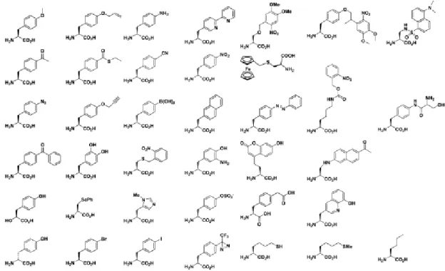
Various nonnatural amino acids with diverse functional groups have been successfully incorporated into proteins (Figure 1-4)39-41. For fluorescent labeling of proteins, proteins can be incorporated with either fluorescent nonnatural amino acids22,42 or nonnatural amino acids carrying biorthogonal chemical groups such as ketones, alkynes, anilines, etc. which are selectively labeled by fluorescent dyes43. Incorporation of nonnatural amino acids allows studies of protein structures and functions as well as site-specific modification of proteins.
It is important to consider types of nonnatural amino acid which are efficiently incorporated into proteins. Hohsaka and coworkers have evaluated the incorporation efficiency
7
of nonnatural amino acids with various side chains in E. coli cell-free translation system. The results reveal that nonnatural amino acids with linearly expanded aromatic groups are more favorable for the translation system than those with widely expand or bend aromatic groups20. Therefore, the side chain of nonnatural amino acids are very important and it should be carefully considered in designing of novel nonnatural amino acids.
Figure 1-4. Examples of nonnatural amino acids that have been incorporated into proteins (J. Am. Chem. Soc. 131: 12497-12515 (2009)).
1-2-3 Fusion with fluorescent proteins
Fluorescent proteins are indispensable tools for fluorescent labeling of proteins. The first discovered fluorescent protein is the Aequorea victoria green fluorescent protein (GFP)2. GFP is encoded by about 240 amino acids which form an 11 -strands barrel with molecular weight about 27 kDa. GFP can be fused to protein of interest and allow spatial, temporal imaging of proteins. Since the discovery of GFP, it has been continuously engineered to improve the brightness as well as stability. On the other hands, many of its derivatives with a wide range of fluorescent colors have been reported3. In addition, circular permuted fluorescent proteins which are found very useful in construction of fluorescent biosensors have been introduced by Tsien group and Miyawaki group4, 44-45.
8
On the other hand, bioluminescent proteins such as luciferase are also commonly used for labeling. Luciferase is a family of photoproteins found in insects, marine organisms and prokaryotes. Luciferase reacts with its substrate luciferin and luminesces without requirement of excitation light. Thus, using luciferase can reduce background signals. To date, numerous studies have been carried out to investigate novel luciferases as well as engineer conventional luciferases to improve their optical properties, such as brightness, photostability, red-shift bioluminescence5.
Alternatively, near-infrared fluorescent proteins derived from bacterial phytochromes have been reported. The bacterial phytochromes utilize biliverdin which is ubiquitous in mammalian tissue as a reactive chromophore. Thus, phytochromes have been suggested as a candidate for fluorescence imaging in mammals. Currently, several phytochrome-based near- infrared fluorescent proteins have been developed, such as IFP1.4, iRFP and Wi-Phy6.
In addition, photoreceptor proteins rhodopsins which are found in proteobacteria and archaea have been used for biosensor construction7,46. Recently, a novel fluorescent protein (UnaG) belongs to the fatty-acid binding protein family in eel has been reported. UnaG emits green fluorescence upon binding to its ligand bilirubin, a heme metabolite. UnaG has been applied for analyzing bilirubin of human clinical samples8. As mentioned above, up to date various fluorescent proteins with a wide range of colors as well as diversity in optical properties has been developed. Fluorescent proteins have become one of the most powerful tools for protein labeling and fluorescent biosensors construction.
1-2-4 Fusion with protein-tags
The most commonly used protein-tags for protein labeling are HaloTag, SNAP-tag and CLIP-tag. HaloTag is a 33 kDa protein derived from Rhodococcus haloalkane dehalogenase (DhaA). DhaA catalyzes a hydrolysis reaction in which it removes halides from aliphatic hydrocarbons by a nucleophilic displacement. HaloTag is a mutant of DhaA in which mutation is introduced to the active site so that catalysis reaction is trapped at the covalent ester bond intermediate between the enzyme and hydrocarbon substrate (Figure 1-5). As a results, various functional groups can be covalently linked to HaloTag. HaloTag was reported to have high affinity to its ligand, the binding of HaloTag and its ligand is rapid and irreversibly. A wide range of HaloTag ligands have been developed and enable its applications in not only protein fluorescence labeling but also protein isolation, purification, analysis of protein functions and interactions47-48.
9
Figure 1-5. (A) Structure of HaloTag and its ligand tunnel with covalently bound TMR ligand.
(B) Structure of TMR ligand (fluorescent ligand) (ACS Chem. Biol. 3(6): 373-382 (2008)).
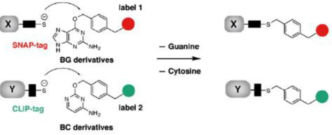
Another approach in protein-tag is SNAP-tag and its variant CLIP-tag. SNAP-tag and CLIP-tag are about 19 kDa proteins derived from human DNA repair protein O6-alkylguanine- DNA alkyltransferase (hAGT). SNAP-tag reacts with its substrates containing O6- benzylguanine (BG) moiety by transferring the alkyl group from substrates to its cysteine residues results in a covalent labeling with functional groups attached on substrate. Similarly, CLIP-tag can be covalently labeled by reacting with O2-benzylcytosine (BC) derivatives (Figure 1-6). The reaction of SNAP-tag and CLIP-tag with their substrates are specific, rapid and irreversible. Variety of fluorescent probes for SNAP-tag and CLIP-tag labeling have been developed. Since SNAP-tag and CLIP-tag are orthogonal, they can be used simultaneously for live cell imaging49-51.
10
Figure 1-6. Labeling of SNAP-tag and CLIP-tag with BG derivatives and BC derivatives, respectively. (Chem. Biol. 15: 128-136 (2008).
11
1-3. Techniques for construction of fluorescent biosensors
Most of fluorescent biosensors have been developed based on Förster (or fluorescence) resonance energy transfer (FRET) and photoinduced electron transfer (PET).
1-3-1. FRET-based biosensors
FRET is a physical phenomenon which involves non-radiative energy transfer between a donor fluorophore and an acceptor fluorophore if the acceptor absorption spectrum is overlapped with the emission spectrum of donor (Figure 1-7A), they are in close distance (typically, less than 10 nm) and their orientations are favorable for dipole-dipole interaction. A Jablonski diagram of FRET is shown in Figure 1-7B. When donor is excited by a light source, its energy is promoted from ground state S0D to excitation state S1D. If the donor is at close proximity with an acceptor and emission energy of donor is match with excitation energy of acceptor, donor can transfer excited energy to acceptor directly. The energy transfer efficiency E is calculated as:
where r is the distance between donor and acceptor, R0 is the Förster distance – the distance at which FRET efficiency is 50%. R0 is calculated based on the following equation:
𝑅06 =9000(𝑙𝑛10)𝜅2𝑄𝐷 128𝜋5𝑁𝑛4 𝐽(𝜆)
where κ2 is the orientation factor that represents the geometric relationship between donor transition dipole and acceptor transition dipole, QD is quantum yield of donor, N is Avogadro’s number, n is the refractive index of the medium, J(λ) is the spectral integral expressing the degree of overlap between donor emission and acceptor absorption spectra52-53.
Thus, the extent of spectral overlap between donor and acceptor, the quantum yield of donor, the distance and relative orientations between them significantly affect FRET efficiency.
𝐸 = 𝑅06 𝑅06+ 𝑟6
12
Figure 1-7. Mechanism of FRET. (A). Spectra of donor emission and acceptor absorption, and spectral overlap (blue filled region) (Principles of Fluorescence Spectroscopy, 3rd edition (Springer, New York) (2006)). (B). Jablonski diagram of energy transfer in FRET (Chem. Rev.
106(5): 1785-1813 (2006)).
Typical FRET-based fluorescent biosensors are consisted of a protein backbone, which can interact with analyte, and tandemly linked two fluorophores at N- and C-termini. Upon interaction with analyte, protein backbone undergoes a conformational change which alters the relative orientation and distance of the labeled fluorophores, as a result, alter FRET efficiency between them. An example for FRET-based fluorescent biosensor is the ATP (adenosine 5’- triphosphate) indicator (ATeam). The bacterial ε subunit of FoF1-ATP synthase was fused with monomeric super-enhanced cyan-emitting fluorescent protein (mseCFP) and monomeric yellow- emitting fluorescent protein (mVenus) at N- and C-termini, respectively. Upon binding of ATP, ε subunit changed it conformation from extended form to retracted form, thus, brought the two fluorophores closer and altered their relative orientation. The indicator showed increase in FRET efficiency in the presence of ATP (Figure 1-8). A series of ATeam for detection of wide range concentrations of ATP from μM to mM were developed54.
Recently, FRET-based biosensor utilizing SNAP-tag and CLIP-tag (CLASH- AChE/HCA) to detect acetylcholine esterase (AChE) inhibitor tacrine has been reported (Figure 1-9). The biosensor was comprised of SNAP-tag and CLIP-tag linked by a rigid 30-proline linker, AChE fused to N-terminus of SNAP-tag, and a human carbonic anhydrase II (HCA) fused to C-terminus of CLIP-tag. The whole biosensor was displayed on cell surface. SNAP- tag was conjugated with a Cy5-labeled BG derivative containing an HCA ligand benzenesulfonamide (SA) and an inhibitor of AChE edrophonium (E). CLIP-tag was labeled with Cy3, which was FRET donor for Cy5. In the absence of tacrine (T), the interaction of edrophonium with AChE prevented the binding of benzenesulfonamide to HCA, thus, the biosensor was in low FRET state. When tacrine was added, the interaction of tacrine with AChE
13
allowed the binding of benzenesulfonamide to HCA and increased FRET efficiency. This strategy was also applied to regulate the activity of enzyme, for example, in this case, HCA can be switched on and off by addition of tacrine55.
Figure 1-8. FRET-based ATP indicator (ATeam). (A). Schematic illustration of ATeam structure in ATP-free and ATP-bound forms. (B). ATP-dependent fluorescent spectral change of ATeam (Proc. Natl. Acad. Sci. USA 106(37): 15651-15656 (2009)).
Figure 1-9. (A). Schematic illustration of CLASH-AChE/HCA. (B). Structure of BG derivative used for labeling of SNAP-tag (Nat. Commun. 6:7830 (2015))
14 1-3-2. Photoinduced electron transfer (PET)
In contrast to FRET which results from a long-distance dipole-dipole interaction of two fluorophores, PET requires van de Waals contact between electron donor (DP) and electron acceptor (AP) molecules, yielding a complex DP+AP- (Figure 1-10A). The complex can return to the ground state without emitting a photon though in rare cases exciplex emission is observed, then, the extra electron on acceptor is returned to donor. Thus, PET usually results in a quenching effect. In PET, excited fluorophore can be either donor or acceptor. Figure 1-10B presents an energy diagram of PET with the excited molecule being the donor. Excitation of donor results in turning an electron from ground state to excited state. Donor can transfer electron to acceptor and forms a complex DP+AP-. The complex returns to the ground state without emitting (quenching), or may emit exciplex emission in rare case52. Thus, the redox potential of donor and acceptor as well as their distance are important for PET efficiency.
In biology studies, nucleic acid such as guanine and amino acids such as tryptophan, methionine, and tyrosine are found to have quenching effect on various fluorophores since they have high tendency of donating electron56-58.
Figure 1-10. Mechanism of photoinduced electron transfer (PET). (A) Molecular orbital schematic for PET. HO is highest occupied orbital, LU is lowest unoccupied orbital. (B) Energy diagram of PET. F is fluorophore, Q is quencher, and νF and νE are emission of fluorophore and exciplex, respectively. (Principles of Fluorescence Spectroscopy, 3rd edition (Springer, New York) (2006)).
Iijima and coworkers have monitored the binding of maltose by site-specific incorporation of BODIPY-linked nonnatural amino acid into maltose binding protein (MBP).
The binding of maltose was detected based on fluorescence quenching by PET. In the absence
A B
15
of maltose, BODIPY dye incorporated at the ligand-binding site was quenched by tryptophan residue. Upon addition of maltose, BODIPY dye was removed from tryptophan residue and the quenching effect was depressed.
They utilized this PET system to generate a fluorescent ratiometric biosensor in combination with FRET (Figure 1-11). MBP was double-labeled with BODIPYFL (FRET donor) at the N-terminus and with BODIPY558 (FRET acceptor) at the ligand-binding site.
FRET occurred regardless the absence and presence of maltose, but BODIPY558 was quenched only in the absence of maltose. Therefore, this biosensor showed significant increase of fluorescence intensity ratio and allow quantitative detection of maltose42.
.
Figure 1-11. (A). Schematic illustration of the detection of maltose based on FRET and fluorescence quenching. (B). Maltose-dependent enhancement of FRET signals. (C). Titration curve of fluorescence ratio (575/515). (D). Fluorescence imaging on a microplate in the presence of maltose with excitation at 488 nm and emission at 520 nm (green) and 605 nm (red) (Chem. Bio. Chem. 10: 999-1006 (2009)).
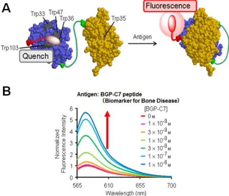
In addition, a new approach in PET-based biosensor, termed “Quenchbody”, has been reported. Quenchbody was developedbased on the antigen-dependent removal of quenching effect on a fluorophore attached to N-terminal domain of an antibody single chain variable region (scFv) (Figure 1-12A). In the absence of antigen, the fluorophore is in close proximity
16
to tryptophan (Trp) residues located at the interface between the variable domains (VH and VL) of scFv, thus, fluorescence from the fluorophore is quenched by Trp via a PET mechanism. In the presence of antigen, this quenching is removed because the tight interaction of VH and VL
prevented the interaction of fluorophore and Trp residues. Therefore, the fluorescence is significantly enhanced and this enhancement is antigen concentration dependent (Figure 1-12B).
Quenchbodies have been proved to have a wide range of application in detection of not only small molecules but also peptides and proteins with high sensitivity9.
Figure 1-12. Fluorescent biosensor based on antibody single chain variable domain (scFv) (Quenchbody). (A) Illustration of Quenchbody in the absence and presence of antigen. (B) Antigen-dependent fluorescent enhancement of Quenchbody (J. Am. Chem. Soc. 133: 17386- 17394 (2011)).
17
1-4. Content of this thesis
While Quenchbody is a valuable biosensor strategy to detect a variety of target molecules, it has several drawbacks. Fluorescence intensity of single labeled Quenchbody increases upon antigen binding to scFv, however, fluorescence intensity also depends on the concentration of Quenchbody. Therefore, if Quenchbody is used under heterogeneous conditions such as inside the cell where local concentration of the biosensor cannot be controlled, it is difficult to distinguish the fluorescence signal is due to binding of antigen or non-uniform concentration of biosensor. In addition, incorporation of fluorescent-labeled nonnatural amino acid into Quenchbody requires the use of chemically synthesized aminoacyl- tRNA and cell-free translation system which also limit its application in live-cell imaging.
In this study, I developed novel types of antibody-based fluorescent and fluorescent ratiometric biosensors utilizing not only fluorescent-labeled nonnatural amino acid but also protein-tags and fluorescent proteins. I demonstrated that the present study successfully improved the disadvantages of Quenchbody and provided a general tool for fluorescent detection of various target molecules not only in vitro but also in vivo.
In chapter 2, N-terminal fluorescently labeled scFv against phosphotyrosine (pTyr) was newly synthesized. This fluorescent-labeled anti-pTyr scFv showed antigen-dependent fluorescent increase upon addition of pTyr-containing peptides (Figure 1-13A). In addition, double labeled scFv by fusion of fluorescent protein was generated. The double-labeled scFv showed FRET between fluorescent protein and labeled fluorophore and fluorescence enhancement of labeled fluorophore upon addition of pTyr peptide without affecting antigen binding affinity (Figure 1-13B).
18
Figure 1-13. (A) Illustration of the synthetic procedure of TAMRA-scFv against pTyr and the detection of pTyr-containing peptide based on antigen-dependent removal of fluorescence quenching effect on TAMRA. (B) Illustration of the synthetic procedure of double labeled scFv(pTyr) in which EGFP was fused to N-terminus of scFv and the detection of pTyr- containing peptide based on FRET from EGFP to TAMRA and antigen-dependent removal of fluorescence quenching effect on TAMRA.
19
In chapter 3, protein-tags (HaloTag, SNAP-tag) and their fluorophore-labeled ligands were used for N-terminal fluorescent labeling of scFvs without using nonnatural amino acid mutagenesis. The resulting fusion proteins showed fluorescence enhancement upon antigen- binding (Figure 1-14A). In addition, type of fluorophores, linker length between fluorophore–
ligand, and orientation of protein-tag to scFv largely affected the fluorescence enhancement.
This result demonstrated that genetically encoded Quenchbody can be obtained by substituting fluorescent nonnatural amino acid by protein-tag. Subsequent fusion of fluorescent protein to protein-tag-scFv generated double labeled biosensors which allowed FRET between fluorescent protein and fluorophore and the antigen-dependent fluorescence enhancement to detect antigen in a ratiometric manner. (Figure 1-14B).
Figure 1-14. (A) Synthetic procedure of fusion of scFv and protein-tag labeled with fluorescent ligand and detection of antigen based on antigen-dependent removal of quenching effect on fluorophore. (B) Synthetic procedure of double labeled scFv with fluorescent protein (FP) and protein-tag, and fluorescence ratio detection of antigen based on FRET from FP to fluorophore and antigen-dependent removal of quenching effect on fluorophore.
20
In chapter 4, I expressed the newly constructed biosensor consisted of fluorescent protein, SNAP-tag, and scFv on surface of mammalian cells (HeLa S3 cells and osteosarcoma U2OS cells) and observed fluorescence of the cells in the absence and presence of antigen (BGP) (Figure 1-15).
Figure 1-15. Detection of BGP secreted by osteosarcoma U2OS cells during differentiation to osteoblasts. Anti-BGP scFv fused with EGFP and SNAP tag was expressed and incorporated on the membrane of U2OS cells. Expressed EGFP-SNAP-scFv is labeled by fluorophore-SNAP ligand. Without BGP, fluorescence ratio of fluorophore/EGFP is low because of quenching of fluorophore. When U2OS cells are induced to differentiate to osteoblast, they secret BGP to extracellular environment. Binding of BGP to EGFP-SNAP-scFv results in fluorescence enhancement of fluorophore, and as consequence, enhancement of fluorescence ratio.
21
1-5 References
(1) Förster T. Zwischenmolekulare Energiewanderung und Fluoreszenz. Annalen der Physik 437: 55-75 (1948)
(2) Shimomura O., Johnson F. H., Saiga Y., J. Cell. Comp. Phys. 59(3): 223-239 (1962) (3) Shaner N. C., Steinbatch P. A., Tsien R. Y. A guide to choosing fluorescent proteins.
Nat. Methods 2(12): 905-909 (2005)
(4) Baird G. S., Zacharias D. A., Tsien R. Y., Circular permutation and receptor insertion within green fluorescent proteins. Proc. Natl. Acad. Sci. USA 96(20): 11241-11246 (1999)
(5) Ozawa T., Yoshimura H., Kim S.B. Advances in fluorescence and bioluminescence imaging. Anal. Chem. 85(2): 590-609 (2013)
(6) Piatkevich K. D., Subach F. V., Verkhusha V. V., Engineering of bacterial phytochromes for near-infrared imaging, sensing, and light-control in mammals.
Chem. Soc. Rev. 42(8): 3441–3452 (2013)
(7) Van de Horst M. A., Hellingwerf K. J. Photoreceptor proteins, “star actors of modern times”: a review of the functional dynamics in the structure of presentative members of six different photoreceptor families. Acc. Chem. Res. 37:13-20 (2004)
(8) Kumagai A., Ando R., Miyatake H., Greimel P., Kobayashi T., Hiabiayashi Y., Shimogori T., Miyawaki A. A bilirubin-inducible fluorescent protein form eel muscle.
Cell 153(7): 1602-1611 (2013)
(9) Abe R., Ohashi H., Iijima I., Ihara M., Takagi H., Hohsaka T., Ueda H.
“Quenchbodies”: Quench-based antibody probes that show antigen-dependent fluorescence. J. Am. Chem. Soc. 133: 17386-17394 (2011)
(10) Takaoka Y., Ojida A., Hamachi I. Protein organic chemistry and applications for labeling and engineering in live-cell systems. Angew. Chem. Int. Ed. 53: 4088-4106 (2013)
(11) Griffin B. A., Adams S. R., Tsien R. Y. Specific covalent labeling of recombinant protein molecules inside live cells. Science 281(5374): 269-272 (1998)
(12) Adams S. R., Campbell R. E., Gross L. A., Martin B. R., Walkup G. K., Yao Y., Llopis J., Tsien R. Y. New biarsenical ligands and tetracysteine motifs for protein labeling in vitro and in vivo: synthesis and biological applications. J. Am. Chem. Soc.
124(21): 6063-6076 (2002)
22
(13) Adams S. R., Tsien R. Y. Preparation of the membrane-permeant biarsenicals FIAsH- EDT2 and ReAsH-EDT2 for fluorescent labeling of tetracysteine-tagged proteins. Nat.
Protoc. 3(9): 1527-1534 (2008)
(14) Noren C. J., Anthony-Cahill S. J., Griffith M.C., Schultz P. G. A general method for site-specific incorporation of unnatural amino acids into proteins. Science 244: 182- 188 (1989)
(15) Anthony-Cahill S. J., Griffth M. C., Noren C. J., Suich D. J., Schultz P. G. Site- specific mutagenesis with unnatural amino acids. Trends, Biochem. Sci. 14: 400-403 (1989)
(16) Bain J. D. Glabe C. g., Dix T.A., Chamberlin A. R. Biosynthetic site-specific incorporation of a non-natural amino acid into a polypeptide. J. Am. Chem. Soc. 111:
8013-8014 (1989)
(17) Hecht S. M., Alford B. L., Kuroda Y., Kitano S. “Chemical aminoacylation: of tRNA’s. J. Biol. Chem. 253: 4517-4520 (1978)
(18) Heckler T. G., Chang L. H. Zama Y., Naka T., Chorghade M. S., Hecht S. M. T4 RNA ligase mediated preparation of novel “chemically misacylated” tRNAPhes.
Biocehmistry 23: 1468-1473 (1984)
(19) Schmied W.H.. Elsässer S.J., Uttamapinant C. Chin J. W. Efficient multisite unnatural amino acid incorporation in mammalian cells via optimized pyrrolysyl tRNA synthetase/tRNA expression and engineered eRF1. J. Am. Chem. Soc. 136(44):
15577-15583 (2014)
(20) Hohsaka T., Kajihara D., Ashizuka Y., Murakami H., Sisido M. Efficient
incorporation of non-natural amino acids with large aromatic groups into streptavidin in in vitro protein synthesizing systems. J. Am. Chem. Soc. 121: 34-40 (1999)
(21) Hohsaka T., Ashizuka Y., Taira H., Murakami H., Sisido M. Incorporation of non- natural amino acids into proteins by using various four-base codons in an Escherichia coli in vitro translation system. Biochemistry 40: 11060-11064 (2001)
(22) Kajihara D., Abe R., Iijima I., Komiyama C., Sisido M., Hohsaka T. FRET analysis of protein conformational change through position-specific incorporation of
fluorescent amino acids. Nature Methods 3(11): 923-929 (2006)
(23) Johnson A.E., Woodward W. R., Herbert E., Menninger J. R., N epsilon-acetyllysine transfer ribonucleic acid: a biologically active analogue of aminoacyl transfer
ribonucleic acids. Biochemistry 15: 569-575 (1976)
23
(24) Crowley K. S., Liao S., Worrell V. E., Reinhart G. D., Johnson A.E. Secretory proteins move through the endoplasmic reticulum membrane via an aqueous, gated pore. Cell 78: 461-471 (1994)
(25) Kreig U. C., Walter P., Johnson A. E. Photocrosslinking of the signal sequence of nascent preprolactin to the 54-kilodalton polypeptide of the signal recognition particle.
Proc. Natl. Accad. Sci. USA 83: 8604-8608 (1986)
(26) Beene D. L., Dougherty D. A., Lester H. A. Unnatural amino acid mutagenesis in mapping ion channel function. Curr. Opin. Neurobiol. 13(3): 264-270 (2003)
(27) Lummis S. C., Beene D. L. Lee L.W., Lester H. A. Broadhurst R. W., Dougherty D.
A. Cis-trans isomerization at proline opens the pore of a neurotransmitter-gated ion channel. Nature 438(7065): 248-252 (2005)
(28) Wang L., Brock A., Herberich B., Schultz P. G. Expanding the genetic code of Escherichia coli. Science 292: 498-500 (2001)
(29) Xie J., Schultz P.G. An expanding genetic code. Methods 36: 227-238 (2005) (30) Polycarpo C. R., Herring S., Bérubé A., Wood J. L., Söll D., Ambrogelly A.
Pyrrolysine analogues as substrates for pyrrolysyl-tRNA synthetase. FEBS Lett.
580(28-29): 6695-6700 (2006)
(31) Yanagisawa T., Ishii R., Fukunaga R., Kobayashi T., Sakamoto K., Yokoyama S.
Multistep engineering of pyrrolysyl-tRNA synthetase to genetically encode
N(epsilon)-(o-azidobenzyloxycarbonyl) lysine for site-specific protein modification.
Chem. Biol. 24; 15(11): 1187-1197 (2008)
(32) Nguyen D.P., Elliott T., Holt M., Muir T. W., Chin J. W. Genetically encoded 1,2- aminothiols facilitate rapid and site-specific protein labeling via a bio-orthogonal cyanobenzothiazole condensation. J. Am. Chem. Soc. 113: 11418-11421 (2011) (33) Mukai T., Kobayashi T., Hino N., Yanagisawa T., Sakamoto K., Yokoyama S.
Adding l-lysine derivatives to the genetic code of mammalian cells with engineered pyrrolysyl-tRNA synthetases. Biochem. Biophys. Res. Commun. 371(4): 818-822 (2008)
(34) Ninomiya K., Minohata T., Nishimura M., Sisido M. In situ chemical aminoacylation with amino acid thioesters linked to a peptide nucleic acid. J. Am. Chem. Soc. 126:
15984-15989 (2004)
(35) Hashimoto N., Ninomiya K., Endo T., Sisido M. Simple and quick chemical aminoacylation of tRNA in cationic micellar solution under ultrasonic agitation.
Chem. Commun. (Camb) 4321-4323 (2005)
24
(36) Taira H., Matsushita T., Kojima K., Shiraga K., Hohsaka T. Comprehensive
screening of amber suppressor tRNAs suitable for incorporation of non-natural amino acids in a cell-free translation system. Biochem. Biophys. Res. Commun. 19;
374(2)304-308 (2008)
(37) Niu W., Schultz P.G., Guo J. An expanded genetic code in mammalian cells with a functional quadruplet codon. ACS Chem. Biol. 19; 8(7): 1640-1645 (2013)
(38) Wang K., Sachdeva A., Cox D. J., Wilf N. W., Lang K., Wallace S., Mehl R. A., Chin J. W. Nature Chemistry 6: 393-403 (2014)
(39) Wang L., Schultz P.G. Expanding the genetic code. Angew. Chem. Int. Ed. 44: 34-66 (2005)
(40) Wu X., Schultz P.G. Synthesis at the interface of chemistry and biology. J. Am.
Chem. Soc. 131: 12497-12515 (2009)
(41) Hohsaka T., Sisido M. Incorporation of non-natural amino acids into proteins. Curr.
Opin. Chem. Biol. 6(6): 809-815 (2002)
(42) Iijima I., Hohsaka T. Position-specific incorporation of fluorescent non-natural amino acids into maltose-binding protein for detection of ligand binding by FRET and
fluorescence quenching. Chem. Bio. Chem. 10: 999-1006 (2009)
(43) Lang K., Chim J. W. Cellular incorporation of unnatural amino acids and biorthogonal labeling of proteins. Chem. Rev. 114: 4764-4806 (2014)
(44) Nagai T., Sawano A., Park E. S., Miyawaki A. Circular permuted green fluorescent proteins engineered to sense Ca2+. Proc. Natl. Acad. Sci. USA 98(6): 3197-3202 (2001)
(45) Nagai T., Yamada S., Tominaga T., Ichikawa M., Miyawaki A. Expanded dynamic range of fluorescent indicators for Ca2+ by circular permuted yellow fluorescent proteins. Proc. Natl. Acad. Sci. USA 101(29): 10554-10559 (2004)
(46) Gong Y. The evolving capabilities of rhodopsin-based genetically encoded voltage indicators. Curr. Opin. Chem Biol. 27: 84-89 (2015)
(47) Los G. V., Encell L.P., McDougall M. G., Hartzell D.D., Karassina N., Zimprich C., Wood M. G., Learish R., Ohana R. F., Urh M., Simpson D., Mendez J., Zimmerman K., Otto P., Vidugiris G., Zhu J., Darzins A., Klaubert D. H., Bulleit R. F., Wood K.V.
HaloTag: A novel protein labeling technology for cell imaging and protein analysis.
ACS Chem. Biol. 3(6): 373-382 (2008)
(48) England C. G., Luo H., Cai W. HaloTag technology: a versatile platform for biomedical application. Bioconjugate Chem. 26: 975-986 (2015)
25
(49) Juilerat A., Gronemeyer T., Keppler A., Gendreizig S.,Pick H., Vogel H., Johnsson K. Direct evolution of O6-alkylguanine-DNA alkyltransferase for efficient labeling of fusion proteins with small molecules in vivo. Chem. Biol. 10: 313-317 (2003)
(50) Keppler A., Gendreizig S., Gronemeyer T., Pick H., Vogel H., Johnsson K. A general method for the covalent labeling of fusion proteins with small molecules in vivo. Nat.
Biotechnol. 21: 86-89 (2003)
(51) Gautier A., Juillerat A., Heinis C., Corrêa Jr.I.R., Kindermann M., Beaufils F., Johnsson K. An engineered protein tag for multiprotein labeling in living cells. Chem.
Biol. 15: 128-136 (2008)
(52) Lakowicz J. R. Principles of Fluorescence Spectroscopy, 3rd edition (Springer, New York) (2006)
(53) Arai Y., Nagai T. Extensive use of FRET in biological imaging. Microscopy (Tokyo) 62(4): 419-428 (2013)
(54) Imamura H., Huynh Nhat K.P., Togawa H., Saito K., Iino R., Kato-Yamada Y., Nagai T., Noji H. Visualization of ATP levels inside single living cells with fluorescent resonance energy transfer-based genetically encoded indicators. Proc.
Natl. Acad. Sci. USA 106(37): 15651-15656 (2009).
(55) Schena A., Griss R., Johnsson K. Madulating protein activity using tethered ligands with mutually exclusive binding sites. Nat. Commun. 6:7830 (2015)
(56) Michalet X., Weiss S., Jӓger M. Single-molecule fluorescence studies of protein folding and conformational dynamics. Chem. Rev. 106(5): 1785-1813 (2006) (57) Torimura M., Kurata S., Yamada K., Yokomaku T., Kamagata Y., Kanagawa T.,
Kurame R., Fluorescence-Quenching phenomenon by photoinduced electron transfer between a fluorescent dye and a nucleotide base. Anal. Sci. 17:155–160 (2001) (58) Knemeyer J. P. Sauer M., Wolfrum J. Inter- and Intramolecular Fluorescence
Quenching of Organic Dyes by Tryptophan. Bioconjugate Chem 14: 1133-1139 (2003)
26
Chapter 2
Antibody-based fluorescent and fluorescent ratio biosensors for detection of phosphotyrosine
2-1 Introduction
Protein phosphorylation is an important post-translational process which affects protein structure and function. In mammalian cells, serine (Ser), threonine (Thr) and tyrosine (Tyr) are phosphorylated in response to extracellular stimuli. For example, in signal transduction the autophosphorylation of Tyr in epidermal growth factor (EGF) receptor upon binding of EGF activates a kinase cascade that relays the signal to the transcriptional apparatus in the nucleus which then triggers cell growth and differentiation. Abnormal protein phosphorylation may cause pathogenesis or even carcinogenesis. Therefore, methods for studies of protein phosphorylation are very important in not only fundamental biology but also biomedical applications.
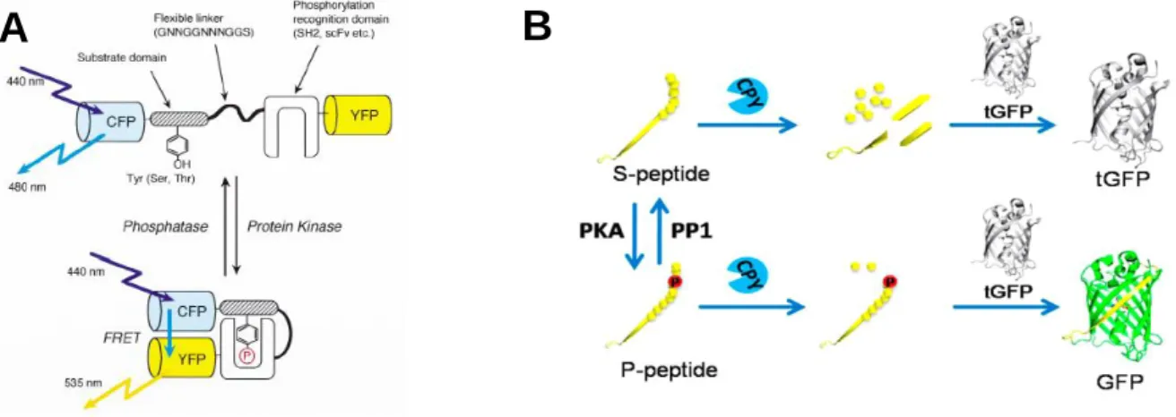
To study protein phosphorylation kinases and phosphatases activities, genetically- encoded fluorescent indicators for protein phosphorylation have been developed. Fluorescence resonance energy transfer (FRET)-based indicators consists of a substrate domain specific for the kinase of interest linked with a phosphorylation recognition domain through a flexible linker and sandwiched by cyan and yellow fluorescent proteins (CFP and YFP, respectively).
Phosphorylation of substrate domain and binding of substrate domain to recognition domain generate a large conformational change of the indicator which alter the distance and/or relative orientation of CFP and YFP, induced increase in FRET signals. In contrast, dephosphorylation of substrate domain decreases FRET signals (Figure 2-1A). The indicators are applicable to real-time monitoring protein phosphorylation and dephosphorylation in live cells1-4. Alternatively, a phosphorylation-mediated assembly of semisynthetic GFP-based biosensor for protein kinases has recently reported. The biosensor consists of S-peptide fused with 10th - strand of GFP (s10) and a kinase substrate peptide. Phosphorylation of the substrate peptide by kinase can protect s10 from cleavage by carboxypeptidase Y (CPY). Binding of s10 to truncated GFP (tGFP, lack of s10) results in an intact GFP which fluoresces. If peptide is not phosphorylated, S-peptide is degraded by CPY and no fluorescence is observed (Figure 2-1B).
The biosensor has demonstrated high sensitivity in analysis of kinase and phosphatase activities in vitro5.
27
Figure 2-1. Genetically-encoded fluorescent indicators for protein phosphorylation.
(A) Schematic illustration of the indicator named “phocus”, the phosphorylation and dephosphorylation of substrate domain was converted to FRET signals between CFP and YFP (Nat. Biotechnol. 20: 287-294 (2002)). (B) Principle of phosphorylation-mediated assembly of semisynthetic GFP-based biosensor for protein kinases (Anal. Chem. 87: 6311-6318 (2015)).
Another method for studying protein kinase is analyzing the phosphorylation of substrate proteins of kinases. Although above indicators successfully monitor kinases activities, they cannot directly detect naturally occurred phosphorylated proteins. Most of strategies for detection of phosphorylated proteins are based on immunoassay recruiting antibodies specific for phosphorylation sites by Western blotting or in situ labeling of phosphorylated molecules by immunohistochemistry on a tissue section6-10. The use of antibodies allows us to detect various phosphorylated antigens; however, it requires tedious and time-consuming process. In addition, it is difficult to detect antigens in a real-time manner.
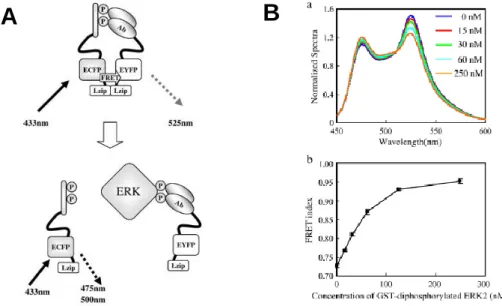
Ohiro and co-workers have reported a fluorescent biosensor for homogeneous competitive immunoassay for detection of phosphorylated protein based on enhanced FRET technology. The enhanced FRET has a characteristic that FRET efficiency is significantly enhanced by interaction of leucine zippers (Lzip) which are tethered with donor and acceptor fluorescent proteins. In this system, the extracellular signal-regulated kinase (ERK) is employed as a model antigen. A synthetic diphosphorylated peptide that mimics the regulatory site of ERK is linked to donor FRET probe (ECFP-Lzip) and an anti-diphosphorylated ERK antibody fragment (Fab) is linked to acceptor FRET probe (EYFP-Lzip). The indicator exhibits high FRET signal in the absence of antigen, but FRET signal decreases in the presence of antigen due to the competitive binding of diphosphorylated ERK to antibody (Figure 2-2A). In vitro
A B
28
assay using this indicator allows detection of diphosphorylated ERK in the range from 15 nM to 250 nM (Figure 2-2B)11. Although this strategy is potentially available for detection of various phosphorylated antigens, the preparation procedure is complicated and it requires careful design of the synthetic phosphorylated peptide so that the binding affinity of antibody- synthetic peptide must be lower than that of antibody-antigen.
Figure 2-2. (A) Principle of the competitive immunoassay for phosphorylated protein antigen.
(B) Assay for diphosphorylated ERK, (a) Fluorescence spectra show decrease in FRET signal upon addition of Glutathione-S-transferase (GST)-fused diphosphorylated ERK, (b) Dose- response curve for GST-diphosphorylated ERK (J. Biosci. Bioeng. 109(1): 15-19 (2010)).
To develop a simple and widely-applicable fluorescent biosensor for protein phosphorylation, the synthesis of fluorescent biosensor utilizing “Quenchbody” (Qbody) technology12 is introduced in this chapter. An anti-phosphotyrosine antibody single-chain variable domain (scFv) was fluorescently labeled at the N-terminus by incorporating a 5(6)- carboxytetramethylrhodamine (TAMRA)-linked nonnatural amino acid in response to a four- base codon CGGG13-14 (Figure 2-3). The biosensor showed fluorescence enhancement upon addition of peptides containing phosphotyrosine (pTyr).
A B
29
Figure 2-3. Schematic illustration of the synthetic procedure of TAMRA-scFv against pTyr and the detection of pTyr-containing antigen based on antigen-dependent removal of fluorescence quenching effect on TAMRA.
In the second part of this chapter, I attempt to construct fluorescent ratio biosensors in combination of FRET and Qbody technologies. Although Qbody is a powerful method for constructing of simple and sensitive biosensor, fluorescence intensity of single labeled Qbody is also dependent on the amount of this biosensor in measuring sample. It is a difficulty in the application of this technique to detection of antigens in the cells where the local concentration of the biosensor cannot be controlled. Therefore, double labeled scFv was developed and the change in fluorescence of this biosensor was based on FRET and fluorescent quenching. The scFv was labeled at C-terminus by TAMRA and fused at either N- and C-terminus with EGFP (enhanced green fluorescent protein) (Figure 2-4) or HaloTag which was subsequently labeled by a 5(6)- RhodamineGreen-linked HaloTag ligand. An expected mechanism for detection of antigen using double labeled scFv is as follows: double labeled scFv exhibits FRET from EGFP or RhodamineGreen (RhG) to TAMRA because emission spectra of EGFP and RhG overlap with absorption spectrum of TAMRA. However, fluorescence of TAMRA is quenched in the absence of antigen. As consequence, the biosensor mainly emits green fluorescence. In the presence of antigen, the quenching effect is eliminated and the biosensor emits both green and red fluorescence.
30
Figure 2-4. Schematic illustration of the synthetic procedure of double labeled scFv against pTyr in which EGFP was fused to N-terminus of scFv and the detection of pTyr-containing antigen based on FRET from EGFP to TAMRA and antigen-dependent removal of fluorescent quenching effect on TAMRA.
31
2-2. Materials and Methods
2-2-1. Materials
KOD-Plus DNA polymerase, Thermo T7 RNA polymerase were purchased from TOYOBO (Osaka, Japan). Primers for PCR were custom synthesized by Invitrogen (Life technologies Japan). QIAquick PCR purification kit, QIAquick gel extraction kit were purchased from QIAGEN (Venlo, Netherlands). pGEMEX1, MagneHis Ni-particles, E. coli S30 extract for linear templates, HaloTag Amine (O2) were from Promega (WI, USA). In- Fusion HD Cloning kit was from Clontech (CA, USA). T4 RNA ligase was obtained from TaKaRa Bio (Otsu, Japan), NdeI, HindIII restriction enzymes, Quick Ligase, Prestained protein marker (7-175 kDa) and T7 RNA polymerase were from New England BioLabs (MA, USA), Zeba desalting spin columns (7K MWCO) were from ThermoFisher Scientific (MA, USA), EGF receptor substrate 2 (Phospho-Tyr5), insulin receptor (1142-1153) (Phospho- Tyr1146,1150,1151) were obtained from GenScript (NJ, USA), pp60src SH2 domain-binding peptide (Ac-pTyr-Glu-Glu-Ile-Glu-OH) was from Bachem (Bubendorf, Switzerland), pFN18A Halo Tag® (WI, Promega) was a kind gift from Prof. Masaru Kawakami (Yamagata University).
2-2-2. Construction scFv(pTyr) genes
cDNA sequences of heavy chain and light chain of anti-pTyr antibody 4G10 was obtained from the National Center for Biotechnology Information USA (NCBI) Nucleotide database15. Accession number for heavy chain is DD058293.1 and accession number for light chain is DD058294.1. The original sequences of anti-pTyr antibody from database are heavy chain (VH): 1365 base pairs and light chain (VL): 645 base pairs. The sequence of scFv against BGP (scFv(BGP)) in Qbody12 was used as reference for designing of scFv against pTyr (scFv(pTyr)). Alignment of amino acid sequences of scFv(BGP) and scFv(pTyr) was performed by “T-Coffee”16-17 and “ESPript 3.0”18. The sequence from base 1 to 354 of VH and the sequence from base 1 to 324 of VL was linked by a (Gly4Ser)3 linker to make scFv(pTyr), All codons encoded for arginine was modified to be different from CGG. The N- and C-termini of scFv(pTyr) were fused with ProX-tag containing a four-base codon (5’-ATG TCT AAA CAA ATC GAA GTA AAC CGGG TCT AAT GAG-3’) and His6-tag, respectively. The scFv(pTyr) gene was custom synthesized by Biomatik (DE, USA), amplified for incorporation of NdeI and HindIII restriction sites respectively to N- and C-termini by PCR with forward primer (5’-GGA ATT CCA TAT GTC TAA ACA AAT CGA AG-3’) and reverse primer (5’- CCC AAG CTT AAT GAT GAT GAT GAT GAT GAG-3’). The PCR product was purified
32
by QIAquick PCR purification kit and digested by the restriction enzymes. The digested DNA fragments were introduced into pre-digested pGEMEX1 between T7 promoter and T7 terminatorby Quick Ligase.
The incorporation of linker (Gly3Ser)3 between ProX-tag and scFv(pTyr) was done by separately amplification of (Gly3Ser)3 and pGEMEX1 containing ProX-scFv(pTyr). After purification, the (Gly3Ser)3 was introduced between ProX-tag and scFv(pTyr) by In-Fusion HD cloning kit yielding ProX-(GGGS)3-scFv(pTyr). The insertion of (Gly3Ser)5linker between ProX-tag and scFv(pTyr) was performed by Quickchange mutagenesis with primers containing (Gly3Ser)2 and ProX-(GGGS)3-scFv(pTyr) as template. The DNA sequences of all constructs were confirmed by DNA sequencing.
The encoding regions of ProX-(GGGS)x-scFv(pTyr) genes were amplified by PCR with forward primer (5’-CCC GCG CGT TGG CCG ATT CA-3’) and reverse primer (5’-CCG GAT AAC CTG GCG TAA TA-3’). The PCR product was purified and transcribed with Thermo T7 RNA polymerase as described before13.
2-2-3. Construction of double labeled scFv(pTyr) genes
The cDNA of EGFP variant (F64L, S65T, Q80R, F99S, M153T, V163A, I167T, and S208L) was amplified by PCR with forward primer (5’-GAA GGA GAT ATA CAT ATG AGT AAA GGA GAA GAA CTT TTC ACT GGA GTT GTC CC-3’) and reverse primer (5’-GCT GCA GCT GGA CCT CTT TGT AGA GCT CATCCA TGC CAT GTG TAA TCC CAG C- 3’) for fusion of EGFP to N-terminus of scFv(pTyr). For the fusion of EGFP to C-terminus of scFv(pTyr), the forward primer (5’-GGT GGC GGT GGC TAG AGT AAA GGA GAA GAA CTT TTC ACT GGA GTT GTC C-3’) and the reverse primer (5’-ATA GAA TAC TCA AGC TTA GTG GTG GTG GTG GTG GTG TTT GTA GAG CTC ATC-3’) were used. The plasmid pGEMEX1 containing scFv(pTyr) linked with amber codon (TAG) at C-terminus through Gly4
or Gly3SerGly4 linker (scFv(pTyr)-G4-TAG or scFv(pTyr)-G3SG4-TAG) was amplified by PCR with forward primer (5’-GAG GTC CAG CTG CAG CAG TCT GGA CCT GAA CTG G-3’) and reverse primer (5’-ATG TAT ATCT CCT TCT TAA AGT TAA ACA AAA TTA TTT C- 3’) for incorporation of EGFP to N-terminus. For incorporation of EGFP to C-terminus, the forward primer (5’-GCT TGA GTA TTC TAT AGT GTC ACC TAA ATC CCA GC-3’) and reverse primer (5’-CTA GCC ACC GCC ACC TGA ACC GCC ACC ACG TTT G-3’) were used. The resulting PCR products were purified by gel extraction from agarose gel. The plasmid and EGFP PCR products were joined by In-Fusion cloning to introduce EGFP to either N- or C-termini of scFv(pTyr). Similar procedure was performed for fusion with HaloTag to
33
make pGEMEX-Halo-scFv(pTyr)-G3SG4-TAG. The forward and reverse primers for amplification of HaloTag were 5’-GAA GGA GAT ATA CAT ATG GCA GAA ATC GGT ACT GGC TTT CCA TTC GAC C-3’ and 5’-CTG CAG CTG GAC CTC GCC GGA AAT CTC GAG CGT CGA CAG CCA GCG CGC G-3’, respectively. The DNA sequences of all constructs were confirmed by DNA sequencing.
2-2-4. Synthesis of RhodamineGreen-linked HaloTag ligand
To a mixture of 50 mM DMSO solution of 5(6)-RhodamineGreen-X-succinimide ester (5(6)-RhG-X-SE) (8 μL, 400 nmol), 100 mM DMSO solution of HaloTag Amine (O2) (2 μL, 200 nmol) and 100 mM NaHCO3aq (4 μL) were added. The mixture was kept on ice for 60 min, and applied to an analytical scale reversed-phase HPLC (XBridge, 2.5 μm, 4.6x20 mm, flow rate 1.5 ml/min, with a linear gradient of 0-100% methanol in 0.38% formic acid, over 10 min) to afford RhG-X-Halo ligand (188 nmol; 94% yield). The product was confirmed by MALDI-TOF MS (calcd. 693.30 for MH+, found 693.34).
Scheme 2-1. Synthetic scheme of RhG-X-Halo ligand
2-2-5. Cell-free translation
The synthesis of TAMRA-X-AF-tRNACCCG by ligating TAMRA-X-AF-pdCpA with a yeast phenylalanine tRNACCCG (lacking the 3’-terminal two nucleotides C and A (-CA)) that contained a CCCG anticodon was carried out as described before13. The reaction mixture (93.6 µL) contained 2.3 nmol tRNACCCG, 6.8 nmol TAMRA-X-AF-pdCpA, 55 mM HEPES-Na (pH 7.5), 1 mM ATP, 15 mM MgCl2, 3.3 mM DTT, 0.002% BSA and 112.4 units T4 RNA ligase.
34
After incubation of 2 hours at 4oC, potassium acetate (pH 4.5) was added to the reaction to final concentration 0.3 M. The TAMRA-X-AF-tRNACCCG was isolated by ethanol precipitation and the pellet was dissolved with 12 µL pre-chilled 1 mM potassium acetate (pH 4.5). TAMRA-X- AF-tRNACUA was prepared in a similar manner using an amber suppressor tRNACUA derived from Mycoplasma capricolum tryptophan tRNA19.
Coding regions of scFv fusions were amplified by PCR and the PCR products were purified by QIAquick PCR purification kit. The transcription to mRNA was performed as described before13.
To incorporate TAMRA-X-AF into scFvs, a 120 µL cell-free translation mixture was prepared with 55 mM HEPES-KOH (pH 7.5), 210 mM GluK, 6.9 mM CH3COONH4, 12 mM (CH3COO)2Mg, 1.2 mM ATP, 0.28 mM GTP, 26 mM phophoenolpyruvate, 1 mM spermidine, 1.9% PEG-8000, 35 µg/ml folinic acid, 0.1 mM of each of the 19 natural amino acid except arginine, 0.01 mM arginine, 2 mM glutathionine oxidized, 0.8 μg/µL mRNA, 2.3 nmol TAMRA-X-AF-tRNACCCG (or TAMRA-X-AF-tRNACUA) and 24 µL E. coli S30 extract. The mixture was incubated at 37oC for 1 hour.
For HaloTag-containing protein, after protein was synthesized by cell-free translation, DMSO solution of RhG-X-Halo ligand was added to the crude translation mixture to final concentration 50 µM and incubated at 25oC for 30 minutes to achieve fluorescent labeling of HaloTag with RhG.
2-2-6. Protein purification
The translation mixture was diluted with buffer A (20 mM sodium phosphate, 0.5M NaCl, 5 mM imidazole, pH 7.5) containing 10M urea to final volume 480 µL and mixed with 48 µL MagneHis Ni-Particles. After shaking for 30 minutes at room temperature, the supernatant was removed and the beads were washed once with buffer A, once with buffer A containing 8 M Urea and followed by three times with buffer A. The His-tagged protein was eluted by buffer B (20 mM sodium phosphate, 0.5M NaCl, 500 mM imidazole, pH 7.5) and was gel-filtrated by Zeba Spin Desalting column (7K MWCO) equilibrated with phosphate buffer saline containing 0.05% Tween (PBS-T). For EGFP-fused scFvs, buffer A containing 4 M urea was used for dilution of the translation mixture and for second wash step.
Samples for SDS-PAGE were taken at each purification step and mixed with SDS- PAGE sample buffer to final volume 20 µL. All samples were heated at 95oC for 5 min before SDS-PAGE. 5 µL of each sample and Prestained protein marker were applied to 15% SDS- PAGE gel. In case of EGFP and Halo-tag fusions, 12% polyacrylamide gel was used. SDS-
35
PAGE gel was visualized by a fluorescence scanner (FMBIO-III; Hitachi Software Engineering, Japan). RhG was excited at 488 nm and visualized at 520 nm, and TAMRA was excited at 532 nm and visualized at 580 nm. Prestained marker was visualized with excitation at 635 nm and detection at 670 nm.
2-2-7. Fluorescence measurement
The purified TAMRA-labeled scFv was diluted in PBS-T buffer to 80 µL with various final concentrations (0 M, 10-9 M, 10-8 M, 10-7 M, 10-6 M, 10-5 M, 10-4 M) of pTyr-containing peptides, EGF receptor substrate 2 (ERS-pTyr), insulin receptor substrate (IRS), and pp60src SH2 domain-binding peptide (Ac-pYEEIE). The mixture was incubated at25oC for 12 hours.
The emission spectra were measured by a Fluorolog-3 instrument (Horiba, Japan) at 25oC. TAMRA was excited at 550 nm and emission was recorded from 565 nm to 700 nm. In case of double labeled scFvs with EGFP, EGFP was excited at 470 nm and emission was scanned from 490 nm to 700 nm. In cases of double labeled scFvs with HaloTag and RhG-X- Halo ligand, RhG was excited at 495 nm and emission was scanned from 510 nm to 700 nm.
Fluorescence intensities of TAMRA, EGFP, and RhG was obtained at 580 nm, 509 nm, and 529 nm, respectively, and fluorescence ratio (accepter/donor) was calculated using these values.
The apparent dissociation constant (Kd) was calculated by fitting the fluorescence intensity or fluorescence ratio values to a sigmoidal dose-response (variable slope) model using Graphpad Prism (Graphpad, CA, USA). All data were normalized by the value in the absence of antigen.