Kyushu University Institutional Repository
Expression of brain-derived neurotrophic factor and its receptor TrkB is associated with poor prognosis and a malignant phenotype in small cell lung cancer
木村, 信一
http://hdl.handle.net/2324/2236059
出版情報:九州大学, 2018, 博士(医学), 課程博士 バージョン:
権利関係:
Contents lists available atScienceDirect
Lung Cancer
journal homepage:www.elsevier.com/locate/lungcan
Expression of brain-derived neurotrophic factor and its receptor TrkB is associated with poor prognosis and a malignant phenotype in small cell lung cancer
Shinichi Kimura
a, Taishi Harada
a,b,⁎, Kayo Ijichi
c, Kentaro Tanaka
a, Renpeng Liu
a, Daisuke Shibahara
a, Yuko Kawano
a, Kohei Otsubo
a, Yasuto Yoneshima
a, Eiji Iwama
a, Yoichi Nakanishi
a, Isamu Okamoto
aaResearch Institute for Diseases of the Chest, Graduate School of Medical Sciences, Kyushu University, Fukuoka, Japan
bDepartment of Respiratory Medicine, Japan Community Healthcare Organization Kyushu Hospital, Kitakyushu, Japan
cDepartment of Anatomic Pathology, Graduate School of Medical Sciences, Kyushu University, Fukuoka, Japan
A R T I C L E I N F O
Keywords:
TrkB
Brain-derived neurotrophic factor (BDNF) Small cell lung cancer (SCLC)
NTRK2gene Prognosis
A B S T R A C T
Objectives:TrkB is a receptor for brain-derived neurotrophic factor (BDNF) and is highly expressed in various cancers, with BDNF-TrkB signaling having been implicated in tumor progression and metastasis. The role of the BDNF-TrkB system in small cell lung cancer (SCLC), a neuroendocrine cancer, has remained unclear, however.
We examined BDNF and TrkB expression in SCLC patients as well as the function of BDNF-TrkB signaling in SCLC cell lines.
Materials and methods: BDNF and TrkB expression in tumor specimens of 58 SCLC patients and 20 non–small cell lung cancer (NSCLC) patients was examined by immunohistochemistry and was scored on the basis of the distribution and intensity of staining. TrkB-overexpressing SCLC (SBC5TrkB) cells were established by retrovirus transduction and were examined for the effects of BDNF on intracellular signaling, cell proliferation, and cell migration in vitro.
Results:The staining score for TrkB in NSCLC and SCLC specimens was 2.80 ± 0.19 and 3.60 ± 0.15, re- spectively, whereas that for BDNF was 1.95 ± 0.32 and 2.76 ± 0.14, respectively. High levels of both TrkB and BDNF expression in SCLC tumors were significantly associated with poor overall survival in multivariate analysis (hazard ratio = 1.821,P= 0.036). BDNF activated AKT and ERK signaling pathways in and promoted the mi- gration of SBC5TrkBcells, and these effects were attenuated by the pan-Trk inhibitor GNF-5837. GNF-5837 also inhibited the proliferation of SBC5TrkBcells in the presence of BDNF.
Conclusion:Coexpression of BDNF and TrkB was associated with poor prognosis in SCLC patients, and BDNF promoted the migration of TrkB-overexpressing SCLC cells. TrkB is thus a potential therapeutic target for SCLC.
1. Introduction
Lung cancer is the leading cause of cancer death. Small cell lung cancer (SCLC) accounts for 15% of lung cancer cases and is classified as a neuroendocrine cancer in the WHO classification of 2015 [1,2].
Compared with non–small cell lung cancer (NSCLC), SCLC is considered to be more aggressive and more likely to metastasize to other organs.
Although SCLC is initially sensitive to chemotherapeutic agents, the disease recurs in most patients. The prognosis of SCLC is thus poor, with an overall 5-year survival rate of only 5% [3].
The tropomyosin receptor kinase (Trk) family of receptor tyrosine
kinases comprises three transmembrane proteins known as TrkA, TrkB, and TrkC. These proteins are encoded by the genesNTRK1,NTRK2, and NTRK3, respectively, and serve as receptors for the neurotrophin family of nerve growth factors. TrkB is composed of an extracellular ligand- binding domain, a transmembrane domain, and a cytoplasmic tyrosine kinase domain. Brain-derived neurotrophic factor (BDNF) is a ligand of TrkB and induces TrkB dimerization and consequent activation of downstream signaling [4,5].
Research on the BDNF-TrkB signaling pathway initially focused on the nervous system, with this pathway being implicated in conditions such as depression, schizophrenia, and Alzheimer’s disease [6–8].
https://doi.org/10.1016/j.lungcan.2018.04.005
Received 24 January 2018; Received in revised form 2 April 2018; Accepted 4 April 2018
⁎Corresponding author at: Research Institute for Diseases of the Chest, Graduate School of Medical Sciences, Kyushu University, 3-1-1 Maidashi, Higashi-ku, Fukuoka, 812-8582, Japan.
E-mail address:[email protected](T. Harada).
Lung Cancer 120 (2018) 98–107
0169-5002/ © 2018 Elsevier B.V. All rights reserved.
T
However, TrkB is also expressed in smooth muscle and airway epithelial cells, cardiomyocytes, and cancer cells [9–11]. We previously showed that TrkB and BDNF are highly expressed in and associated with the prognosis of NSCLC, and that a Trk inhibitor attenuated TrkB-depen- dent NSCLC cell migration and proliferation in the presence of BDNF [12,13]. The relation between SCLC and TrkB has remained largely uninvestigated, however. We have therefore now examined the ex- pression of TrkB and BDNF in SCLC as well as the possible role of BDNF- TrkB signaling in SCLC cell lines.
2. Materials and methods 2.1. Patients and specimen collection
We retrospectively analyzed specimens from 58 SCLC patients who had undergone surgical resection or bronchoscopic biopsy between January 2000 and March 2015 as well as from 20 NSCLC patients who had undergone surgical resection between January and July 2010 at Kyushu University Hospital. Specimens werefixed in neutral-buffered formaldehyde and processed for histopathologic and
immunohistochemical evaluation. The characteristics of the 58 SCLC patients are summarized inTable 1. Clinicopathologic factors, including age, sex, history, TNM staging, and SCLC staging, were evaluated.
Tumor histological subtype and pathological stage were determined according to the WHO classification of 2004 and UICC guidelines for TNM classification, respectively. The histological diagnosis was con- firmed by two pathologists. This study was approved by the ethics committee of Kyushu University.
2.2. Immunohistochemistry
Immunohistochemistry was performed with rabbit polyclonal anti- bodies to TrkB (sc-8316; Santa Cruz Biotechnology, Santa Cruz, CA, USA) and to BDNF (sc-20981, Santa Cruz Biotechnology). Paraffin- embedded sections of surgically resected or biopsy specimens were routinely depleted of paraffin and rehydrated. The sections were in- cubated overnight at 4 °C with primary antibodies to TrkB (1:100 di- lution) or to BDNF (1:50 dilution), and immune complexes were de- tected with biotin-conjugated secondary antibodies and streptavidin- conjugated peroxidase (Histofine SAB-PO kit; Nichirei, Tokyo, Japan).
Table 1
Relation of TrkB and BDNF expression status to various clinical factors in SCLC patients.
Factor Total patients
(n= 58)
TrkB BDNF TrkB + BDNF
Positive (n= 33)
Negative (n= 25)
Pvalue Positive (n= 38)
Negative (n= 20)
Pvalue Positive (n= 27)
Negative (n= 31)
Pvalue
Age (years)
< 70 28 14 14 0.3055 13 15 0.0027* 11 17 0.2829
≥70 30 19 11 25 5 16 14
Sex
Male 44 25 19 0.98 28 16 0.5892 21 23 0.75
Female 14 8 6 10 4 6 8
PS
0–1 38 21 17 0.7292 17 21 0.5213 12 21 0.7025
≥2 20 12 8 10 10 10 10
T status
T1 (T1a + T1b) 15 9 6 0.9413 11 4 0.8023 9 6 0.4049
T2 (T2a + T2b) 20 12 8 13 7 9 11
T3 12 6 6 8 4 6 6
T4 11 6 5 6 5 3 8
N status
N0 6 3 3 0.7807 3 3 0.8515 3 3 0.8182
N1 3 2 1 2 1 2 1
N2 29 15 14 19 10 12 17
N3 20 13 7 14 6 10 10
M status
M0 31 16 15 0.3839 20 11 0.8635 14 17 0.8201
M1 (M1a + M1b) 27 17 10 18 9 13 14
Brain metastasis
Positive 12 8 4 0.4428 10 2 0.1448 7 5 0.3582
Negative 46 25 21 28 18 20 26
TNM stage
I 6 3 3 0.5904 3 3 0.3823 3 3 0.7258
II 1 0 1 0 1 0 1
IIIA + IIIB 24 13 11 17 7 11 13
IV 27 17 10 18 9 13 14
SCLC stage
LD 28 15 13 0.6213 18 10 0.8488 12 16 0.5856
ED 30 18 12 20 10 15 15
Treatment
Surgery 8 3 5 0.5792 5 3 0.4389 3 5 0.5251
Chemotherapy +radiation
16 10 6 8 8 6 10
Chemotherapy 30 17 13 22 8 15 15
None 4 3 1 3 1 3 1
* P< 0.05 (chi-square test). LD and ED, limited and extensive disease, respectively.
Peroxidase activity was revealed by staining with 3,3′-diaminobenzi- dine. Normal brain sections were examined as positive controls for TrkB and BDNF. Negative control reactions in which primary antibodies were omitted were performed in parallel and did not show substantial background staining. All immunohistochemical staining was evaluated by Pathology Institute Corp. (Toyama, Japan) without knowledge of the patients’ clinical records. Staining for TrkB and BDNF was weak or nonexistent in normal lung and bronchus tissue, with bronchial smooth muscle of normal lung manifesting weak staining for both antigens (data not shown). Tumor cells with brown staining of the cell mem- brane or cytoplasm were regarded as positive. We classified staining as 0 for negative, 1 for weak, 2 for moderate, or 3 for intense, and the percentage of positive tumor cells as 1 for 1% to 50% or 2 for≥51%.
The two scores for each sample were added to give afinal score of 0, 2, 3, 4, or 5. This scoring system was described previously [14,15]. Al- though some of the tumor specimens analyzed were relatively old, we detected no significant correlation between sample collection time and staining score (data not shown).
2.3. Cell culture
H82, H69, H446, H146, H526, HCC827, H1975, H2228, H1299, H441, A549, and Beas-2B cells were obtained from American Type Culture Collection (Manassas, VA, USA). SBC5 and H3122 cells were obtained as previously described [16,17]. Amphopack293 cells were obtained from Clontech (Mountain View, CA, USA). The various cell lines were cultured under a humidified atmosphere of 5% CO2at 37 °C in RPMI 1640 medium (Gibco, Carlsbad, CA, USA) or Dulbecco’s modified Eagle’s medium (DMEM, Gibco), each supplemented with 10% fetal bovine serum (FBS).
2.4. RT and real-time PCR analysis
Total RNA was extracted from cells with the use of an RNeasy Mini Kit (Qiagen,Valencia, CA, USA) and was subjected to reverse tran- scription (RT) with the use of PrimeScript RT Master Mix (Takara Bio, Shiga, Japan). The resulting cDNA was then subjected to real-time polymerase chain reaction (PCR) analysis with the use of SYBR Premix Ex Taq II (Takara Bio) and a Thermal Cycler Dice Real Time System II (Takara Bio). The PCR primers (forward and reverse, respectively) in- cluded those for TrkB (5′-TGTGTGGCGGAAAATCTTG-3′and 5′-GGTT TGGGGTTGCCTTTCAC-3′) and 18S rRNA (5′-ACTCAACACGGGAAACC TCA-3′and 5′-AACCAGACAAATCGCTCCAC-3′). The amount of TrkB mRNA was normalized by that of 18S rRNA.
2.5. Plasmid construction and viral infection
SBC5 and Beas-2B cells were infected with retroviruses according to standard protocols. The plasmids pDONR223-NTRK2 (Addgene
#23883) and pcDNA-DEST40 (Thermo Fisher Scientific, Waltham, MA, USA) were used as Gateway entry and destination vectors, respectively.
Human TrkB cDNA was subcloned into the pQCXIP retroviral vector (Takara Bio) with the use of an In-Fusion HD Cloning Kit (Clontech), and retroviruses encoding TrkB were generated by transfection of Amphopack293 cells with the resulting construct with the use of the Xfect Transfection Reagent (Takara Bio). Cells were infected with TrkB retroviruses in the presence of polybrene (Sigma-Aldrich, St. Louis, MO, USA) at 4 mg/ml. Stably infected cells were obtained by selection with puromycin (Invitrogen, Carlsbad, CA, USA) at 50 ng/ml.
2.6. Immunoblot analysis
Cultured cells were lysed in SDS sample buffer, the lysates were fractionated by SDS-polyacrylamide gel electrophoresis, and the sepa- rated proteins were transferred to a polyvinylidene difluoride mem- brane. The membrane was incubated overnight at 4 °C with primary
antibodies to phosphorylated TrkB (#4619, diluted 1:1000; Cell Signaling Technology, Danvers, MA, USA), to TrkB (sc-8316, diluted 1:500; Santa Cruz Biotechnology), toβ-actin (#4970S, diluted 1:1000;
Cell Signaling Technology), to phosphorylated AKT (#4060S, diluted 1:1000; Cell Signaling Technology), to AKT (#9272S, diluted 1:1000;
Cell Signaling Technology), to phosphorylated ERK (#4370S, diluted 1:1000; Cell Signaling Technology), or to ERK (#9102S, diluted 1:1000;
Cell Signaling Technology). Immune complexes were detected with horseradish peroxidase–conjugated goat antibodies to rabbit im- munoglobulin G (GE Healthcare UK, Amersham, UK), Pierce Western Blotting Substrate Plus (Thermo Fisher Scientific), and the ChemiDoc XRS+ system (Bio-Rad, Hercules, CA, USA).
2.7. Cell proliferation assay
Cells were seeded in 24-well plates at a density of 40,000 per well and were cultured for 72 h in the absence or presence of BDNF (Sigma- Aldrich) or GNF-5837 (Selleck Chemicals, Houston, TX, USA) in 500μl of RPMI 1640 medium. Cell number was then determined with the use of a Luna automatic cell counter (Logos Biosystems, Gyeonggi, South Korea).
2.8. Wound closure assay
Retrovirus-infected SBC5 or Beas-2B cells were plated in six-well plates and maintained in RPMI 1640 or DMEM, respectively, each supplemented with 1% FBS. After the cells had achieved 80%–90%
confluence, a linear scratch wound was introduced into the cell monolayer with a 100-μl micropipette tip. The cells were washed with phosphate-buffered saline to remove debris and then incubated for 20 h with fresh medium containing (or not) BDNF or GNF-5837. Cell mi- gration into the wound area was evaluated from photographs taken with a digital camera. The width of the wound was measured with the use of Image J software (NIH, Bethesda, MD, USA), and the extent of wound closure was calculated as: 100% × [(wound width at 0 h)−(wound width at 20 h)/(wound width at 0 h)].
2.9. Transwell assay of cell migration
Cell migration was assayed with the use of 24-well Transwell chambers fitted with filters with a pore size of 8.0μm (Corning, Corning, NY, USA). Cells (5 × 10⁴ in 100μl of RPMI 1640 supple- mented with 1% FBS) were added to the upper chamber of each well, and the lower chamber wasfilled with 650μl of RPMI 1640 supple- mented with 10% FBS, with or without BDNF (50 ng/ml) or 300 nM GNF-5837. The chambers were incubated for 24–68 h at 37 °C, after which cells attached to eachfilter werefixed and stained with hema- toxylin (Muto Pure Chemicals, Tokyo, Japan). The upper surface of the filter was scraped twice with cotton swabs to remove nonmigrating cells, and the number of migrating cells was then counted with the use of a BZ-8100 microscope (Keyence, Osaka, Japan).
2.10. Statistical analysis
Survival curves were plotted with the Kaplan-Meier method and were analyzed with the two-sided log-rank test. Survival times were measured from the date offirst visit to death or last follow-up. The Cox proportional hazard model was applied for determination of univariate and multivariate hazard ratios (HRs) for the study parameters.
Significant variables in univariate analysis were subjected to multi- variate analysis. Data for cell proliferation, wound closure, and cell migration are presented as means ± SEM and were analyzed with Student’st-test. All statistical analysis was performed with JMP soft- ware version 13 (SAS Institute, Cary, NC, USA). APvalue of < 0.05 was considered statistically significant.
S. Kimura et al. Lung Cancer 120 (2018) 98–107
100
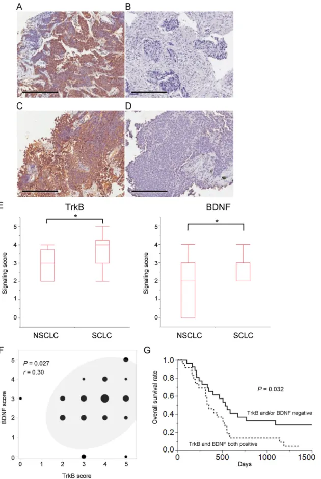
Fig. 1.Immunohistochemical analysis of TrkB and BDNF expression in SCLC specimens. (A–D) Representative positive (A,C) and negative (B,D) staining for TrkB (A,B) or BDNF (C,D) in tumor cells. Scale bars, 200μm. (E) Summary of TrkB and BDNF staining scores for all 58 SCLC patients and 20 NSCLC patients. Box and whisker plots show maximum, third-quartile, median,first-quartile, and minimum values. *P <0.05 (Student’st-test). (F) Scatter plot showing the correlation between TrkB and BDNF staining scores for SCLC specimens. The size of each circle indicates the number of specimens corresponding to the staining scores. The 95%
confidence ellipse is also shown, and Pearson’s correlation coefficient is indicated byr. (G) Kaplan-Meier survival curves for SCLC patients who did not undergo surgical resection classified as positive for both TrkB and BDNF expression (n= 24) or not (n= 26). ThePvalue was calculated with the log-rank test.
3. Results
3.1. TrkB and BDNF expression in SCLC specimens
We examined the expression of TrkB and BDNF in SCLC and NSCLC specimens by immunohistochemical analysis, with representative images of positive and negative staining being shown inFig. 1A–D. The expression scores (means ± SEM) for TrkB in NSCLC and SCLC were 2.80 ± 0.19 and 3.60 ± 0.15, respectively, whereas those for BDNF were 1.95 ± 0.32 and 2.76 ± 0.14, respectively (Fig. 1E). Both TrkB and BDNF scores were thus significantly (P <0.05) higher in SCLC than in NSCLC. Moreover, TrkB and BDNF scores were significantly correlated in SCLC patients (Fig. 1F).
For classification of SCLC patients as positive or negative for TrkB or BDNF expression, we assigned cutoffvalues of 4 and 3, respectively, for expression scores on the basis of the corresponding median values for SCLC. Among the total of 58 SCLC patients, 33 (56.9%) were positive for TrkB and 38 (65.5%) were positive for BDNF. Clinical factors for SCLC patients according to TrkB and BDNF expression status are shown inTable 1. Only age (< 70 or≥70 years) and BDNF expression status manifested a significant association, with clinical factors including sex, performance status (PS), T status, N status, M status, brain metastasis at the time of diagnosis, clinical stage, SCLC stage (extensive or limited disease), and treatment not differing between patients positive or ne- gative for TrkB or BDNF expression. For the 50 SCLC patients who did not undergo surgical resection, overall survival did not differ sig- nificantly between those positive or negative for TrkB or BDNF ex- pression (Supplementary Fig. S1). However, among these 50 patients, overall survival was significantly (P= 0.032) shorter for those positive for both TrkB and BDNF expression than for the remaining individuals (Fig. 1G).
Univariate analysis with the Cox proportional hazard model showed that TrkB positivity (HR = 1.455, P= 0.213) and BDNF positivity (HR = 1.895,P= 0.059) tended to be associated with shorter overall survival among all SCLC patients, although these associations were not statistically significant. Furthermore, patients positive for both TrkB and BDNF expression had a significantly poorer overall survival (HR = 2.021,P= 0.023). Multivariate analysis with factors found to be significant in the univariate analysis revealed that positivity for both TrkB and BDNF expression was an independent prognostic factor for overall survival [HR = 1.821, 95% confidence interval (CI) = 1.039–3.213,P= 0.036] (Table 2). SCLC stage was also a sig- nificant independent factor for overall survival.
3.2. Endogenous TrkB expression in SCLC and NSCLC cell lines
To investigate further the relevance of TrkB in SCLC, wefirst ex- amined TrkB expression in SCLC and NSCLC cell lines. RT and real-time PCR analysis revealed that the abundance of TrkB mRNA was higher in SCLC cell lines than in NSCLC cell lines (Fig. 2A). Immunoblot analysis also showed that TrkB was expressed at a higher level in SCLC cell lines than in NSCLC lines (Fig. 2B). These data were thus consistent with our
immunohistochemical analysis of clinical specimens. We also examined RNA sequencing data forNTRK2andBDNFin 48 SCLC cell lines listed in the Cancer Cell Line Encyclopedia [18,19]. In contrast to the corre- lation detected between TrkB and BDNF expression in our SCLC spe- cimens (Fig. 1F), however, the expression levels ofNTRK2andBDNFin these SCLC cell lines were not significantly correlated (Supplementary Fig. S2).
To investigate whether TrkB might promote SCLC tumor progres- sion, we generated SCLC cells that stably overexpress TrkB. SBC5 cells, which are derived from an SCLC patient and express TrkB at a lower level compared with some of the SCLC cell lines examined (Fig. 2), were thus infected with a retrovirus encoding TrkB or with the corresponding empty virus to yield SBC5TrkBand SBC5emptycells, respectively. TrkB is thought to regulate the growth and survival of cells through activation of phosphatidylinositol 3-kinase (PI3K)–AKT and extracellular sig- nal–regulated kinase (ERK) signaling pathways [20]. Immunoblot analysis showed that treatment of SBC5TrkBcells with BDNF induced phosphorylation of TrkB as well as that of the downstream signaling molecules AKT and ERK, and these effects of BDNF were attenuated by the pan-Trk inhibitor GNF-5837 (Fig. 3A). Such BDNF-induced phos- phorylation of TrkB, AKT, and ERK was not observed in SBC5emptycells, consistent with the relatively low abundance of endogenous TrkB in SBC5 cells. To examine BDNF-TrkB signaling in normal bronchial epi- thelial cells, we studied the immortalized human bronchial epithelial cell line Beas-2B. BDNF also induced the phosphorylation of TrkB, AKT, and ERK in Beas-2BTrkBcells in a manner sensitive to inhibition by GNF- 5837, whereas such effects were not apparent in Beas-2Bempty cells (Supplementary Fig. S3).
We next examined the effect of BDNF on the proliferative capacity of TrkB-overexpressing SCLC cells. BDNF had no significant effect on the proliferation of SBC5emptyor SBC5TrkB cells, although GNF-5837 significantly inhibited that of SBC5TrkBcells in the presence of BDNF (Fig. 3B). In contrast, BDNF increased the number of Beas-2BTrkBcells in a manner sensitive to inhibition by GNF-5837 (Supplementary Fig. S4).
GNF-5837 alone had no effect on the proliferation of either TrkB- overexpressing or control SBC5 or Beas-2B cells. These data thus sug- gested that BDNF-TrkB signaling might influence the phenotype of SCLC by mechanisms in addition to promotion of cell proliferation, such as stimulation of cell migration and invasion.
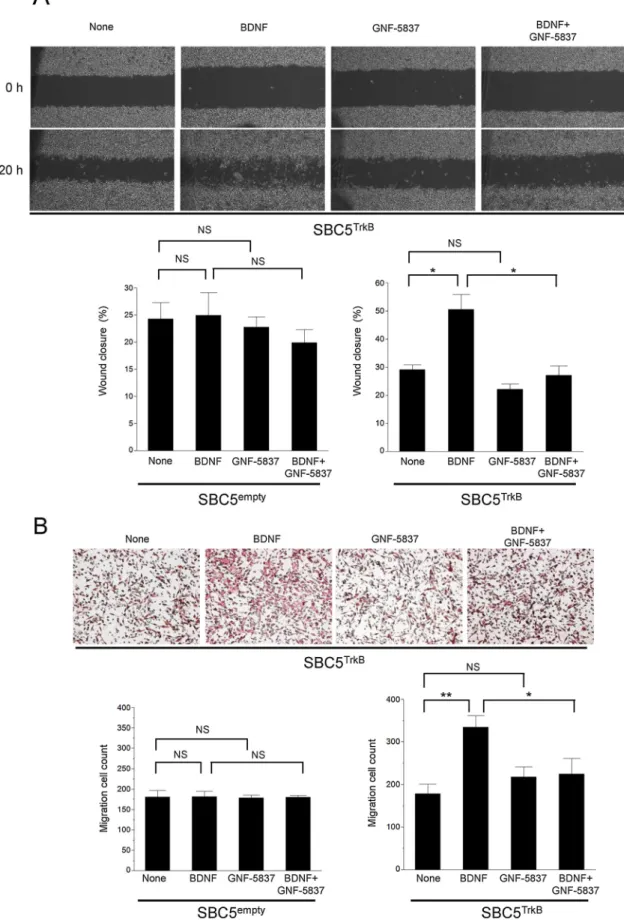
We therefore examined the effect of BDNF on cell migration in TrkB- overexpressing SCLC cells. Wefirst performed a scratch wound healing assay and found that BDNF promoted the migration of SBC5TrkBcells in this assay and that this effect was inhibited by GNF-5837. Such an effect of BDNF was not apparent in SBC5emptycells, and the Trk inhibitor alone did not affect the migration of either SBC5TrkBor SBC5emptycells (Fig. 4A, Supplementary Fig. S5A). We obtained results with Beas- 2BTrkBand Beas-2Bemptycells similar to those obtained with the corre- sponding SBC5 cells (Supplementary Fig. S6).
To confirm the results obtained with the wound healing assay, we examined cell migration in a Transwell assay. Again, BDNF significantly increased the number of migrating cells for SBC5TrkBand Beas-2BTrkB cells in a manner sensitive to inhibition by GNF-5837, whereas it had no
Table 2
Univariate and multivariate analyses of clinicopathologic factors for overall survival in all 58 SCLC patients according to the Cox proportional hazard method.
Factor Univariate Multivariate
Hazard ratio 95% CI Pvalue Hazard ratio 95% CI Pvalue
TrkB positivity 1.455 0.807–2.673 0.213
BDNF positivity 1.895 0.978–3.978 0.059
Both TrkB and BDNF positivity 2.021 1.101–3.742 0.023* 1.821 1.039–3.213 0.036*
Sex 0.937 0.486–1.950 0.853
Age (> 70 years) 1.443 0.804–2.629 0.219
SCLC stage (ED vs. LD) 4.534 2.360–9.119 < 0.0001* 2.920 1.630–5.331 < 0.0003*
* P <0.05.
S. Kimura et al. Lung Cancer 120 (2018) 98–107
102
effect on the migration of SBC5emptyor Beas-2Bempty cells (Fig. 4B, Supplementary Figs. S5B and S7). Both assays of cell migration thus suggested that the poor prognosis of TrkB- and BDNF-expressing SCLC tumors might be related to an enhanced capacity for cell migration and invasion.
4. Discussion
TrkB has been shown to be highly expressed in many malignancies and associated with poor prognosis [5,21]. The interaction of BDNF with TrkB triggers the activation of Ras-Raf-ERK, PI3K–AKT–mTOR (mechanistic target of rapamycin), and phospholipase C–γ (PLC- γ)–protein kinase C (PKC) signaling pathways and facilitates tumor progression through promotion of cell invasion, metastasis, angiogen- esis, and chemotherapy resistance [4,22]. The frequency of TrkB ex- pression in NSCLC has been found to range from 24% to 86.7% by immunohistochemistry [23–25]. High expression levels of BDNF and TrkB as revealed by immunohistochemistry in NSCLC tumors were
found to be associated with reduced overall survival and progression- free survival [13]. A previous study of 99 patients with squamous cell lung carcinoma revealed that the frequency of patients with a high expression level of TrkB as determined by immunohistochemistry was 43% and that the overall survival of these patients was significantly shorter than that of those with a low expression level [26]. In the present study, we found that the frequency of SCLC patients with high expression levels of BDNF or TrkB was 65.5% and 56.9%, respectively.
Although it is difficult to compare ourfindings with those of previous studies because of differences in methodology, we did confirm that the expression of BDNF and TrkB was significantly higher in SCLC than in NSCLC by analysis of both clinical specimens and cell lines. We also found that BDNF expression in SCLC patients was associated with age, although the reason for this association is unclear. The serum con- centration of BDNF was previously shown to be negatively correlated with age in healthy elderly individuals [27], whereas it was sig- nificantly increased in patients with chronic obstructive pulmonary disease or asthma [28,29]. We found no association between smoking Fig. 2.Expression of TrkB in SCLC cell lines. (A) RT and real-time PCR analysis of TrkB mRNA abundance in the indicated SCLC and NSCLC cell lines. Data are means ± SEM from three independent experiments. *P= 0.0184 for comparison of overall mean values between SCLC and NSCLC lines (Student’st-test). (B) Immunoblot analysis of TrkB andβ-actin (loading control) in NSCLC and SCLC cell lines. Data are representative of three independent experiments.
history (pack years) and BDNF expression in the SCLC patients of the present study (data not shown).
We also confirmed that BDNF induced TrkB phosphorylation and the activation of downstream signaling pathways in TrkB-over- expressing SCLC cells. The inhibitory effect of GNF-5837 on the acti- vation of downstream signaling was only partial, however. It is thus possible that bypass signaling such as that triggered by the p75
neurotrophin receptor (p75NTR) and Sortilin, which together are thought to function as a receptor for BDNF in addition to TrkB [30], may have limited the effect of GNF-5837. The reversible nature of GNF- 5837 action might also have contributed to the incomplete inhibition of downstream signaling.
The BDNF-TrkB pathway has been shown to promote the migration and proliferation of brain tumor, NSCLC, gastric cancer, and gall Fig. 3.Effects of BDNF on TrkB signaling and cell proliferation in TrkB-overexpressing SBC5 cells. (A) SBC5 cells infected with a retrovirus encoding TrkB (SBC5TrkB) or with the corresponding empty virus (SBC5empty) were incubated with or without the Trk inhibitor GNF-5837 (300 nM) for 1 h and then in the additional absence or presence of BDNF (25 ng/ml) for 20 min, after which cell lysates were subjected to immunoblot analysis with antibodies to total or phosphorylated (p) forms of TrkB, AKT, or ERK. Data are representative of three independent experiments. (B) Relative cell number for SBC5TrkBand SBC5emptycells incubated for 72 h in the absence or presence of BDNF (25 ng/ml) or GNF-5837 (300 nM). Data are means ± SEM from three independent experiments. *P <0.05; NS, not significant (Student’st-test).
S. Kimura et al. Lung Cancer 120 (2018) 98–107
104
Fig. 4.Effects of BDNF on migration of TrkB-overexpressing SBC5 cells. (A) SBC5TrkBand SBC5emptycells were subjected to a scratch wound healing assay in the presence of BDNF (50 ng/ml) or GNF-5837 (300 nM) for 20 h, after which the extent of wound closure was determined. Representative phase-contrast images of SBC5TrkBcell monolayers immediately and 20 h after wounding are shown. Quantitative data are means ± SEM from three independent experiments. *P< 0.05 (Student’st-test). (B) SBC5TrkBand SBC5emptycells were incubated in a Transwell apparatus with BDNF (50 ng/ml) or GNF-5837 (300 nM) in the lower chamber.
After 68 h, the number of cells that had migrated to the lower side of thefilter was determined. Representative images of migrated SBC5TrkBcells stained with hematoxylin are shown. Quantitative data are means ± SEM from three independent experiments. *P< 0.05, **P< 0.01 (Student’st-test).
bladder cancer cells [26,31–38]. We also found that the migration ac- tivity of SBC5TrkB cells was enhanced by BDNF, whereas that of the corresponding cells infected with the empty retrovirus was not. This effect of BDNF was attenuated by GNF-5837, whereas the Trk inhibitor had no effect on cell migration in the absence of BDNF. These results thus indicate that both BDNF and TrkB are required for stimulation of SCLC cell migration in vitro, and they are consistent with the coex- pression of the receptor and ligand detected in SCLC specimens by immunohistochemistry. We found that BDNF had no effect on the proliferation of SBC5TrkBcells, although GNF-5837 inhibited the pro- liferation of these cells in the presence of BDNF. BDNF was previously shown not to affect the proliferation of gall bladder cancer cells ex- pressing TrkB, whereas depletion of TrkB by RNA interference inhibited cell proliferation [38]. One possible explanation for these results is that ligands other than BDNF, such as neurotrophin-3 or neurotrophin-4, produced by cancer cells stimulate cell proliferation via TrkB. In con- trast to SBC5TrkBcells, we found that BDNF stimulated both the mi- gration and proliferation of Beas-2BTrkBcells. Together, our results thus suggest that the BDNF-TrkB pathway may preferentially promote the migration rather than proliferation of SCLC cells.
Oncogenic NTRK gene fusions have recently been detected in var- ious tumor types. Preclinical and clinical studies have shown that Trk inhibitors have antitumor activity for such cancers, with some such agents now being under investigation in phase 2 clinical trials [5,39–43]. Larotrectinib, a highly selective Trk inhibitor, thus induced a marked response in patients with NTRK fusion–positive cancers, with an overall response rate of 75% [44]. Although NTRK gene fusion or mutation has been detected at a low frequency in NSCLC [45,46], no such gene alterations have yet been reported for SCLC. The relevance of Trk inhibitors to the treatment of SCLC is thus currently unknown. We have now shown that the pan-Trk inhibitor GNF-5837 attenuated TrkB- dependent SCLC cell migration and proliferation in the presence of BDNF. Further study is necessary to determine whether TrkB is a po- tential therapeutic target in SCLC.
In conclusion, we have shown that BDNF and TrkB are expressed in SCLC tumors and that such expression is associated with a poor prog- nosis. Furthermore, BDNF-TrkB signaling plays an important role in the migration of SCLC cells expressing TrkB at a high level. Our results thus provide a basis for further investigations of TrkB-targeted therapy in lung cancer patients.
Conflicts of interest
The authors declare no conflicts of interest.
Acknowledgments
This study was supported by a research fund of Research Institute for Diseases of the Chest, Graduate School of Medical Sciences, Kyushu University. The authors thank A. Sato for technical assistance.
Appendix A. Supplementary data
Supplementary data associated with this article can be found, in the online version, athttps://doi.org/10.1016/j.lungcan.2018.04.005.
References
[1] A.F. Gazdar, P.A. Bunn, J.D. Minna, Small-cell lung cancer: what we know what we need to know and the path forward, Nat. Rev. Cancer 17 (12) (2017) 725–737.
[2] W.D. Travis, E. Brambilla, A.G. Nicholson, Y. Yatabe, J.H.M. Austin, M.B. Beasley, L.R. Chirieac, S. Dacic, E. Duhig, D.B. Flieder, K. Geisinger, F.R. Hirsch, Y. Ishikawa, K.M. Kerr, M. Noguchi, G. Pelosi, C.A. Powell, M.S. Tsao, I. Wistuba, The 2015 World Health Organization classification of lung tumors: impact of genetic clinical and radiologic advances since the 2004 classification, J. Thorac. Oncol. 10 (9) (2015) 1243–1260.
[3] R.L. Siegel, K.D. Miller, A. Jemal, Cancer statistics 2015, CA Cancer J. Clin. 65 (1) (2015) 5–29.
[4] A. Tajbakhsh, A. Mokhtari-Zaer, M. Rezaee, F. Afzaljavan, M. Rivandi, S.M. Hassanian, G.A. Ferns, A. Pasdar, A. Avan, Therapeutic potentials of BDNF/
TrkB in breast cancer; current status and perspectives, J. Cell. Biochem. 118 (9) (2017) 2502–2515.
[5] Y.B. Khotskaya, V.R. Holla, A.F. Farago, K.R. Mills Shaw, F. Meric-Bernstam, D.S. Hong, Targeting TRK family proteins in cancer, Pharmacol. Ther. 173 (2017) 58–66.
[6] T. Numakawa, N. Adachi, M. Richards, S. Chiba, H. Kunugi, Brain-derived neuro- trophic factor and glucocorticoids: reciprocal influence on the central nervous system, Neuroscience 239 (2013) 157–172.
[7] O. Schulte-Herbruggen, M.C. Jockers-Scherubl, R. Hellweg, Neurotrophins: from pathophysiology to treatment in Alzheimer's disease, Curr. Alzheimer Res. 5 (1) (2008) 38–44.
[8] G. Shoval, A. Weizman, The possible role of neurotrophins in the pathogenesis and therapy of schizophrenia, Eur. Neuropsychopharmacol. 15 (3) (2005) 319–329.
[9] Y.S. Prakash, R.J. Martin, Brain-derived neurotrophic factor in the airways, Pharmacol. Ther. 143 (1) (2014) 74–86.
[10] W. Hartman, M. Helan, D. Smelter, V. Sathish, M. Thompson, C.M. Pabelick, B. Johnson, Y.S. Prakash, Role of hypoxia-induced brain derived neurotrophic factor in human pulmonary artery smooth muscle, PLoS One 10 (7) (2015) e0129489.
[11] V. Sathish, S.K. Vanoosten, B.S. Miller, B. Aravamudan, M.A. Thompson, C.M. Pabelick, R. Vassallo, Y.S. Prakash, Brain-derived neurotrophic factor in ci- garette smoke-induced airway hyperreactivity, Am. J. Respir. Cell Mol. Biol. 48 (4) (2013) 431–438.
[12] T. Harada, Y. Yatabe, M. Takeshita, T. Koga, T. Yano, Y. Wang, G. Giaccone, Role and relevance of TrkB mutations and expression in non-small cell lung cancer, Clin.
Cancer Res. 17 (9) (2011) 2638–2645.
[13] K. Okamura, T. Harada, S. Wang, K. Ijichi, K. Furuyama, T. Koga, T. Okamoto, K. Takayama, T. Yano, Y. Nakanishi, Expression of TrkB and BDNF is associated with poor prognosis in non-small cell lung cancer, Lung Cancer 78 (1) (2012) 100–106.
[14] J. Fukuoka, T. Fujii, J.H. Shih, T. Dracheva, D. Meerzaman, A. Player, K. Hong, S. Settnek, A. Gupta, K. Buetow, S. Hewitt, W.D. Travis, J. Jen, Chromatin re- modeling factors and BRM/BRG1 expression as prognostic indicators in non-small cell lung cancer, Clin. Cancer Res. 10 (13) (2004) 4314–4324.
[15] A. Yoshizawa, J. Fukuoka, S. Shimizu, K. Shilo, T.J. Franks, S.M. Hewitt, T. Fujii, C. Cordon-Cardo, J. Jen, W.D. Travis, Overexpression of phospho-eIF4E is asso- ciated with survival through AKT pathway in non–small cell lung cancer, Clin.
Cancer Res. 16 (1) (2010) 240–248.
[16] K. Ota, K. Azuma, A. Kawahara, S. Hattori, E. Iwama, J. Tanizaki, T. Harada, K. Matsumoto, K. Takayama, S. Takamori, M. Kage, T. Hoshino, Y. Nakanishi, I. Okamoto, Induction of PD-L1 expression by the EML4-ALK oncoprotein and downstream signaling pathways in non-small cell lung cancer, Clin. Cancer Res. 21 (17) (2015) 4014–4021.
[17] K. Ota, T. Harada, K. Otsubo, A. Fujii, Y. Tsuchiya, K. Tanaka, I. Okamoto, Y. Nakanishi, Visualization and quantitation of epidermal growth factor receptor homodimerization and activation with a proximity ligation assay, Oncotarget 8 (42) (2017) 72127–72132.
[18] J. Barretina, G. Caponigro, N. Stransky, K. Venkatesan, A.A. Margolin, S. Kim, C.J. Wilson, J. Lehár, G.V. Kryukov, D. Sonkin, A. Reddy, M. Liu, L. Murray, M.F. Berger, J.E. Monahan, P. Morais, J. Meltzer, A. Korejwa, J. Jané-Valbuena, F.A. Mapa, J. Thibault, E. Bric-Furlong, P. Raman, A. Shipway, I.H. Engels, J. Cheng, G.K. Yu, J. Yu, P. Aspesi, M. de Silva, K. Jagtap, M.D. Jones, L. Wang, C. Hatton, E. Palescandolo, S. Gupta, S. Mahan, C. Sougnez, R.C. Onofrio, T. Liefeld, L. MacConaill, W. Winckler, M. Reich, N. Li, J.P. Mesirov, S.B. Gabriel, G. Getz, K. Ardlie, V. Chan, V.E. Myer, B.L. Weber, J. Porter, M. Warmuth, P. Finan, J.L. Harris, M. Meyerson, T.R. Golub, M.P. Morrissey, W.R. Sellers, R. Schlegel, L.A. Garraway, The Cancer Cell Line Encyclopedia enables predictive modelling of anticancer drug sensitivity, Nature 483 (2012) 603.
[19] B.D. Yard, D.J. Adams, E.K. Chie, P. Tamayo, J.S. Battaglia, P. Gopal, K. Rogacki, B.E. Pearson, J. Phillips, D.P. Raymond, N.A. Pennell, F. Almeida, J.H. Cheah, P.A. Clemons, A. Shamji, C.D. Peacock, S.L. Schreiber, P.S. Hammerman, M.E. Abazeed, A genetic basis for the variation in the vulnerability of cancer to DNA damage, Nat. Commun. 7 (2016) 11428.
[20] V.K. Gupta, Y. You, V.B. Gupta, A. Klistorner, S.L. Graham, TrkB receptor signalling:
implications in neurodegenerative psychiatric and proliferative disorders, Int. J.
Mol. Sci. 14 (5) (2013) 10122–10142.
[21] D.P. Radin, P. Patel, BDNF: an oncogene or tumor suppressor? Anticancer Res. 37 (8) (2017) 3983–3990.
[22] B. Ricciuti, M. Brambilla, G. Metro, S. Baglivo, R. Matocci, M. Pirro, R. Chiari, Targeting NTRK fusion in non-small cell lung cancer: rationale and clinical evi- dence, Med. Oncol. 34 (6) (2017) 105.
[23] A. Ricci, S. Greco, S. Mariotta, L. Felici, E. Bronzetti, A. Cavazzana, G. Cardillo, F. Amenta, A. Bisetti, G. Barbolini, Neurotrophins and neurotrophin receptors in human lung cancer, Am. J. Respir. Cell Mol. Biol. 25 (4) (2001) 439–446.
[24] S. Zhang, D. Guo, W. Luo, Q. Zhang, Y. Zhang, C. Li, Y. Lu, Z. Cui, X. Qiu, TrkB is highly expressed in NSCLC and mediates BDNF-induced the activation of Pyk2 signaling and the invasion of A549 cells, BMC Cancer 10 (1) (2010) 43.
[25] J. Terry, A. De Luca, S. Leung, G. Peacock, Y. Wang, W.M. Elliot, D. Huntsman, Immunohistochemical expression of neurotrophic tyrosine kinase receptors 1 and 2 in lung carcinoma: potential discriminators between squamous and nonsquamous subtypes, Arch. Pathol. Lab. Med. 135 (4) (2011) 433–439.
[26] K. Ozono, Y. Ohishi, H. Onishi, K. Nakamura, J. Motoshita, M. Kato, R. Nakanishi, M. Nakamura, Y. Oda, Brain-derived neurotrophic factor/tropomyosin-related ki- nase B signaling pathway contributes to the aggressive behavior of lung squamous
S. Kimura et al. Lung Cancer 120 (2018) 98–107
106
cell carcinoma, Lab. Invest. 97 (11) (2017) 1332–1342.
[27] A.A. Ziegenhorn, O. Schulte-Herbruggen, H. Danker-Hopfe, M. Malbranc, H.D. Hartung, D. Anders, U.E. Lang, E. Steinhagen-Thiessen, R.T. Schaub, R. Hellweg, Serum neurotrophins–a study on the time course and influencing fac- tors in a large old age sample, Neurobiol. Aging 28 (9) (2007) 1436–1445.
[28] P. Stoll, U. Wuertemberger, K. Bratke, C. Zingler, C.J. Virchow, M. Lommatzsch, Stage-dependent association of BDNF and TGF-β1 with lung function in stable COPD, Respir. Res. 13 (2012) 116.
[29] G.C. Muller, P.M. Pitrez, A.L. Teixeira, P.S. Pires, M.H. Jones, R.T. Stein, M.E. Bauer, Plasma brain-derived neurotrophic factor levels are associated with clinical severity in school age children with asthma, Clin. Exp. Allergy 40 (12) (2010) 1755–1759.
[30] I. Khalin, R. Alyautdin, G. Kocherga, M.A. Bakar, Targeted delivery of brain-derived neurotrophic factor for the treatment of blindness and deafness, Int. J. Nanomed. 10 (2015) 3245–3267.
[31] Y. Okugawa, K. Tanaka, Y. Inoue, M. Kawamura, A. Kawamoto, J. Hiro, S. Saigusa, Y. Toiyama, M. Ohi, K. Uchida, Y. Mohri, M. Kusunoki, Brain-derived neurotrophic factor/tropomyosin-related kinase B pathway in gastric cancer, Br. J. Cancer 108 (1) (2013) 121–130.
[32] S. Odate, K. Nakamura, H. Onishi, M. Kojima, A. Uchiyama, K. Nakano, M. Kato, M. Tanaka, M. Katano, TrkB/BDNF signaling pathway is a potential therapeutic target for pulmonary large cell neuroendocrine carcinoma, Lung Cancer 79 (3) (2013) 205–214.
[33] P.A. Forsyth, N. Krishna, S. Lawn, J.G. Valadez, X. Qu, D.A. Fenstermacher, M. Fournier, L. Potthast, P. Chinnaiyan, G.T. Gibney, M. Zeinieh, P.A. Barker, B.D. Carter, M.K. Cooper, R.S. Kenchappa, p75 neurotrophin receptor cleavage by alpha- and gamma-secretases is required for neurotrophin-mediated proliferation of brain tumor-initiating cells, J. Biol. Chem. 289 (12) (2014) 8067–8085.
[34] R. Gotz, M. Sendtner, Cooperation of tyrosine kinase receptor TrkB and epidermal growth factor receptor signaling enhances migration and dispersal of lung tumor cells, PLoS One 9 (6) (2014) e100944.
[35] S. Lawn, N. Krishna, A. Pisklakova, X. Qu, D.A. Fenstermacher, M. Fournier, F.D. Vrionis, N. Tran, J.A. Chan, R.S. Kenchappa, P.A. Forsyth, Neurotrophin sig- naling via TrkB and TrkC receptors promotes the growth of brain tumor-initiating cells, J. Biol. Chem. 290 (6) (2015) 3814–3824.
[36] Z. Hua, X. Gu, Y. Dong, F. Tan, Z. Liu, C.J. Thiele, Z. Li, PI3 K and MAPK pathways mediate the BDNF/TrkB-increased metastasis in neuroblastoma, Tumor Biology 37 (12) (2016) 16227–16236.
[37] B. Chen, Y. Liang, Z. He, Y. An, W. Zhao, J. Wu, Autocrine activity of BDNF induced by the STAT3 signaling pathway causes prolonged TrkB activation and promotes human non-small-cell lung cancer proliferation, Sci. Rep. 6 (2016) 30404.
[38] M. Kawamoto, H. Onishi, K. Ozono, A. Yamasaki, A. Imaizumi, S. Kamakura, K. Nakano, Y. Oda, H. Sumimoto, M. Nakamura, Tropomyosin-related kinase B mediated signaling contributes to the induction of malignant phenotype of gall- bladder cancer, Oncotarget 8 (22) (2017) 36211–36224.
[39] R.C. Doebele, L.E. Davis, A. Vaishnavi, A.T. Le, A. Estrada-Bernal, S. Keysar, A. Jimeno, M. Varella-Garcia, D.L. Aisner, Y. Li, P.J. Stephens, D. Morosini, B.B. Tuch, M. Fernandes, N. Nanda, J.A. Low, An oncogenic NTRK fusion in a pa- tient with soft-tissue sarcoma with response to the tropomyosin-related kinase in- hibitor LOXO-101, Cancer Discov. 5 (10) (2015) 1049–1057.
[40] M. Russo, S. Misale, G. Wei, G. Siravegna, G. Crisafulli, L. Lazzari, G. Corti, G. Rospo, L. Novara, B. Mussolin, A. Bartolini, N. Cam, R. Patel, S. Yan, R. Shoemaker, R. Wild, F. Di Nicolantonio, A.S. Bianchi, G. Li, S. Siena, A. Bardelli, Acquired resistance to the TRK inhibitor entrectinib in colorectal cancer, Cancer Discov. 6 (1) (2016) 36–44.
[41] R.A. Okimoto, T.G. Bivona, Tracking down response and resistance to TRK in- hibitors, Cancer Discov. 6 (1) (2016) 14–16.
[42] M.J. Fuse, K. Okada, T. Oh-Hara, H. Ogura, N. Fujita, R. Katayama, Mechanisms of resistance to NTRK inhibitors and therapeutic strategies in NTRK1-Rearranged cancers, Mol. Cancer Ther. 16 (10) (2017) 2130–2143.
[43] C. Rolfo, L. Raez, New targets bring hope in squamous cell lung cancer: neuro- trophic tyrosine kinase gene fusions, Lab. Invest. 97 (11) (2017) 1268–1270.
[44] A. Drilon, T.W. Laetsch, S. Kummar, S.G. DuBois, U.N. Lassen, G.D. Demetri, M. Nathenson, R.C. Doebele, A.F. Farago, A.S. Pappo, B. Turpin, A. Dowlati, M.S. Brose, L. Mascarenhas, N. Federman, J. Berlin, W.S. El-Deiry, C. Baik, J. Deeken, V. Boni, R. Nagasubramanian, M. Taylor, E.R. Rudzinski, F. Meric- Bernstam, D.P.S. Sohal, P.C. Ma, L.E. Raez, J.F. Hechtman, R. Benayed, M. Ladanyi, B.B. Tuch, K. Ebata, S. Cruickshank, N.C. Ku, M.C. Cox, D.S. Hawkins, D.S. Hong, D.M. Hyman, Efficacy of larotrectinib in TRK fusion–positive cancers in adults and children, N. Engl. J. Med. 378 (8) (2018) 731–739.
[45] L. Ding, G. Getz, D.A. Wheeler, E.R. Mardis, M.D. McLellan, K. Cibulskis, C. Sougnez, H. Greulich, D.M. Muzny, M.B. Morgan, L. Fulton, R.S. Fulton, Q. Zhang, M.C. Wendl, M.S. Lawrence, D.E. Larson, K. Chen, D.J. Dooling, A. Sabo, A.C. Hawes, H. Shen, S.N. Jhangiani, L.R. Lewis, O. Hall, Y. Zhu, T. Mathew, Y. Ren, J. Yao, S.E. Scherer, K. Clerc, G.A. Metcalf, B. Ng, A. Milosavljevic, M.L. Gonzalez- Garay, J.R. Osborne, R. Meyer, X. Shi, Y. Tang, D.C. Koboldt, L. Lin, R. Abbott, T.L. Miner, C. Pohl, G. Fewell, C. Haipek, H. Schmidt, B.H. Dunford-Shore, A. Kraja, S.D. Crosby, C.S. Sawyer, T. Vickery, S. Sander, J. Robinson, W. Winckler, J. Baldwin, L.R. Chirieac, A. Dutt, T. Fennell, M. Hanna, B.E. Johnson, R.C. Onofrio, R.K. Thomas, G. Tonon, B.A. Weir, X. Zhao, L. Ziaugra, M.C. Zody, T. Giordano, M.B. Orringer, J.A. Roth, M.R. Spitz, I.I. Wistuba, B. Ozenberger, P.J. Good, A.C. Chang, D.G. Beer, M.A. Watson, M. Ladanyi, S. Broderick, A. Yoshizawa, W.D. Travis, W. Pao, M.A. Province, G.M. Weinstock, H.E. Varmus, S.B. Gabriel, E.S. Lander, R.A. Gibbs, M. Meyerson, R.K. Wilson, Somatic mutations affect key pathways in lung adenocarcinoma, Nature 455 (7216) (2008) 1069–1075.
[46] A. Marchetti, L. Felicioni, G. Pelosi, M.D. Grammastro, C. Fumagalli, M. Sciarrotta, S. Malatesta, A. Chella, F. Barassi, F. Mucilli, P. Camplese, T. D'Antuono, R. Sacco, F. Buttitta, Frequent mutations in the neurotrophic tyrosine receptor kinase gene family in large cell neuroendocrine carcinoma of the lung, Hum. Mutat. 29 (5) (2008) 609–616.