九州大学学術情報リポジトリ
Kyushu University Institutional Repository
疎水性相互作用に基づく細胞膜修飾により細胞を機 能化する分子の開発
畠中, 渉
http://hdl.handle.net/2324/2236204
出版情報:Kyushu University, 2018, 博士(工学), 課程博士 バージョン:
権利関係:
博士論文
疎水性相互作用に基づく細胞膜修飾により 細胞を機能化する分子の開発
平成31年3月
九州大学大学院 工学府 材料物性工学専攻 片山研究室 畠中 渉 指導教官: 片山 佳樹
第一章 ... 1
序論... 1
1. 1 細胞膜修飾による細胞の機能化 ... 1
1. 1. 1 遺伝子工学的手法による膜修飾 ... 1
1. 1. 2 化学的手法による膜修飾 ... 1
1. 2 疎水性相互作用に基づく細胞膜修飾... 2
1. 2. 1 細胞膜外葉の修飾 ... 3
1. 2. 2 細胞膜内葉の修飾 ... 11
1. 2. 3 細胞膜貫通型の修飾 ... 13
1. 3 本論文の目的および構成 ... 15
1. 4 参考文献 ... 16
第二章 ... 19
細胞膜表面への安定な分子提示を可能にするペプチド型膜アンカーの開発 ... 19
2. 1 緒言 ... 19
2. 1. 1 ペプチド型膜アンカー ... 19
2. 1. 2 本研究の目的および戦略 ... 19
2. 2 実験 ... 20
2. 2. 1 試薬 ... 20
2. 2. 2 Fmoc固相合成法によるペプチド型膜アンカーの合成 ... 20
2. 2. 3 細胞培養... 21
2. 2. 4 細胞へのペプチド型膜アンカーの修飾 ... 21
2. 2. 5 ペプチド型膜アンカーの溶解性 ... 22
2. 2. 6 ペプチド型膜アンカーの細胞上での安定性 ... 23
2. 2. 7 ペプチド型膜アンカーの濃度依存的な修飾量および細胞生存率の変化 ... 23
2. 2. 8 ペプチド型膜アンカーの細胞内局在 ... 23
2. 2. 9 リサイクリング経路によるペプチド型膜アンカーの再提示 ... 23
2. 2. 10 レセプターとして機能するペプチド型膜アンカーの合成... 24
2. 2. 11 細胞表面におけるレセプター/リガンド認識... 25
2. 3 結果と考察 ... 26
2. 3. 1 Fmoc固相合成法によるペプチド型膜アンカーの合成 ... 26
2. 3. 2 細胞へのペプチド型膜アンカーの修飾 ... 27
2. 3. 3 ペプチド型膜アンカーの溶解性 ... 30
2. 3. 4 ペプチド型膜アンカーの細胞上での安定性 ... 32
2. 3. 5 ペプチド型膜アンカーの濃度依存的な修飾量および細胞生存率の変化 ... 33
2. 3. 6 ペプチド型膜アンカーの細胞内局在 ... 35
2. 3. 7 リサイクリング経路によるペプチド型膜アンカーの再提示 ... 36
2. 3. 8 レセプターとして機能するペプチド型膜アンカーの合成... 38
2. 3. 9 細胞表面におけるレセプター/リガンド認識... 39
2. 4 結語 ... 42
2. 5 参考文献 ... 43
第三章 ... 44
膜貫通タンパク質の機能模倣を目指した膜貫通分子の開発 ... 44
3. 1 緒言 ... 44
3. 1. 1 膜貫通タンパク質の機能 ... 44
3. 1. 2 膜貫通状態を達成する膜修飾法 ... 45
3. 1. 3 膜貫通分子の設計 ... 46
3. 1. 4 本研究の目的および戦略 ... 48
3. 2 実験 ... 49
3. 2. 1 試薬 ... 49
3. 2. 2 Fmoc固相合成法によるペプチドの合成 ... 49
3. 2. 3 ペプチドの溶解性 ... 51
3. 2. 4 細胞培養... 52
3. 2. 5 細胞上でのペプチドの挙動 ... 52
3. 2. 6 ペプチドの細胞内局在 ... 52
3. 2. 7 膜電位阻害条件下でのペプチドの細胞内局在の変化 ... 52
3. 2. 8 ペプチド濃度に依存した細胞生存率への影響 ... 53
3. 2. 9 人工脂質二重膜を使用したペプチドの膜透過能の評価 ... 53
3. 2. 10 膜貫通分子の細胞表面におけるレセプター/リガンド認識 ... 53
3. 3 結果と考察 ... 55
3. 3. 1 Fmoc固相合成法によるペプチドの合成 ... 55
3. 3. 2 ペプチドの溶解性 ... 57
3. 3. 3 細胞上でのペプチドの挙動 ... 59
3. 3. 4 ペプチドの細胞内局在 ... 60
3. 3. 5 膜電位阻害条件下でのペプチドの細胞内局在の変化 ... 61
3. 3. 6 ペプチド濃度に依存した細胞生存率への影響 ... 61
3. 3. 7 人工脂質二重膜を使用したペプチドの膜透過能の評価 ... 62
3. 3. 8 膜貫通分子の細胞表面におけるレセプター/リガンド認識 ... 65
3. 4 結語 ... 69
3. 5 参考文献 ... 70
第四章 ... 71
内葉上に分子提示する膜貫通分子のクリック反応による合成とその機能評価 ... 71
4. 1 緒言 ... 71
4. 1. 1 クリック反応を用いた複雑な分子の合成 ... 71
4. 1. 2 本研究の目的および戦略 ... 73
4. 2 実験 ... 74
4. 2. 1 試薬 ... 74
4. 2. 2 クリック反応用分子の合成 ... 74
4. 2. 3 膜貫通分子のクリック反応による合成 ... 77
4. 2. 4 膜貫通分子の溶解性 ... 78
4. 2. 5 細胞培養... 78
4. 2. 6 細胞上での膜貫通分子の挙動 ... 78
4. 2. 7 mCherry-eDHFRの発現 ... 79
4. 2. 8 mCherry-eDHFR発現細胞膜内葉上でのレセプター/リガンド認識 ... 79
4. 3 結果と考察 ... 80
4. 3. 1 クリック反応用分子の合成 ... 80
4. 3. 2 膜貫通分子のクリック反応による合成 ... 82
4. 3. 3 膜貫通分子の溶解性 ... 84
4. 3. 4 細胞上での膜貫通分子の挙動 ... 84
4. 3. 5 mCherry-eDHFR発現細胞膜内葉上でのレセプター/リガンド認識 ... 87
4. 4 結語 ... 90
4. 5 参考文献 ... 91
第五章 ... 92
結論... 92
謝辞... 95
第一章
序論
1. 1 細胞膜修飾による細胞の機能化
細胞膜は、主に両親媒性のリン脂質とコレステロールから成る厚さ約5 nmの二重膜であ り、受容体、イオンチャネルや接着タンパク質を含む様々なタンパク質で満たされている。
そのため細胞膜は、細胞の内外を分けるバリアとしての働きだけでなく、細胞内へのシグナ ル伝達、細胞内外への基質の輸送や細胞間のコミュニケーションといった様々な機能発現の 場となっている 1。近年、細胞膜を種々の方法で修飾することで細胞機能の制御や新たな機 能を付与する、細胞膜修飾による細胞の機能化が盛んに研究されている2-4。細胞を機能化す る膜修飾法は、二つの手法に大別される5。一つ目が遺伝子工学的手法であり、二つ目が化学 的手法である。
1. 1. 1 遺伝子工学的手法による膜修飾
本手法は、目的のタンパク質をコードした遺伝子を細胞に導入することで、人工的に細胞 膜にタンパク質を発現させる一般的な方法である 6。物理的に一時的な細孔を細胞膜に開け DNA を導入する方法やカチオン性の脂質と DNA を複合体化させて細胞に導入する方法が ある。後者はキットとして市販されており、遺伝子工学的に細胞膜を改変する場合に簡便に 実行できる。遺伝子工学的手法の利点は、タンパク質を細胞膜上に永続的に導入できる点で あり、導入したタンパク質によるシグナル伝達の誘起など細胞の高度な機能化が実現する。
例えば、細胞傷害性の免疫細胞(T細胞)にがん細胞特異的なキメラ抗原受容体(CAR)を 発現させ治療を行うCAR-T 療法は、特定のがんに対して効果を発揮、実用化されている7。 一方、改変手段がタンパク質の発現に限られ、また目的のタンパク質をコードした遺伝子の 設計や発現条件の最適化など研究者に技量を要求する。さらに、遺伝子導入による細胞表現 系への影響、つまり細胞元来の機能を損なう危険性がある点には注意を要する。
1. 1. 2 化学的手法による膜修飾
本手法は、化学結合を利用して細胞膜表面に分子を導入することで修飾を行う方法である。
分子を膜タンパク質へ共有結合で導入する方法、正電荷に調整した分子を負電荷を帯びた細 胞表面に静電相互作用により結合させる方法や分子に疎水基を修飾して細胞膜に対して疎水 性相互作用により修飾する方法などが報告されている(Figure 1-1)3, 4。化学的手法では、
あらゆる分子を細胞に添加するだけの簡便な操作で短時間に修飾できるという汎用性と利便
性を備えている。また、遺伝子工学的手法で問題視される細胞表現型への影響も少ない。し かし、共有結合での分子提示であれば修飾量がタンパク質の発現量に依存し、また修飾によ りタンパク質の機能を損なう危険性がある。静電相互作用による修飾は、強い細胞毒性が懸 念されている。その点、疎水性相互作用に基づく膜修飾は、分子を膜に修飾するため修飾量 の制限や細胞毒性の心配が少ない点で優れている。また、膜内葉にも分子を提示できるため、
多くの分子が疎水性相互作用によって細胞膜上に修飾され、その物性評価や細胞の機能化が 報告されている。
Figure 1-1. 代表的な細胞膜の化学的な修飾方法
1. 2 疎水性相互作用に基づく細胞膜修飾
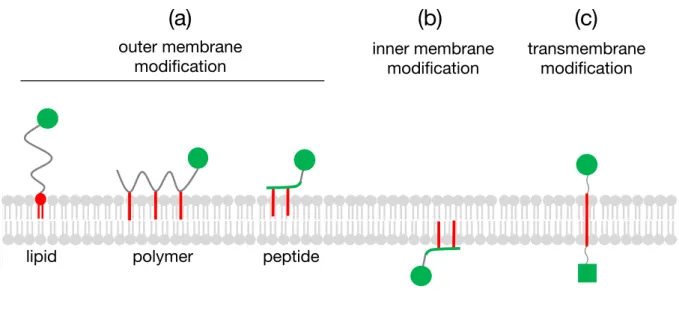
これまでに報告されている疎水性相互作用に基づいた膜修飾分子を(Figure 1-2)にまと めた。膜修飾分子を大別すると、分子提示の対象が細胞膜外葉または内葉の分子および膜貫 通型の三種類に分類することができる(Figure 1-2(a), (b))。
共有結合
+ + + +
- - - -
静電相互作用 疎水性相互作用
課題 細胞表面における寿命
消失原因 細胞内取込み 脱離 細胞内取込み
脱離 細胞内取込み
細胞種依存性 大 小 小
細胞傷害性 小 大 小
分子の提示位置 細胞表面 膜外葉・内葉
Figure 1-2. (a) 膜外葉上に分子を提示する各種膜修飾分子、(b) 膜内葉上に分子を提示する膜 修飾分子、(c) 細胞膜外、内葉上に分子を提示する膜貫通型の膜修飾分子
1. 2. 1 細胞膜外葉の修飾
膜修飾分子による膜外葉の修飾(Figure1-2(a))は、これまでに多くの研究が報告されてお り、タンパク質8-10、ペプチド11およびDNA12,13などが提示可能と示されてきた。提示でき る分子の多様性に加え、細胞機能への摂動や細胞毒性の低さから、疎水性相互作用に基づい て機能化された細胞が再生医療分野へ応用されている。再生医療とは、自家の幹細胞や人工 多能性幹細胞を用いて炎症や機能低下した組織を回復させる治療法である。再生医療におけ る細胞の投与形態として、細胞懸濁液14や細胞/マトリックス混合物15、スフェロイド16、細 胞シート17があり、いずれも組織機能の回復に一定の効果を示している(Figure 1-3)5。
Figure 1-3. 再生医療における細胞の投与形態5
inner membrane
modification transmembrane modification outer membrane
modification
lipid polymer peptide
(a) (b) (c)
4
中でも、疎水性相互作用に基づいた細胞の機能化により細胞接着を誘導し、細胞を組織化 することで治療に貢献する研究が報告されている。Iwataらは、糖尿病治療に向けた膵島移 植で問題となる免疫応答を、膜修飾分子を介した膵島表面への細胞の被覆により回避する戦 略を報告している 18。彼らは、PEG 修飾脂質型膜修飾分子を介して線維芽細胞である
HEK293細胞膜上にビオチンついでストレプトアビジンを導入した。そこにPEG修飾脂質
型膜修飾分子を介してビオチンを修飾した膵島を加えた結果、HEK293細胞を膵島表面に固 定化できた(Figure 1-4)18。膵島細胞は正常に機能し、培養数日後には膵島の表面が細胞層 で完全に覆われた。このことから、膜修飾分子による細胞の機能化が細胞集合体の形成に応 用できることが示された。彼らは、同様の戦略を肝臓細胞にも適用し効果があることを示し ている19。また彼らは、相補的なDNAを有する一対の膜修飾分子を異なる細胞膜上に修飾 するという戦略を用いた異種細胞間の接着にも成功している20。そして、胚葉体への適用に より再生医療への応用の可能性を示している。
Figure 1-4. (a) ビオチンを修飾した脂質型膜修飾分子、(b) ビオチン修飾脂質型膜修飾分子を
介した細胞表面へのストレプトアビジンの修飾、(c) 膵島細胞を別の細胞で覆う戦略の模式 図18
Reprinted with permission from Biomaterials, 2009, 30, 2270. Copyright 2009 Elsevier Ltd.
1,2-Dipalmitoyl-sn-glycerol-3-phosphatidylethanolamine (DPPE) was purchased from NOF Corporation (Tokyo, Japan). Dichloromethane; chloroform;N,N0-dime- thylformamide (DMF); diethyl ether; N,N0-dicyclohexylcarbodiimide (DCC);D- biotin; and streptavidin fromStreptomyces avidinwere purchased from Nacalai Tesque (Kyoto, Japan). Fluorescein isothiocyanate (FITC) and Hoechst 33342 were purchased from Dojindo Laboratories (Kumamoto, Japan). FITC-streptavidin was purchased from Zymed Laboratories (South San Francisco, CA). CellTracker!, the green fluorescent probe for the cell; Alexa 488-labeled goat anti-guinea pig IgG;
minimum essential medium (MEM); HEPES buffer solution; Hanks’ balanced salt solution; and Medium 199 were purchased from Invitrogen (Carlsbad, CA). Fetal bovine serum (FBS) was purchased from BioWest (Miami, FL). Phosphate-buffered saline (PBS) was purchased from Nissui Pharmaceutical Co., Ltd. (Tokyo, Japan). The enzyme-linked immunosorbent assay (ELISA) kits for the insulin assay were purchased from Shibayagi Co., Ltd. (Gunma, Japan). Tissue-Tek was purchased from Sakura Fine Technical Co., Ltd. (Tokyo, Japan). Goat normal serum and polyclonal guinea pig anti-insulin were purchased from Dako (Glostrup, Denmark). The 10%
formalin solution and Triton X-100 were purchased from Wako Pure Chemical (Osaka, Japan).
2.2. Synthesis of biotinylated PEG-conjugated DPPE
The NH2–PEG–lipid was synthesized from DPPE and NHS–PEG–Boc, which carries an activated ester (NHS) that reacts with an amino group on DPPE, and a protective group (Boc) for the amino group, as reported previously[10]. Briefly, NHS–PEG–Boc(175 mg) and DPPE (21 mg) were dissolved in 5 mL dichloromethane solution and stirred for 3 days at room temperature (RT). Then, a solution of TFA (2 mL) was added and stirred for 20 min at 4!C to remove theBocgroups. The crude product was purified by precipitation with diethyl ether. After chloroform extraction
was obtained as a white powder (10 mg, yield 40%). Biotin–PEG–lipid1H NMR (CDCl3, 400 MHz,dppm): 0.88 (t, 6H, –CH3), 1.25 (br, 52H, –CH2–, DPPE), 3.18 (d, 2H, C–CH2–S, biotin), 3.24 (q, 1H, S–CH(–C)–C, biotin), 3.64 (br, 460H, PEG), 4.52 (m, 2H, C–CH(–C)–N, biotin), 5.15 (s, 2H, C–NH–C, biotin).
2.3. Encapsulation of islets with HEK293 cells
HEK293 (human endoderm kidney cell line) cells were obtained from the Health Science Research Resources Bank (Tokyo, Japan). The HEK293 cells were maintained in MEM medium supplemented with 10% fetal bovine serum, 100 U/mL penicillin, and 0.1 mg/mL streptomycin at 37!C under 5% CO2. The cells were collected by centrifugation (180g, 5 min, RT) after treatment with trypsin. The cells were sus- pended in Hanks’ balanced salt solution and then the medium exchanged. A cell pellet (4"106cells) was obtained after centrifugation. After the addition of biotin–
PEG–lipid solution (200mL, 500mg/mL) to the cell suspension, the suspension was incubated for 30 min with gentle agitation at RT. The cells were then suspended in 10 mL Hanks’ balanced salt solution and collected by centrifugation (180g, 5 min, 25!C, twice) to obtain biotin–PEG–lipid-modified cells. Then, streptavidin (100mL, 100mg/mL) was added to the cell suspension and the suspension incubated for 30 min with gentle agitation at 4!C. The cells were then suspended in 10 mL Hanks’
balanced salt solution and collected by centrifugation (180g, 5 min, 4!C, twice) to obtain streptavidin-immobilized cells.
Islets were isolated from the pancreas of female Syrian hamsters (7–8 weeks old, Japan SLC, Inc., Shizuoka, Japan) using the collagenase digestion method. The islets were cultured for 7 days after isolation to remove or sediment cells damaged during the isolation procedure. The islets were maintained in culture medium (Medium 199 with 10% FBS, 8.8 mMHEPES buffer, 100 units/mL penicillin, 100mg/mL streptomycin, and 8.8 U/mL heparin). A biotin–PEG–lipid solution was added to the islets sus- 14
O O O
a O
O P O OO
NH
14
O
O n
NCH
O S
HN NH O
Biotin-PEG-lipid streptavidin
Biotin-PEG-lipid-modified islets
streptavidin-immobilized HEK293
b
c
Scheme 1.(a) Chemical structure of biotin–PEG-conjugated DPPE (biotin–PEG–lipid). (b) Schematic illustration of the interaction between streptavidin and biotin–PEG–lipid at the lipid bilayer cell membrane. Biotin–PEG–lipid has hydrophobic acyl chains and is incorporated into the cell surface by anchoring into the lipid bilayer. Streptavidin was immobilized on the cell surface by anchoring to biotin–PEG–lipid. (c) Scheme for the immobilization of streptavidin-immobilized HEK293 cells on the surface of biotin–PEG–lipid-modified islets.
After mixing streptavidin-immobilized HEK293 cells and biotin–PEG–lipid-modified islets, they were cultured in medium at 37!C on a culture dish. During culture, HEK293 cells were spread and grown on the cell surface to cover the whole surface.
Y. Teramura, H. Iwata / Biomaterials 30 (2009) 2270–2275 2271
また、再生医療の中でも細胞懸濁液の投与は、血中投与も可能な簡便かつ低侵襲な方法で あるという大きな利点がある。しかし、投与した細胞の標的部位への生着率は一般的に 3%
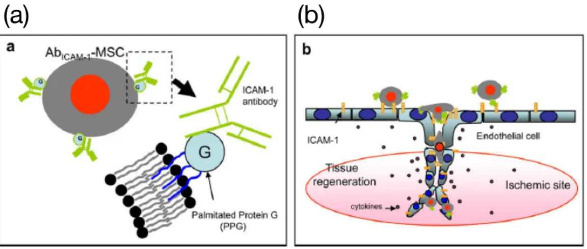
以下ときわめて低いことが問題として認識されている21。そのため、膜修飾分子による細胞 の機能化により、標的組織への細胞の選択性を高める戦略が有効と考えられる。例えば Ko らは、間葉系幹細胞(MSC)表面にパルミトイル化したプロテインG(PPG)を介して炎症 状態の血管内皮細胞に高発現している細胞間接着分子-1(ICAM-1)に対する抗体を修飾した 細胞を調製した(Figure 1-5(a), (b))22。この細胞を用いることで炎症状態の血管内皮細胞へ のMSCの送達にin vitroで成功している。またBullらは、虚血性心筋梗塞後に心筋細胞上 に高度に発現するstromal cell-derived factor 1(SDF-1)を認識するCXCケモカイン受容 体4(CXCR4)をMSC上に脂質型膜修飾分子を介して修飾した23。この細胞は、in vitroに
おいてSDF-1に応答した遊走を示した。これらの研究から、疎水性相互作用に基づいた細胞
の機能化がMSCの投与による再生医療へ応用できる可能性が示された。
Figure 1-5. (a) PPGを介して抗体を修飾したMSCの模式図、(b) 抗体修飾MSCの患部への 送達の模式図22
Reprinted with permission from Biomaterials, 2009, 30, 3702. Copyright 2009 Elsevier Ltd.
(c)
(a) (b)
6
KoやBullらにより疎水性相互作用に基づいて機能化された細胞が細胞の投与という治療 法へ応用できる可能性が示されたものの、その実現には至っていない。この原因として、分 子の提示時間(寿命)の短さが挙げられる20。特に、血中ではせん断応力による物理的な脱 離が懸念されるため、安定した分子の提示が機能化した細胞の体内投与に求められると考え られる22。これは、共有結合や転移酵素の処理により膜タンパク質を修飾した細胞では効果 的な細胞の送達が in vivoで達成されていることからも推察される 24,25。寿命の短さという 弱点を克服することで、その利点から疎水性相互作用による膜修飾の応用範囲が広がり、よ り簡便な再生医療の実現に繋がると期待される。そのため、この寿命を延ばす方法が研究さ れている。
疎水性相互作用により細胞膜上に修飾された分子の寿命を左右する要因として、分子の膜 からの脱離およびエンドサイトーシスによる取り込みが挙げられる。そこで、分子の寿命を 延ばす方法として、三つの戦略が報告されている。それらは、(1)膜修飾分子上の疎水基の 数を増やす方法、(2)疎水基の疎水性を高くする方法により膜からの脱離を抑制する戦略、
および(3)取り込まれた分子をリサイクリングエンドソームにより再提示させるという戦略 である。一つ目の戦略では、Nagamuneらが脂質型膜修飾分子の疎水基が二つの分子の方が 一つのものよりも細胞膜上での半減期が長いことを報告している(Figure 1-6)26。彼らは、
オレイル基を一または二本有する膜修飾分子にPoly(ethylene glycol)(PEG)を修飾した脂 質型膜修飾分子を用意した。それらを細胞に修飾後、膜上での分子の寿命をラベルされてい る蛍光分子の蛍光強度から評価した。
Figure 1-6. 異なる数の疎水基をもつ脂質型膜修飾分子26
Reprinted with permission from Biotechnol. Prog., 2004, 20, 897. Copyright 2004 John Wiley & Sons, Inc.
der iva t ives, glycosyl ph osph a t idyl in osit ol (GP I) a n ch or (16, 17), a n d ch ela t or lipid a n ch or , n it r ilot r ia cet ic a cid dit et r a decyla m in e (NTA-DTDA) (18-20). H owever , t h ese lipid a n ch or in g m et h ods h a ve som e dr a wba cks. F or exa m ple, a ch olest er ol a n ch or t en ds t o a ssem ble in t h e r a ft r egion of t h e cell m em br a n e, r esu lt in g in in t er fer en ce wit h cell sign a lin g. St ea r yl, ch olest er ol, a n d ch ela t or lipid a n ch or s wit h ou t a h ydr oph ilic lin ker t en d t o for m m i- celles, r esu lt in g in slow in cor por a t ion in t o cells. Th e GP I a n ch or m et h od r equ ir es pr epa r a t ion of t h e r ecom bin a n t pr ot ein wit h t h e GP I a n ch or , a n d t h is t a kes a lon g per iod of time and uses complicated procedures for the construc- tion of the expression vector a nd production a nd purifica - t ion of t h e r ecom bin a n t pr ot ein .
In t h is st u dy, we focu sed on developin g a n ea sier a n d m or e r a pid m et h od for a n ch or in g of su bst a n ces in t o t h e m em br a n es of va r iou s cells. We em ployed a n oleyl ch a in a s a lipid a n ch or , beca u se it wa s t h ou gh t t h a t su ch a n u n sa t u r a t ed lipid ch a in a n ch or m a y be a n ch or ed in t h e cell m em br a n e wit h ou t a ffect in g t h e r a ft a n d it s sign a l- in g. Th e oleyl ch a in it self, h owever , is difficu lt t o u t ilize in a qu eou s m edia beca u se of it s wa t er in solu bilit y. Th u s, t h e oleyl ch a in wa s bou n d t o a m ph iph ilic poly(et h ylen e glycol) (P E G) for solu biliza t ion in a qu eou s solu t ion s. Th is der iva t ive wa s design a t ed Biocom pa t ible An ch or for Mem br a n e (BAM). We r epor t h er ein t h a t t h e BAM cou ld r a pidly a n d ea sily a n ch or som e su bst a n ces in clu din g pr ot ein s (st r ept a vidin , en h a n ced gr een flu or escen ce pr o- t ein (E GF P ), a n d a n t ibody) in t o t h e m em br a n es of va r iou s livin g cells wit h ou t cyt ot oxicit y, wh ich wa s eva lu a t ed by la ct a t e deh ydr ogen a se (LDH ) r elea se t est .
Ma te ria ls a n d Me th o d s
Ma te ria ls . Th e followin g r ea gen t s wer e pu r ch a sed:
E Z-Lin k Biot in -P E O-Am in e (P ier ce, IL), flu or escein - labeled streptavidin, fluorescein-labeled biotin and 5-(ami- nomethyl)-fluorescein (Molecular Probes Inc., OR), strepta- vidin (Wa ko Ch em ica l Co., Tokyo, J a pa n ), goa t a n t i-GST a n t ibody (Am er sh a m P h a r m a cia Biot ech In c., NJ ), ph os- ph a t e-bu ffer ed sa lin e (P BS), Du lbecco’s m odified E a gle m ediu m (DME M) a n d RP MI1640 cu lt u r e m ediu m (NIS- SUI P h a r m a ceu t ica l Co., Lt d., Tokyo, J a pa n ), a n d fet a l bovin e ser u m (F BS) (Asa h i Tech n o Gla ss Co., Tokyo, J a pa n ).
B AM Re a g e n ts . Oleyl-O-poly(et h ylen e glycol)-su cci- n yl-N-h ydr oxy-su ccin im idyl est er s, design a t ed Biocom - pa t ible An ch or s for Mem br a n e (BAMs), wer e syn t h esized wit h differ en t n u m ber s of et h ylen e oxide (E O) u n it r e- pea t s (BAM40 a n d BAM90) in t h e P E G m oiet y, dioleyl- ph osph a t idylet h a n ola m in e (DOP E )-P E G-NH S est er wit h 80 u n it s of P E G (DOP E -BAM80) a n d m et h oxy-poly- (et h ylen e glycol)-su ccin yl-N-h ydr oxy-su ccin im idyl est er wit h 110 u n it s of P E G (P E G110) (F igu r e 1). Th ese r ea gen t s wer e gift s fr om NOF Cor por a t ion (Tokyo, J a - pa n ).
Ce ll Cu ltu re . Mou se fibr obla st NIH 3T3 cells wer e pr opa ga t ed in DME M su pplem en t ed wit h 10% F BS, 20 m M H E P E S, 2.05 m M glu t a m in e, a n d 30µg/m L ka n a - m ycin . Con foca l m icr oscopy wa s u sed t o im a ge t h e NIH 3T3 cells cu lt u r ed in gla ss-bot t om ed cu lt u r e dish es (35 m m u n coa t ed dish es, n o. 0 cover slip; Ma t Tek Co., Ash la n d, MA) a t 37 °C a n d 5% CO2.
Mou se m yeloid 32D a n d m ou se pr oB Ba /F 3 cells wer e cu lt u r ed in RP MI1640 m ediu m su pplem en t ed wit h 10%
F BS, 2 n g/m L in t er leu kin -3, 2.05 m M glu t a m in e, a n d 30 µg/m L ka n a m ycin .
P re p a ra tio n o f Ta g g e d En h a n c e d Gre e n F lu o re s - c e n c e P ro te in (tEGF P ). Th e gen es for E GF P a n d t h e C dom a in of pr ot ein G wer e fu sed, a n d t h e r ecom bin a n t fu sion pr ot ein , t E GF P , wa s expr essed inE sch erich ia coli J M109 a n d pu r ified fr om t h e cell lysa t e u sin g IgG a ffin it y ch r om a t ogr a ph y a s descr ibed pr eviou sly (21).
P re p a ra tio n o f Glu ta th io n e -S-Tra n s fe ra s e -EGF P fu s io n p ro te in (GS T-EGF P ).Th e gen e en codin g E GF P wa s pr epa r ed fr om pE GF P (Clon t ech , CA) by st a n da r d P CR. Th e a m plified fr a gm en t s wer e digest ed wit h B am - H I a n dS alI. F or t h e expr ession of GST-fu sion pr ot ein s, t h e B amH I-S alI fr a gm en t en codin g E GF P wa s clon ed in t o t h e m u lt iple clon in g sit es of pGE X 5X3 (Am er sh a m P h a r m a cia Biot ech , NJ ) bet ween t h e B amH I a n d S alI sit es t o give t h e pla sm id pGE X-E GF P .E . coli TG-1 wa s t r a n sfor m ed wit h t h e pla sm ids a n d select ed on LB a ga r pla t es con t a in in g 0.05 m g/m L a m picillin . F or a ll su bse- qu en t cu lt iva t ion s, LB m ediu m con t a in in g 0.05 m g/m L a m picillin wa s u sed. Two lit er s of su per br ot h m ediu m wa s in ocu la t ed wit h t h e st r a in s h a r bor in g pGE X-E GF P a n d cu lt u r ed a t 25 °C. At a n OD600 of a bou t 0.5, IP TG wa s a dded (a t a fin a l con cen t r a t ion of 0.075 m M) t o in du ce t h e expr ession of t h e fu sion pr ot ein , a n d t h e cells wer e t h en cu lt u r ed a t 16 °C for a fu r t h er 18 h . H a r vest ed cells wer e r esu spen ded in son ica t ion bu ffer (50 m M Tr is- H Cl pH 8.0; 50 m M Na Cl; 2 m M E DTA) a n d disr u pt ed by sonica tion (a t 4 °C, for 10 min) with a Bra nson Sonifier 250 (Son ic P ower Co., USA). Th e su per n a t a n t wa s obt a in ed by cen t r ifu ga t ion a t 15,000gfor 20 m in a t 4 °C, a n d a pplied t o a Glu t a t h ion e-Seph a r ose (Am er sh a m P h a r m a cia Biot ech ) colu m n u sin g a st a n da r d pr ot ocol.
Th e colu m n wa s wa sh ed wit h P BS, a n d t h e bou n d proteins were eluted with PBS containing 10 mM reduced glu t a t h ion e. F r a ct ion s em it t in g gr een flu or escen ce wer e F ig u re 1. (a ) Th e st r u ct u r e of a biocom pa t ible a n ch or for m em br a n e (BAM) wit h a sin gle lipid a n ch or . Th e poly(et h ylen e glycol) oleyl et h er is su ccin yla t ed a t t h e h ydr oxyl en d of P E G a n d m odified wit hN-h ydr oxysu ccin im ide (NH S) a t t h e su ccin yl PEG end. A BAM with NHS is amino-reactive and can bind most pr ot ein s. In a ddit ion , BAMs com posed of P E G ch a in s of va r iou s len gt h s (n ) 40 a n d 90) wer e syn t h esized t o com pa r e t h eir per for m a n ce a s a n ch or in g r ea gen t s. (b) Th e st r u ct u r e of a BAM wit h a dou ble lipid a n ch or . Dioleylph osph a t idylet h a n ola m in e (DOP E ) wa s pegyla t ed a n d su ccin yla t ed a t t h e h ydr oxyl en d of P E G a n d m odified wit h N-h ydr oxysu ccin im ide (NH S) a t t h e su ccin yl P E G en d.
898 Biotechnol. Prog.,2004, Vol. 20, No. 3
(a)
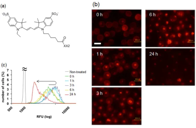
二つ目の戦略では、Iwata らが、より長い脂肪酸を有する脂質型膜修飾分子を使用するこ とで細胞表面上での分子の寿命が延びることを明らかにしている(Figure 1-7)27。本研究で 彼らは、1,2-dipalmitoyl-sn-glycero-3-phosphatidylethanolamine(DPPE)を基準に、1,2- dimyristoyl-sn-glycero-3-phosphatidylethanolamine(DMPE)および 1,2-distearoyl-sn- glycero-3-phosphatidylethanolamine(DSPE)を用意し、それぞれに分子量5 kのPEGを 修飾した脂質型膜修飾分子を用意した。そして、それらを細胞に修飾した後に、膜上での分 子の寿命および脱離した分子の定量をラベルされている蛍光分子の蛍光強度から評価した。
また、ここで示された結果と同様の報告が他の研究グループからなされている28,29。
Figure 1-7. 各脂肪酸が修飾された脂質型膜修飾分子27
Reprinted with permission from ACS Appl. Mater. Interfaces., 2010, 2, 1514. Copyright 2010 American Chemical Society
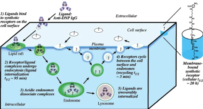
三つ目の戦略では Peterson らが、リガンドにコレステロールを修飾した脂質型膜修飾分 子を細胞に修飾し、エンドソームによる取り込み後のリサイクリングエンドソームによる分 子の膜外葉への再提示の評価を報告している30。彼らの膜修飾分子は、リサイクリングエン ドソーム経路により3分程度で細胞膜上に再提示されることがわかった(Figure 1-8)30。リ サイクリングエンドソームによる膜上への分子の再提示は、細胞が受容体など天然のタンパ ク質を再利用する場合に利用される31。そのため、再提示機構を膜修飾分子に応用すること で、天然のタンパク質並みの寿命(葉酸受容体の半減期は24時間32)が期待できる。実際彼 らの膜修飾分子は、その半減期が20時間であることが示されている。
(a)
Figure 1-8. (左)リガンド修飾コレステロールがエンドサイトーシスにより取り込まれ、リ サイクリングエンドソームによって膜上に再提示される経路および(右)リガンド修飾コレ ステロールの構造30
Reprinted with permission from J. Am. Chem. Soc., 2004, 126, 16379. Copyright 2004 American Chemical Society
ここまでをまとめると、細胞膜外葉を修飾する分子の寿命向上を考えた場合、膜修飾分子 に導入する疎水基の数およびその疎水性を上げることによって、膜からの脱離を抑制し寿命 を延ばすことができると考えられる。また、エンドサイトーシスにより分子が取り込まれて もリサイクリングエンドソームによる膜上への再提示を利用することで、寿命が延びると期 待できる。以上の戦略を膜修飾分子の設計に落とし込む場合、ここまでに取り上げたリン脂 質を中心とする脂質型の基体では、その設計の自由度に限界があると考えられる。
そこで、提示分子の寿命を延ばすことを目標として、様々な種類の疎水基を任意の数、簡 便に導入可能な基体としてポリマーやペプチドを採用した研究が報告されている(Figure 1-
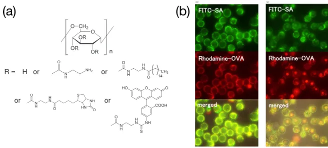
2 polymer and peptide)。例えば、我々の研究室では、複数の疎水性分子をデキストラン主鎖
上に導入可能なポリマー型膜修飾分子を用いて、細胞膜上にストレプトアビジン(SA、53
kDa)を介してオボアルブミン(OVA、45 kDa)が安定に提示できることを報告した(Figure
1-9(a),(b))33。このようなポリマー型の膜修飾分子は、他にも報告されている34,35。
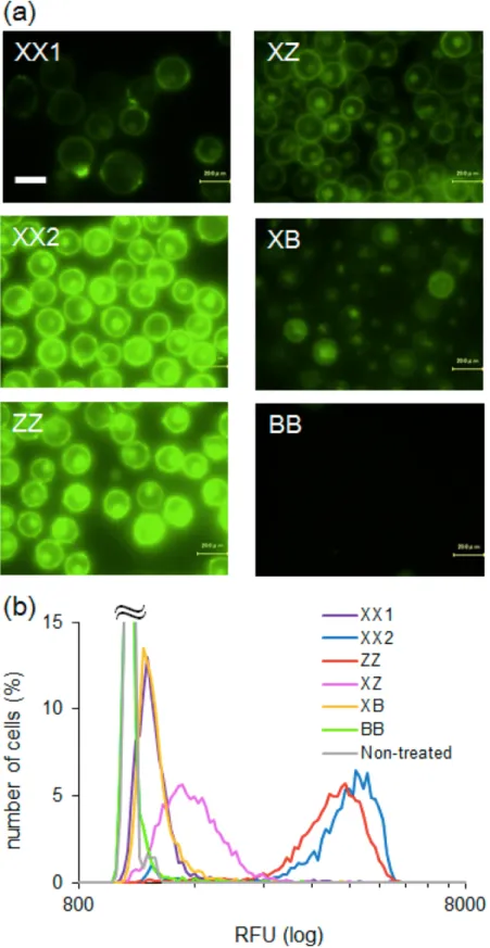
Figure 1-9. (a) デキストランを主鎖とするポリマー型膜修飾分子、(b) ポリマー型膜修飾分子 により膜上に修飾されたSA(緑)およびOVA(赤)(左:修飾直後、右:培養1時間後)33
Reprinted with permission from J. Control. Release, 2014, 177, 27. Copyright 2013 Elsevier Ltd.
streptomycin, and 0.25μg/mL amphotericin B (all from Gibco Invitrogen Co., Grand Island, NY, USA). Cells were harvested in a humidified atmo- sphere containing 5% CO2at 37 °C.
2.8. Modification of cells with polymer
K562 cells (1×10⁶) were collected by centrifugation. After removal of the supernatant, the cells were washed with Dulbecco's phosphate- buffered saline (DPBS). Then, 50μL of polymer (0.1 mg/mL, containing 1.8 nmol of biotin) dissolved in DPBS was added to the cell suspension and incubated for 30 min with gentle agitation at 4 °C. After cell surface modification, DPBS (2 mL) was added to the mixture and the cells were collected by centrifugation. These procedures were repeated twice to remove unbound polymer. The cells were then incubated in 1 mL of RPMI-1640 medium containing 10% FBS, 100 U/mL pen- icillin, 100μg/mL streptomycin, and 0.25μg/mL amphotericin B at 37 °C under 5% CO2. Fluorescence intensities of the polymer on the cell surface were estimated with a BZ-8000fluorescence mi- croscope (Keyence) and a Tali™Image-Based Cytometer (Life Technologies).
2.9. Cellular uptake of SA and SA/OVA complex
K562 cells were modified with polymer 2 or 3 according to the same procedures described above. Then, Cy3-SA (0.07 nmol) was added to the polymer 2-modified K562 cell (1×10⁵) dispersion in RPMI-1640 medium (100μL) and was gently agitated. FITC-SA was mixed with rho- damine- and biotin-modified OVA at a 1:1 ratio to prepare the SA/OVA complex. The resulting complex (0.07 nmol) was added to the polymer 3-modified K562 cell (1×10⁵) dispersion in RPMI-1640 medium (100μL) and was gently agitated.
As for DSPE-PEG-biotin and PE-biotin, they were modified on K562 cells according to the same procedures with polymer 2. They werefirst dissolved in DMSO and diluted with DPBS to prepare solu- tions for the cellular modification. The biotin concentration of DSPE- PEG-biotin and PE-biotin in the solutions for the cellular modifica- tion was adjusted to be the same with the solution of polymer 2 (1.8 nmol).
2.10. Cytotoxicity evaluation of polymer
Cytotoxicity of the polymer was evaluated using a WST-8 kit (Dojindo, Kumamoto, Japan). K562 cells were modified with different polymer concentrations (0.01, 0.1 mg/mL) and placed in a 96-well plate at an initial density of 7500 cells/well and incubated for 4 h at 37 °C. To measure cell viability, 10μL of WST-8 was added to each well and the cells were incubated for another 4 h at 37 °C. The WST-8 formazan absorbance at 450 nm was measured by a microplate spec- trum reader AD200 (Beckman Coulter) and expressed as a percentage relative to non-modified cells.
2.11. Effect of endocytosis inhibitor
Cytochalasin D is known to specifically inhibit actin polymerization, and can therefore inhibit endocytosis. K562 cells (2×10⁶cells/mL) were collected by centrifugation. After removal of the supernatant, 10μL of cytochalasin D (10μM) was added to the cell suspension and incubated for 30 min with gentle agitation at room temperature and washed with DPBS. Then, 50μL of polymer (0.1 mg/mL) was added to the cytochalasin D-treated cells and incubated for 30 min with gentle agitation at 4 °C. The cells were washed, incubated for 24 h at 37 °C under 5% CO2, and examined with afluorescence microscope.
3. Results and discussion 3.1. Synthesis of polymers
Chart 1 depicts the structure of the biotinylated polymer that was synthesized by adding ethylenediamine to dextran using carbonyldiimidazole. Then other groups (palmitoyl, biotin, and FITC), were added to the dextran by amide or thiourethane bond formation with the amine group of a modified ethylenediamine. Three polymers (1–3) were obtained, and the content of each group per glucose unit is summarized inTable 1.
3.2. Polymer modification on cell surface
Polymer 1 was attached to K562 cells by mixing the polymer solu- tion with a cell dispersion in PBS for 30 min at a low temperature (4 °C) to avoid endocytosis.Fig. 2A shows thefluorescence image of the cells right after modification with the polymer. Thefluorescence from FITC labeling of polymer 1 was observed exclusively from the cell surface, and was not observed when the cells were mixed with palmitoyl-free dextran (data not shown). These results indicate that polymer 1 was successfully incorporated onto the cell surface via hydro- phobic anchoring by palmitoyl groups in the cell membrane. The num- ber of polymer chains attached to the cell surface was determined from thefluorescence intensity.Fig. 2B plots the number of the polymer chains present per single cell as a function of the polymer concentration used for the modification. The number of polymer chains increased with polymer concentration until saturation at 15×106chains/cell at concentrations above 0.1 mg/mL. Because the average size of the cells was 8μm, the polymer density on the cell surface was estimated to be 54 nm2/chain. Assuming random coil conformations of the polymer chains with a hydrodynamic radius of 5 nm[18]on the cell surface, the surface area occupied by one polymer chain is 79 nm2/chain. Thus there is good agreement between these densities, indicating that the
R = H or or
or or
Chart 1.Structure of biotinylated polymers.
Table 1
Contents of each component in the biotinylated polymers.
Polymer Content (mol%) Number of palmitoyla
Ethylenediamine Palmitoyl Biotin FITC
1 16 5.4 – 0.6 13
2 5.7 10 5.0 0.3 25
3 18 6.0 8.1 – 15
aNumber of palmitoyl groups per one polymer chain.
K. Tobinaga et al. / Journal of Controlled Release 177 (2014) 27–33 29
endocytosis into cells with membranes modified with polymer 3.Fig. 8B shows thefluorescence images after mixing the SA/OVA complex with the cells. The SA (FITC) and OVA (rhodamine)fluorescence images indi- cated colocalization on the cell surface, verifying that the SA/OVA com- plex recognized the biotin-modified on the cell surface. After 1 h of incubation at 37 °C, strongfluorescence of SA/OVA complex was observed inside the cells, demonstrating the successful uptake of the SA/OVA complex via the biotinylated polymer-mediated endocytosis.
As a negative control, we examined the cellular uptake of biotin-non- modified OVA based on the same procedures as the above-mentioned biotin-modified OVA (Fig. S5). Rhodamine'sfluorescence resulting from OVA detected neither on the cell surface nor inside of the cell.
These results clearly show that the cellular uptake of OVA was mediated by SA which functions as a crosslinker between the cell surface biotin and OVA.
We further examined the effect of the amount of the cell surface- modified polymer 3 on the cellular uptake of OVA. Polymer 3 was modified on the K562 cell surface with varying concentrations. To the modified cell was added SA/OVA complex and incubated for 1 h at 37 °C for endocytosis. Thefluorescence intensity inside of the cells which is originated from the taken up OVA was plotted against the poly- mer concentration inFig. 9. The cellular uptake of OVA proportionally increased with the polymer concentration. Thus to raise the amount of cellular uptake of OVA, the modification of polymer 3 on the cell surface should be higher.
4. Conclusions
We presented a method for the intracellular delivery of pro- teins based on the biotinylated polymer-mediated endocytosis. The bi- otinylated polymers were modified with multiple palmitoyl groups and stably bound to the cell surface via multivalent anchoring in the cell membrane. Biotin molecules presented on the cell surface by the polymer recognized SA and the resulting SA/biotinylated poly- mer complex was taken up rapidly by the cells via the endocytotic pathway. The endocytosis was not affected by the presence of 10%
FBS. The biotinylated polymers enabled the uptake of an OVA/SA
complex. Thus, the biotinylated polymer-mediated endocytosis was found to be potentially applicable to any kinds of proteins.
Acknowledgment
We thank thefinancial support of a Grant-in-Aid for Young Scientists (B) (23700539) from the Ministry of Education, Culture, Sports, Science and Technology of Japan.
Fig. 7.(A) Chemical structures of synthetic receptors. (B) Comparison of cellular uptake by polymer 2 with commercial reagents and other synthetic receptors.
Fig. 9.Effect of polymer 3 concentration used for the K562 surface modification on the fluorescence intensity inside of the cell resulting from the taken up OVA.
Fig. 8.Complex formation between biotin-modified OVA and SA for cellular uptake (A).
Cellular uptake of SA/OVA complex by K562 modified with polymer 3 in serum containing medium right after (B) and 1 hour after (C) the complex addition.
32 K. Tobinaga et al. / Journal of Controlled Release 177 (2014) 27–33
(a) (b)
ポリマー以外の基体としてペプチドが用いられるが、その理由として天然の膜結合型のタ ンパク質は、細胞膜内葉への安定した提示のためにペプチドを基体とした疎水性のアンカー を使用することが報告されているからである36。そのような天然のタンパク質の例としてSrc ファミリープロテインキナーゼやGタンパク質がある。それらのタンパク質は、自身のアミ ノ酸配列の N または C 末端に疎水性アンカーが二つ導入された状態で膜上に安定に提示さ れる 36。この知見に基づき Silviusらは、短いペプチド上に疎水基を二つ導入したペプチド が一つの分子よりもリポソーム膜上でより安定であることを見出している(Figure 1-10)37。 本研究で彼らは、上記のような翻訳後修飾によって脂質が修飾されるアミノ酸配列を模した ペプチドに種々の疎水基を導入し、それらペプチド型の膜修飾分子のリポソーム上での安定 性を評価している。この研究から、ペプチドが膜修飾分子の基体として利用できることが示 された。
Figure 1-10. 疎水基が導入されたペプチドの構造37
Reprinted with permission from Biochemistry, 1995, 34, 3813. Copyright 1995 American Chemical Society
(a) (b)
1. 2. 2 細胞膜内葉の修飾
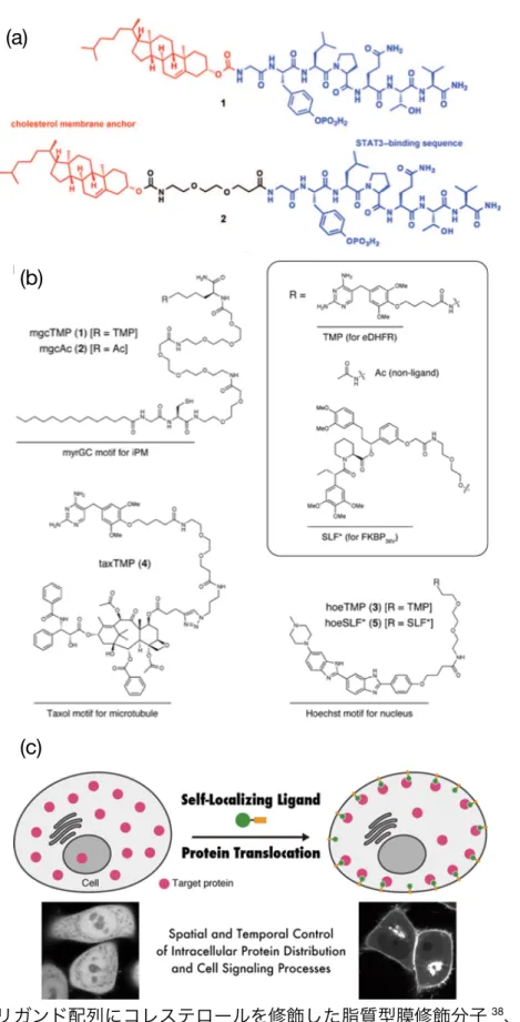
細胞膜内葉に分子を提示し細胞質内のタンパク質の局在を制御することで細胞機能を制御 する試みが報告されている(Figure1-2(b))。膜内葉上への分子の修飾は、細胞内のタンパク 質の動きを制御することで細胞機能を制御できる点で従来にはない作用機序の薬が開発され る可能性がある。Gunning らは、細胞の分化や増殖に関与する signal transducer and activator of transcription 3(STAT3)特異的に結合するリガンド配列(GpYLPQTV-NH2) にコレステロールを修飾した分子を膜内葉に提示することで、細胞質内に存在する STAT3 の細胞核への移行を阻害した(Figure 1-11(a))38。興味深いことに、リガンドとコレステロ ールの間にエチレングリコール(EG)のスペーサーを導入すると、STAT3の膜への結合量 が大きく減少した。この原因として彼らは、スペーサーの効果によって分子が折りたたまれ、
STAT3の認識に関与できる分子の量が減少したためと推察している。Tsukijiらは、膜内葉
に局在化するモチーフに大腸菌由来ジヒドロ葉酸還元酵素(eDHFR)特異的に結合する trimethoprim(TMP)を修飾した分子を5 µMの濃度で細胞に添加することで、10分とい う短時間で細胞内に発現したeDHFRの細胞膜内葉への局在を確認している(Figure 1-11(b))
39。本研究で用いられた局在化モチーフは、Lyn キナーゼの N 末端に存在する翻訳後修飾 によってパルミトイル基が修飾される配列(MyrGC)40(Myr:ミリスチン酸)である。こ の配列中のCys側鎖のシステインがゴルジ体表面でパルミトイル化され、膜内葉へ移行し局 在化することがわかっている。しかし、このようなペプチドが細胞に添加された後どのよう にして細胞質に移行するかは分かっていない。
Figure 1-11. (a) リガンド配列にコレステロールを修飾した脂質型膜修飾分子38、(b) TMPを 修飾した脂質型膜修飾分子39、(c) TMPによるeDHFRの膜内葉への局在化39
Reprinted with permission from Angew. Chem. Int. Ed., 2011, 50, 6248. Copyright 2011 John Wiley & Sons, Inc.
Reprinted with permission from J. Am. Chem. Soc., 2013, 135, 12684. Copyright 2013 American Chemical Society
(a)
(b)
(c)
1. 2. 3 細胞膜貫通型の修飾
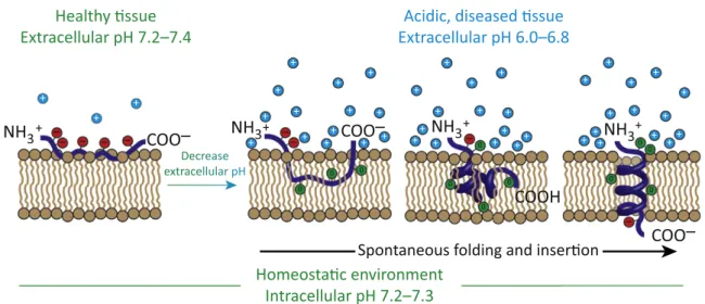
細胞膜上で膜貫通状態をとる分子(Figure1-2(c))として、Engelmanらはバクテリオロド プシンの膜貫通領域(配列下線部)とその両端にある二つのフランキング配列から成るペプ チ ド ( 配 列 :AEQNPIYWARYADWLFTTPLLLLDLLLVDADEGT, pH Low Insertion Peptide: pHLIP)を報告している41。このペプチドは、がん細胞表面など酸性pH条件(<
7.0)に応答してC端側のフランキング配列中の二つのAsp側鎖がプロトン化され、疎水性
を高めることでC末端側から膜に挿入され、最終的に膜貫通修飾を達成する(Figure 1-12)
41。本ペプチドの利点は、その機構が不明であるものの、方向を制御して膜に挿入され、膜貫 通状態が達成されることである。そのため、N末端に分子を修飾することで膜外葉に分子を 提示できる 42,43。また、C末端に分子を修飾することで膜内葉に分子を提示でき、上皮成長 因子受容体(EGFR)の二量体化の阻害が可能であることが報告されている 44。さらに、分 子をジスルフィド結合でC末端に修飾することで、膜内葉上で分子が放出される設計が可能 であり、蛍光試薬45、環状ペプチド46,47そしてmicro RNA48などがpHLIPを介して細胞内 部へと送達されている。しかし pHLIP は、酸性条件でのみ膜修飾が達成される点や pH の 変化に過敏で Asp 側鎖の脱プロトン化により即座に膜貫通状態が解かれる点 49で膜修飾分 子として使い勝手が良いとは言い難い。また細胞膜に対して方向を制御して挿入される機構 が不明であるため、本ペプチドへの分子の修飾にはその機能を損なわないように配慮する必 要がある。
pHLIP以外にも、イオンチャネルの機能50を模倣した分子51やGPCR52のように細胞外 部の刺激を内部へと伝達する機能に着目した分子が報告されている53。これらの分子は、脂 質人工二重膜を再構成する段階で分子を組み込むことで膜上に修飾するという点で疎水性相 互作用に基づいた膜修飾分子として分類した。しかし、これらの分子は脂質人工二重膜上で のみその機能が報告されており、生細胞膜上での機能発現には至っていない。それは、二つ の理由によると推察される。一つ目は、膜貫通を達成する多くの分子が水溶性に乏しいため、
細胞培養液に添加した段階で凝集し、細胞膜に対する分子の導入が困難という点である。二 つ目は、pHLIP のように分子を細胞膜に対して挿入方向を制御して修飾できない点である。
実際、人工膜上で機能する膜貫通型の膜修飾分子の多くは、その構造を対称にすることで、
膜上での方向に依らず機能する分子設計となっている 51,53。膜を貫通した状態での分子の修 飾は、膜貫通タンパク質の機能を模倣できる可能性が十分にあり、細胞膜上に導入できるよ うになれば細胞膜外葉や内葉のみを分子で修飾するよりも高度な細胞の機能化が可能になる と考えられる。
14
Figure 1-12. pHLIPの細胞膜上での挙動41
Reprinted with permission from Trends Biotechnol., 2017, 35, 653. Copyright 2017 Elsevier Ltd.
ItisimportanttodistinguishbetweenthepHLIPfamily,whichcomprisesmembrane-inserting peptides,andcell-penetratingpeptides(CPPs)oramphipathicpore-formingpeptides.
CPPsand pore-formingpeptides have entirely different mechanisms of action anddifferent biologicalperformance;theyare notpartof thepHLIP family.
TargetingExtracellularAcidity
Targetingcancercellsbyexploitingextracellularacidificationshouldbecomparedwithother targetingapproaches,whicharecommonlybasedonthepresenceofgrowthfactorsortumor antigens. A problem in targeting these kinds of specific features is that the cancer cell population, both between tumors and within an individual tumor, is heterogeneous and adaptable[43].The potentialfor successfully treating tumors that do notexpress or exhibit limited expression of the targeted growth factor or antigen is low and, even in treatment- responsive tumors, minority populations of cancer cells can undergo clonal selection and eventuallyregrowintoatreatment-resistantphenotype.Subsequenttreatmentswilleitherhave limitedefficacyorbeentirelyineffective.Additionally,thesetypesoftargetingmethodhavethe potentialtoseverelydamagehealthytissueswherethegrowthfactororantigenmightalsobe present. Therefore, targeting tumor acidity, a physical characteristic that is found not only across an entire tumor, but also in tumors of all sizes, including metastases, could have advantages over targeting methods based on characteristics that are found only in some tumorsor somecancercell populations[44,45].
Acidityisproducednotonlyby canceroustissue,butalso byanytissuethatisexperiencing hypoxia:thelack of oxygenin thetissue triggers cellsto switchto a highrate of useof the anaerobic, glycolytic energy production pathway, consequently resulting in acidification.
RNApolymeraseII:akeyenzyme thatcatalyzestranscription.
Seminaphtharhodafluor(SNARF):
afluorescentdyewithtwopH- dependentemissionpeaks,makingit possibletouseSNARFtomeasure pHthroughspectrographyor ratiometricimaging.
Tissuefactor(TF):aproteinthatis exposedtothebloodupontissue damage,triggeringbloodclot formation.
Transmembranehelix:the formationbyapolypeptideofahelix thatspansthelipidbilayerofa cellularmembrane.
Triple-negativebreastcancer:any breastcancerthatdoesnotexpress theestrogenreceptor,progesterone receptor,andepidermalgrowth factorreceptor2(HER2/neu).Triple- negativebreastcancerisdifficultto treatduetotherelianceoftypical chemotherapiesontheexpressionof oneormoreofthesereceptors.
Tubulin:afamilyofproteinsthat polymerizeintomicrotubules,which arepartofthecytoskeletonand contributetostructuralstability, cytoplasmictransport,cell movement,andcelldivision.
Tumorinvasion:thespreadof cancercells(metastasis)fromthe primarytumorsitetoadjacenttissue orblood.
Healthy !ssue Extracellular pH 7.2–7.4
NH3+ NH3+ NH3+ NH3+
+ +
+
+ +
+ +
+ + ++
+
+ +
+
+ +
+ + + +
+
+ +
+ +
++ ++
+ +
+ +
+ +
+ ++ + ++
+ + +
– – – – –
– –
–
COO– COO–
COO–
COOH
Decrease extracellular pH
Acidic, diseased !ssue Extracellular pH 6.0–6.8
0 0
0 0 0
0 0
0
00
0
Spontaneous folding and inser!on Homeosta!c environment
Intracellular pH 7.2–7.3
–
Figure2.MechanismofpH(Low)InsertionPeptide(pHLIP1)InsertionintotheCellularMembrane.Whenthe pHLIP(blue)encountershealthytissuewheretheextracellularpHisaroundpH7.4,theprotonatableresiduesofthepHLIP (redcircles)remaindeprotonatedandnegativelycharged,andthepeptideresidesatornearthehydrophilicsurfaceofthe cellularmembrane.Weaklyboundtothemembrane,thepHLIPiswashedfromthemembranevianormalperfusionand continuesto circulatethrough the body.However, cancer cellsproduce excessacidity asa consequenceof their malfunctioningmetabolismsandoverexpressionofcertainsurfaceproteins,andpumptheseacidicbyproductsoutofthe cellinteriortomaintaincomfortableconditionsinsidethecell,resultingintheacidificationoftumortissue.WhenthepHLIP encounterstumortissue,itsensesthelowextracellularpHatthecancercellsurface[i.e.,theconcentrationofprotons (cyancircles)atthesurfaceofthecellularmembraneishigh],andtheprotonatableresiduesandnegativelychargedC- terminalcarboxylgroupofthepHLIPbecomeneutrallycharged(greencircles).Theprotonationleadstoanincreaseinthe overallhydrophobicityofthepHLIP,increasingtheaffinityofthepeptidetothehydrophobiccoreofthecellularmembrane andtriggeringthepHLIPtospontaneouslyfoldintoahelixandinsertacrossthemembrane,resultingintheformationofa transmembranehelix.WhentheC-terminalprotonatableresidueandcarboxylgrouparethenexposedtothenormal intracellularpHofthecell,theyaredeprotonated, againbecomingnegativelycharged,andanchorthepHLIPinthe membrane.
1. 3 本論文の目的および構成
本章では、細胞の機能化を目的とした細胞膜修飾法の中でも、疎水性相互作用に基づいた 細胞膜修飾分子についてまとめた(Figure 1-2)。細胞膜内葉上への分子の修飾(Figure 1-2(b)) は、細胞内のタンパク質の動きを制御することで細胞機能を制御できる点で、従来にない作 用機序の薬が開発される可能性を秘めている。細胞膜外葉上への分子の修飾(Figure 1-2(a))
は、最も研究されており、脂質型のみならずポリマー型やペプチド型の膜修飾分子が報告さ れている。その応用が再生医療分野で期待されものの、特に投与形態として細胞懸濁液を用 いる場合、分子の膜上での寿命がボトルネックとなり実用には至っていないと考えられる。
そのため、分子の寿命を延ばす戦略を取り入れ、細胞投与を見据えた膜修飾分子の設計およ び評価が望まれる。
また、細胞膜を貫通した状態での分子の修飾(Figure 1-2(c))は、細胞膜内外に分子を提 でき、膜貫通タンパク質の機能を模倣した高度な細胞の機能化が期待できる。pHLIPで膜貫 通の修飾が達成されているものの、低pH条件に限られる。環境に依存することなく方向を 制御して膜を貫通し、その状態を維持できる分子が必要と考えられる。
以上より本論文では、膜修飾による細胞の機能化法として有望な化学的な手法、中でも細 胞機能への摂動が最も少ない疎水性相互作用に基づいた膜修飾による細胞の機能化を達成す る分子の開発をテーマに、以下の二点を報告する。一点目は、細胞投与を見据えて提示分子 の膜上での寿命を延ばすことを目的とした、ペプチド型膜修飾分子(ペプチド型膜アンカー)
の開発とその詳細な評価および応用についてである(二章)。二点目は、膜貫通状態での膜修 飾を実現する分子の開発とその修飾法(三章)および細胞膜を貫通した状態で細胞内外に分 子を提示可能な膜修飾分子の開発とその効率的な合成方法(四章)についてである。