九州大学学術情報リポジトリ
Kyushu University Institutional Repository
食道癌発育様式におけるp53蛋白発現の意義
野添, 忠浩
https://doi.org/10.11501/3163987
出版情報:Kyushu University, 1999, 博士(医学), 論文博士 バージョン:
権利関係:
ONCOLOGY REPORTS 5: 1119-1123. 1998
Significance of p53 protein expression in growth pattern of esophageal squamous cell carcinoma
TADAHIRO NOZOE, HIROYUKI KUW ANO, YASUSHI TOH, MASA YUKI WATANABE, MASA YUKI KITAMURA and KEIZO SUGIMACHI
Department of Surgery II, Faculty of Medicine, Kyushu University, Fukuoka, 812, Japan Received May 4, 1998; Accepted July 6, 1998
Abstract. The significance of intraepithelial carcinoma concomitant with esophageal squamous cell carcinoma during carcinogenesis and progression of the tumor has been discussed diversely. The purpose of the current study was to elucidate the relation between p53 protein expression and the growth pattern of the squamous cell carcinoma of the esophagus with attention to coexistence of intracpithelial carcinoma. Seventy cases with squamous cell carcinoma of the esophagus surgically resected without preoperati vc adjuvant therapy, including 49 cases with i ntraepi thelia!
carcinoma contiguous to the invasive le ion, were analyzed immunohistochemically for p53 expression. Positive immuno
reactivity of p53 was found in 36 (51.4%) of 70 cases. The frequency of p53 protein expression in cases with intra
epithehal carcinoma (65.3%; 32/49) was significantly higher than that ( 19.0%; 4/21) in cases without intraepithelial carcinoma (p<O.OO l). The value of invasion coefficient, which indicates a ratio of the area of invasive cancerous lesion occupied in the whole lesion, in the cases with p53 protein expression was significantly smaller than that in the cases without p53 protein expression (p<0.001). In conclusion, p53 protein expression was found to be significantly related to the coexistence and spreading of intraepithelial carcinoma contiguous to squamous cell carcinoma of the esophagus.
Introduction
Regarding the significance of intraepithelial carcinoma concomitant with squamous cell carcinoma of the esophagus, a variety of investigations have already been made. We also previously reported that in squamous cell carcinoma of the esophagus, the more the main cancerous lesion progressed, the less frequently the incidence of intraepithelial carcinoma
Correspondence to: Dr Tadahiro Nozoe, Depatment or Surgery II, Faculty or Medicine, Kyushu University, 3-1-1, Maidashi, Higashi Ward, Fukuoka 812 Japan
Key words: esophageal carcinoma, p53 expre sian, intraepithelial carcinoma, growth pattern
conc omitant with the invasive lesion occurred, which indicated that, in squamous cell carcinomas of the esophagus, the intraepithelial components may originate from field carcinomatous transformation rather than from an intra
epithelial spread from the main lesions, and also lent support to the concept of field carcinogenesis in the esophagus ( l ,2).
On the other hand, Mandard et al (3) and Soga et al ( 4) stressed with the histopathological investigations that intraepithelial carcinomatous lesions concomitant with esophageal squamous cell carcinoma were the results of intraepithelial spread from the invasive carcinoma. However, no biochemical or genetical investigations have been reported regarding this phenomenon in carcinomas of the esophagu · .
The mutation of p53 gene is the most common genetic abnormality in human cancers (5,6), and the genetic alterations·
have been reported to be considered as an early event in carcinomas of the esophagus (7 ,8), head and neck (9, 1 0), or lung ( 11, 12), on the other hand, a late event in carcinomas of the colon (13), Liver (14), or brain (15), during the multistage of the carcinogenesis. And it has been reported to be closely related to the progression or invasion in adenocarcinoma of colon and rectum ( 16), transitional cell carcinoma of urinary bladder (17), hepatocellular carcinoma (18), and oral squamous cell carcinoma (19).
In the current study, we evaluated the relation between p53 protein expression and intraepithelial carcinoma which is contiguously concomitant with invasive squamous cell carcinoma of the esophagus to observe the significance of p53 protein expression in the growth pattern of squamous cell carcinoma of the esophagus.
Materials and methods
Subjects. Seventy cases of esophageal squamous cell carcinoma, surgically resected without preoperative adjuvant therapy in the Second Department of Surgery, Kyushu University Hospital, were investigated in this study. Of these cases, 49 coexisted with intracpithclial carcinoma contiguous to invasive cancerous lesions, and the other 21 did not.
Histopathologic investigations. The resected specimens were fixed in 10% fom1alin, cut into 4-mm thick pieces, and then embedded in paraffin. For all ections, hematoxylin and eosin staining was done and histopathologic investigation was performed.
1120 NOZOE et (1{: p53 EXPRESSION AND GROWTH PATTERN OF ESOPHAGEAL CARCINOMA
8 q � 1? 13
� intraepithelial carcinoma
� invasive carcinoma
---
...... ---:---
·---·:---·-·-··· · · · ·· · · ----'---- ----------------
�---
'"'1""'--·--- - - -
---.--------------- --·---:---
•••••• J ... .
Figure I. Culculntion of invasion coefficient (I C). A, The resected specimen of a reprensentntivc case of esophageal carcinoma. The cancerous lesion was clearly ob ervcd after applying Lugol's solution. B, The map of lesions with esophageal carcinoma was drawn as n ground figure. The area of cancerous lesion was calculated as a geometrical value. In this en e, the value of area of invasive lesions was 910 mm2 a the sum of areas occupied by the invasive cancerous lesion, and the value of area of the entire cancerous lesion was 4,970 mm2, the invasion coefficient (!C) of this case was 18.3%.
Criteria of intraepithelial carcinoma. To diagnose intra
epithelial carcinoma, we used the criteria of Suckow et al (20):
i) absence of cellular differentiation with variations in size and shape, and hyperchromatism of the nuclei with increased mitotic activity; ii) the aforementioned changes must involve the entire thickness of the epithelium and may involve submucous grands and ducts; and iii) intact basement membrane. Excluding cases of multiple intraepithelial carcinoma, carcinomatous lesions associated with intra
epithelial portions of continuous malignant changes were analyzed.
Immunohistochemistry. For p53 immunostaining, the avidin
biotin-peroxidase complex method was used. 4 ).1m-thick sections sliced from paraffin-embedded specimen were prepared on the slideglass pre-coated with poly-L-lysin. PAb 1801 (Ab-2; Oncogene science, Inc., Manha set, NY), mouse monoclonal p53 antibody, which detects both wild and mutant types of human p53 protein, was used in this study.
After removing paraffin in xylene, washing in ethanol inactivating endogenous peroxidase activity in methanol with 0.3% H202, exposure to microwaves for 20 min, and blocking cross-reactivity with the preimmune 10% rabbit serum (Histofine SAB-PO (M) Kit; Nichirei Co., Tokyo, Japan) for 30 min, the samples were incubated with primary antibody in
Table I. Clinicopathological factors of esophageal carcinomas with regard to p53 protein expression.
p53 protein expression
Clinicopathological Positive Negative p-value
factors (n=36) (n=34)
Location of tumors
Upper 5 2
Middle 18 23 NS
Lower 13 9
Differentiation
We !I 2 3
Moderately 23 19 NS
Poorly 11 12
Depth f invasion
muscularis mucosa 4 0
submucosa 16 13 NS
muscularis propria 8 5
adventitia 8 16
Lymph node metstasis
Negative 18 19 NS
Positive 18 15
Lymphatic inva ion
Negative 17 22 NS
Positive 19 12
Venous invasion
Negative 25 24 NS
Positive 11 10
NS, not significant.
a moist chamber overnight. After washing in phosphate
buffered saline (PBS), biotinylated rabbit anti-mouse immuno
globulin was applied, and then the samples were incubated in a moist chamber for 30 min at room temparature. After washing in PBS, the samples were incubated with peroxidase
conjugated streptavidin for 30 min at room temparature.
After washing in PBS, the localization of p53 protein was visualized with diaminobenzidine tetrahydrochloride.
Culculation of invasion coefficient. From the histopathologic investigation of hematoxylin-eosin staining, the lesions of subjective cases with esophageal squamous cell carcinomas were drawn as ground figures (Fig. 1). The cancerous lesions which progressed over epitheli urn of the esophagus were treated as invasive lesions. Then the invasion coefficient (IC), which is considered to be an indicator reflecting the spread of intraepithelial carcinoma, was calculated as a proportion of area occupied by the invasive lesion for all the cancerous lesions (IC = area of invasive lesion/area of whole cancerous lesions).
ONCOLOGY REPORTS 5: 1119-1123, 1998 1121
Table II. p53 protein expression in esophageal carcinomas A with contiguous intraepithelial carcinoma.
Intraepithelial carcinoma
Invasive carcinoma Total
p53 ( +) p53 (-)
p53 ( +)
32 0 32 p53 (+), p53 positive; p53 (-), p53 negative.
p53 (-)
0 17 17
Total
32 17 49
Table III . p53 protein expression and coexistence of contiguous intraepithelial carcinoma.
Coexistence of intra
epithelial carcinoma
Positive(%) Negative(%) Total
p53 protein expression Positive
(36 cases)
32 (65.3)a 4 (19.0}1 36
Negative (34 cases)
17 (34.7) 17 (81.0) 34 Values in parentheses are percentages. ap<O.OO 1.
Total
49 ( 100) 21 (tOO) 70
Statistical analysis. Chi-square test was used to investigate the relations between p53 protein expression and the coexistence of intraepithelial carcinoma concomitant with carcinoma of the esophagus as well as between p53 protein expression and the clinicopathologic features. Mann-Whitney test was used to compare the data of IC. Any differences with a p
values of lower than 0.05 were considered to be significant.
Results
Relationsh i p between p53 pro tein expression and c linicopatho logic backgrounds. Among 70 cases with esophageal squamous cell carcinoma, 36 (51.4%) showed positive immunoreactivity. There was no statistical differences between cases with and without p53 protein expression relating to clinicopathologic features (Table I).
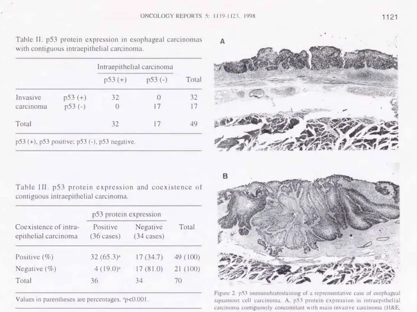
p53 protein expression. Among 49 cases coexisting with intraepithelial carcinoma, 32 had p53 protein expression in both intraepithelial and invasive carcinoma, and in the other 17 cases, p53 protein expression was found in neither intraepithelial carcinoma nor invasive cancerous lesion. In addition, among cases with contiguous in traepi the! ial carcinoma there was no case with different imunoreactivity in intraepithelial and invasive carcinomas (Table II). The immunohistochemical findings of a representative case with intraepithelial carcinoma contiguously concomitant with
.... �����'-·
.�,..�
"!!?}� ---�, .. ·Y.�-
-�"''" ?;.' '"/
7:.,.�· .
.·.
�
Figure 2. p53 immunohistostaining of a reprensentativc case of esophageal squamou cell carcinoma. A, p53 protein expression in intraepithelial carcinoma contiguou ly concomitant with main inva ivc carcinoma (H&E, original magnification x42). B, p53 protein expression in an invasive carcinoma (H&E, original magnification x42). In both lesions p53 protein was localized only in the nuclei of the cancer cell .
invasive lesion of esophageal squamous cell carcinoma are demonstrated in Fig. 2. In both lesions p53 protein was localized only in the nuclei of cancer cells.
Relationship between p53 pro tein expression and the coexistence of intraepithelial carcinoma. In the 49 cases coexisting with intraepithelial carcinoma contiguous to invasive cancerous lesions, 32 (65.3%) had p53 protein expression in both the intraepithelial carcinoma and the invasive cancerous lesion. On the other hand, in the 21 cases without intraepithelial carcinoma contiguos to invasive cancerous lesion, only four (19.0%) demonstrated p53 protein
expression. There was a significant statistical difference
between these proportions (p<O.OOl) (Table III).
Relationship between p53 protein expression and invasion coefficient. In 21 cases without intraepithelial carcinoma contiguously concomitant with invasive lesion, all values of IC were treated a 100%. In the 34 cases without p53 protein expression, the mean IC was 88.7±19.1 %. On the other hand, in 36 cases with p53 protein expression, the mean IC was 53.4±28.2%, and the proportion was smaller than that of the former 34 case without p53 protein expre sion
1122 NOZOE era!: p53 EXPRESSION AND GROWTH PATTERN OF ESOPHAGEAL CARCINOMA
(%) 100
90 80 70 60 50 40 30 20 10 0
IC
p53 Positive (n=36)
p<0.0001
p53 Negative (n=34)
Figure 3. The relation between p53 protein expression and invasion coefficient (IC). The mean IC in 36 cases with p53 protein expression was 53.4±28.2%, which was significantly smaller than that (88.7±19.1 %) in 34 cases without p53 protein expression (p<O.OOOJ ).
(%) 100
90 80 70 60 50 40 30 20 10 0
IC
p53 Positive (n=32)
0 0
B�
0
p<0.001
p53 Negative (n=17)
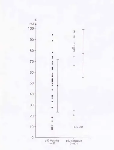
Figure 4. The relation between p53 protein accumulation and invasion coefficient ([C) among cases with intraepithelial carcinoma contiguosly concomitant with invasive carcinoma. The mean IC in 32 cases with p53 protein accumulation was 47.6±24.2%, which was significantly smaller than that (77 .3±21.9%) in 17 cases without p53 protein accumulation (p<O.OO I).
(p<O.OOO l) (Fig. 3). In the investigation restricted to cases with intraepithelial carcinoma contiguosly concomitant with invasive lesion, the mean IC (47.6±24.2%) in 32 cases with p53 protein accumulation was also significantly smaller than the mean IC (77 .3 ±21.9%) in 17 cases without p53 protein expression (p<O.OOI) (Fig. 4).
Discussion
The existence of intraepithelial carcinoma contiguously con com i tan t with invasive cancerous lesion has b een reported to be a relatively frequent event in the squamous cell carcinoma of the esophagus ( 1-4).
The significnce of p53 gene alterations in squamous cell carcinoma of the esophagus remains controversial. Benett et al (8) reported that the mutations of p53 gene occur in esophageal dysplasia and were concerned with the growth advantage of cancer cells in esophageal squamous cell carcinoma. Sarbia et al (21) suggested that there was no significant relation between the overexpression of p53 gene and the prognosis or clinicopathologic features of patients with esophageal carcinoma. The purpose of this study was to elucidate the relation between p53 protein expression and the growth pattern of the esophageal carcinoma with special attention to the coexistence of intraepithelial carcinoma.
In our study, the ratio of p53 protein expression in esophageal squamous cell carcinoma was 5 I .4% (36170), which was generally consistent with the values in previous reports (22,23). The proportion (65.3%; 32/49) of cases with p53 protein expression in cases coexisting with intra
epithelial carcinoma contiguous to invasive cancerous lesion was found to be significantly larger than that ( 19.0%; 4/21) in the cases without intraepithelial carcinoma. These data thus suggested that p53 protein expression is considered to be closely related to the formation of intraepithelial carcinoma contiguously concomitant with squamous cell carcinoma of the esophagus. Moreover the IC was adopted to investigate the relation bet ween p53 protein expression and growth pattern of esophageal squamous cell carcinoma. The significance of IC expressed that carcinomas with a larger IC value had a trend of profound invasion, while on the other hand, carcinomas with smaller IC value had a trend of intraepithelial spread. The value of the IC in cases with p53 protein expression was significantly smaller than that in cases without p53 protein expression. These data thus suggested that squamous cell carcinomas of the esophagus with p53 protein expression have significant relation to the i ntraepi thelia! spreading type growth, while carcinomas without p53 expression have significant relation to the profound penetrating type growth.
Concerning the relation between p53 protein expression and growth pattern of carcinoma, Oiwa et al (24) reported that there was a significant relation between p53 expression and the penetrating type growth pattern of gastric adeno
carcinomas. Our data showed that p53 expression was significantly related to intra-epithelial spreading type growth of esophageal squamous cell carcinoma. The functions of p53 protein on the growth of carcinomas have not been fully elucidated, however, it may mediate the growth pattern of squamous cell carcinoma of the esophagus.
ONCOLOGY REPORTS 5: I I I 9- I 123, 1998 1123
In conclusion, p53 protein expression was found to be significantly related to the coexistence and spread of intra
epithelial carcinoma contiguous to squamous cell carcinoma of the esophagus and it is thus considered that the intraepithelial spreading type growth of squamous cell carcinoma of the esophagus may be potentially mediated by p53 mutation.
Acknowledgments
The authors thank Brian T. Quinn for helpful comments on the manuscript.
References
1. Kuwano H, Matsuda H, Matsuoka H, Kai H Okudaira Y and Sugimachi K: Intra-epithelial carcinoma conc'omitant with eso
phageal squamous cell carcinoma. Cancer 59: 783-787, 1987.
2. Kuwano H, Nagamatsu M, Ohno S, Matsuda H, Mori M and Sugimachi K: Coexistence of intraepithelial carcinoma and grandular differentiation in esophageal squamous cell carcinoma.
Cancer 62: I 568-1572, 1988.
3. Mandard AM, Tourneux J, Jignoux M, Blanc L, Segal P and Mandar? JC: /�1 situ carcinoma of the esophagus: macroscopic study With particular reference to the Lugol test. Endoscopy 12:
51-57,1980.
4. Soga J, Tanaka 0, Sasaki K, Kawaguchi M and M uto T:
Superficial spreading carcinoma of the esophagus. Cancer 50:
1641-1645,1982.
5. Nigro JM, Baker SJ, Preisinger AC, Jessup JM, Hostetter R, Cleary K, Eigner SH, Davidson , Baylin S, Devilee P, Glover T, Collins FS, Weston A, Modali R, Harris CC and Yogelstein B: Mutations in the p53 gene occur in diverse human tumour types. Nature 342: 705-708, 1989.
6. Holls�ein �· Sidransky D, Yogelstein B and Harris CC: p53 mutatiOns 111 human cancers. Science 253: 49-53, 1991.
7. Casson AG, Mukhopadhyay T, Cleary KR, Ro JY, Levin B and Roth JA: p53 gene mutations in Barrett's epithelium and esophageal cancer. Cancer Res 51: 4495-4499, 1991.
8. Bennett WP, Hollstein MC, Metcalf RA, Welsh JA, He A, Zhu .sM, Kusters I, Resau JH, Trump BF, Lane DP and Harr.Is CC: p53 mutation and protein accumulation during multistage human esophageal carcinoaenesis. Cancer Res 52:
6092-6097, 1992. 0
9. Dolcetti R, Doglioni C, Maestro R, Gasparotto D, Barzan L,
?astore A, Roman.elli M and Boiocchi M: p53 over expression IS a� early event In the development of human squamous cell carcmoma of the larynx-genetic and prognostic implictions. Int J Cancer 52: 178-182, 1992.
10. Chung KY, Mukhopadhyay T, Kim J, Casson A, Ro JY, Geop�ert
�·
Hong WK and Roth JA: Discordant p53 gene mutatiOns. 111 pnmary head and neck cancers and corresponding second pnmary cancers of the upper aerodigestive tract. Cancer Res 53: 1676-1683, 1993 .11. Sundaresan V, Ganly P, Hasleton P, Rudd R, Sinha G, Bleehen NM and Rabbitts P: p53 and chromosome-3
abnormalities, characteristic of malignant luna tumours are detectable in preinvasive lesions of the bronchu
�
. Oncoge�e 7:1989-1997, 1992.
12. Sozzi G, Miozzo M, Donghi R, Pilotti S , Cariani CT, Pa Lorino U, Dellaporta G and Pierotti MA: Deletions of 17p and p53 mutation in preneoplastic lesions of the lung. Cancer Res 52: 6079-6082, 1992.
13.Baker SJ, Preisinger AC, Jessup JM, Para keva C, Markovitz S, WilsOI: JKV, Ham.ilton S and Vogelstein B: P53 gene mutations occur 111 combination with 17p allelic deletions as late event in colorectal tumorigenesis. Cancer Re 50: 7717-7722, 1990.
14. Murak�mi Y� Hayashi K, Hirohashi S and Sekiya T:
Aberrat10na o1 the tumor suppressor p53 and retinoblastoma genes in human hepatocellular carcinomas. Cancer Res 5 I:
5520-5525, ] 991.
I 5. Sidransky D, Mikkelsen T, Schwechheimer K, Rosenblum ML, Cavenee W and Yogelstein B: Clonal expansion of p53 mutant cells is associated with brain tumor progression. Nature 355:
846-84 7' 1992.
16. Vogel tein B, Fearon ER, Hamilton SR, Kern SE, Preisinger AC, Leppert M, Nakamura Y, White R, Smits AMM and Bas JL:
Genetic alteration during colorectal tumor development. N Eng!
J Med 319: 525-532, 1988.
17. Esrig D, Spruck CHIll, ichols PW, Chaiwun B, Steven K.
Groshen S, C�en SC, Skinner DG, Jones PA and Cote RJ: p53 nuclear protem accumulatiOn correlates with mutations in the p53 gene, tumor grade, and stage in bladder cancer. Am J Pathol 143: 1389-1397, 1993.
18. Tanaka S, Toh Y, Adachi E, Matsumata T, Mori R and Sugimachi K: Tumor progression in hepatocellular carcinoma may be mediated by p53 mutation. Cancer Res 53: 2884-2887
1993. '
19. Warnakulasuriya KAAS and Johnson NW: Expression of p53 mutant nuclear phosphoprotein in oral carcinoma and potentially malignant oral lesions. J Oral Pathol Med 21:
404-408, 1992.
20. Suckow EE, Yokoo H and Brock DR: Intraepithelial carcinoma concomitant with esophageal carcinoma. Cancer 15: 733-740
1962. '
21. Sarbia M, Porschen R, Borchard F, Horstmann 0, Willers R and Gabbert HE: p53 protein expres ion and prognosis in squamous cell carcinoma of the esophagus. Cancer 74: 2218-2223, 1994.
22. Toh Y, Kuw ano H, Sonoda K, Saeki H, Kawaguchi H, Kitamura K, akashima H and Sugimachi K: Correlation bet wee� re�uced p21 WAF I /CfP I expression and abnormal p53 expressiOn Ill esophageal carcinomas. lnt J Oneal 11: 703-708 1997.
, 23. Chaves P, Pereira AD, Pinto A, Oliveira AG, Queimado L, G Ioria L, Cardoso P, Mira FC and So are J: p53 protein immunoexpression in esophageal squamous cell carcinoma and adjacent epithelium. J Surg Oncol 65: 3-9, 1997.
24. Oiwa H, Maehara Y, Ohno S, Sakaguchi Y, Ichiyoshi Y and Sugimachi K: Growth pattern and p53 overexpression in patients with early gastric cancer. Cancer 75 (Suppl.): 1454-1459, 1995.
.--�-�-�=--=-=---".=-=-=-------=-,..---=-=�---�---