Acta Med. Nagasaki 45 : 71-74
Case Report
A Case of Ductal Dysplasia of the Pancreas A Possible Prerequisite for Pancreatic Cancer
Susumu EGUCHI 1), Minoru NISHIHARA 1), Takuo SHINOZAKI 1), Tadayoshi MORIYAMA 1), Sadayuki OKUDAIRA 2), Takashi KANEMATSU 2)
1) Department of Surgery, Nakatsushima Hospital (former National Tsushima Hospital) 2) Second Department of Surgery, Nagasaki University School of Medicine
The precise pathobiology of precursor lesions, which de- velop into pancreatic adenocarcinoma, remains controver- sial. Recently, we encountered a patient with dysplastic le- sion of the pancreas; a case in which a precursor lesion of the pancreatic carcinoma may have been documented. The patient was a 73-year-old female with epigastric discomfort.
As part of a general check-up, she underwent abdominal ul- trasound, which revealed the pancreatic duct in the pancre- atic body to be slightly indented. Computed tomography (CT) scan revealed mild dilatation of the main pancreatic duct and a small low-density area (less than 1 cm in diameter). Endo- scopic retrograde cholangiopancreatography (ERCP) showed the irregularity in the body of the main pancreatic duct.
Under general anesthesia, resection of the distal portion of the pancreas was performed with splenic conservation. Path- ological examination revealed focal hyperplastic epithelium of the pancreatic duct with moderate dysplasia. Expression of the proliferating cell nuclear antigen (PCNA) in the le- sion was observed, indicating a slightly proliferative na- ture. Mutant p53 protein was slightly expressed in the le- sion. As is seen in this case, ductal hyperplasia of the pancreas might represent precursor lesions, and constitute part of a continuous development spectrum evolving into ductal adenocarcinoma of the pancreas with accumulation of genetic alterations.
Key Words: ductal hyperplasia of the pancreas, precursor le- sion of the pancreatic cancer
Address Correspondence: Susumu Eguchi, M.D.
Second Department of Surgery, 1-7-1 Sakamoto, Nagasaki, 852- 8501 Japan
TEL: +81-95-849-7316 FAX: +81-95-849-7319
Introduction
Although the precise pathobiology of precursor le- sions that develop into pancreatic adenocarcinoma re- mains controversial, elucidation of the mechanisms of tumorigenesis might possibly provide for earlier detec- tion, prevention, and treatment. Thus far, infiltrating adenocarcinoma of the pancreas has been characterized at the molecular level, though little is currently known about the early events in the development of this neo- plasm or about the role of precursor lesions in tumor development"). The identification of the precursor le- sions to infiltrating adenocarcinoma of the panreas is especially important because most infiltrating cancers of the pancreas already have spread beyond the pan- creas by the time the lesions are detected clinically.
Recently, we encountered a patient with pancreatic hyperplasia for whom partial resection of the pancreas was performed. Here we describe the case and discuss an hypothesis regarding tumorigenesis.
Case report
The patient was a 73-year-old female with epigastric discomfort, who was referred to our hospital in 1998 due to hyperglycemia. As a part of general check up, she underwent abdominal ultrasound, which revealed the pancreatic duct in the pancreatic body, having an appearance similar to a string of beads. No particular family history existed. She had no history of alcohol abuse nor was she a heavy smoker. On admission, the patient's physical examination revealed no particular abnormal findings. In addition, count of blood ball cells, biochemistry (Amylase 105 IU/1), and tumor markers
(CA19-9 27U/ml, Span-I 50U/ml) except for Dupan-2 (190 U/ml) all showed normal data. Both her pancre- atic exocrine (PFD test: 43.5%) and endocrine (75g oral glucose tolerance test was diabetes mellitus pattern)
function were poor. CT scan revealed mild dilatation of the main pancreatic duct and a small low-density area (less than 1 cm in diameter; Fig. 1). Preoperative ERCP showed the irregularity in the body of the main pancreatic duct (Fig. 2). Magnetic resonance imaging
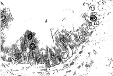
showed slight dilatation of the main pancreatic duct and a small mass lesion in the body. Angiography re- vealed no abnormalities. Due to a preoperative diagno- sis of possible pancreatic cancer, under general anes- thesia, resection of the distal portion of the pancreas was performed with splenic conservation. Pathological examination revealed focal hyperplastic epithelium of the pancreatic duct with dysplasia (Fig. 3 A, B, C). In
Fig 1. CT scan revealed mild dilatation of the main pancre- atic duct and a small low-density area less than 1 cm
in diameter (arrow).
Fig 2. Preoperative ERCP showed the irregularity in the body of the main pancreatic duct.
Fig 3. Pathological examination revealed focal hyperplastic
epithelium of the pancreatic duct with some degree of dysplasia. (A:100x, B:200x, C:400x)
addition, pyloric gland hyperplasia in the lesion was seen and slight inflammation was present in the whole resected specimen. The final histological diagnosis was
ductal hyperplasia with moderate dysplasia of the pancereas. Expression for proliferating nuclear antigen
(PCNA) was observed in the nucleus of the lesion (Fig. 4). Immunohistochemically, mutant p53 protein was expressed in the lesion. Also, both CA 19-9 and Carcinoembryonic antigen (CEA) protein were slightly expressed in the lesion. Post operative course was un- eventful and the patient was discharged home. There has been no evidence of recurrence of the disease and she has been symptom-free for 1 year at this writing.
Fig 4. Slight expression of the proliferating nuclear antigen (PCNA) was observed in the lesion. (200x)
Discussion
In the present case, we may have detected a precur- sor lesion of pancreatic carcinoma, which can be hardly detected clinically. In the resected specimen, slight ex- pression of PCNA was observed in the lesion, indicating a slightly proliferative nature. Generally, aside from de novo development, it has been presumed that the de- velopment spectrum of hyperplasia - dysplasia - carci- noma in situ exists, although it is relatively difficult to prove in vivo 3, 4). Thus far, several reports have been published stressing the involvement of ductal hyper- plasia in infiltrating adenocarcinoma of the pancreas, although the papillary hyperplasia - atypical hyperplasia
(dysplasia) - to papillary adenocarnoma sequence has been relatively well investigated with regard to mucin producing adenocarcinoma 5, 5). Thus, although a puta- tive development spectrum evolving into ductal adeno- carcinoma of the pancreas was advocated, hyper or dysplastic lesions of -the pancreas are barely detected, though this may have been accomplished in the present
case.
Mutant p53 protein was slightly expressed in the le- sion. In a previous report, it was shown that mutant p53 protein was expressed at 0% in normal duct, 55%
in hyperplastic lesions, 67% in dysplastic lesions and 80% in adenocarcinomas'). It has also been reported that K-ras mutation was highly related to carcinogenesis of pancreatic cancer, although it was not investigated in our case"). This finding implies a putative develop- ment spectrum evolving into ductal adenocarcinoma of the pancreas with accumulation of genetic alteration.
Ductal hyperplasia and dysplasia of the pancreas might represent precursor lesions, in a fashion similar to that seen in colorectal tumorigenesis with accumulating progressive genetic alterations such as p53 and K-ras'.
From a different perspective, it is presumed that pancreatic hyperplasia might be a different entity from
hyperplasia of other organs such as gastrointestinal tract. Brat et al. stressed that the term "pancreatic hyperplasia" should be replaced by the more specific term "pancreatic intraepithelial neoplasia" since pancre- atic hyperplasia can develop into infiltrating adenocar- cinoma just as adenoma in the colorectum 10'. In their cases, adenocarcinoma developed long-term after detec- tion of the atypical papillary hyperplasia. The differ- ence of meaning of "hyperplasia" should be elucidated, in time, by further investigation with more detailed genetic clarification.
As seen in our case, chronic inflammation and/or ductal epithelial papillary hyperplasia may play a role in the pathogenesis of the tumor. However, thus far, the relationship between chronic pancreatitis and ductal neoplasma of the pancreas is still under discussion 11.12) In any event, the identification of the precursor lesions to infiltrating adenocarcinoma of the pancreas is espe- cially important because most infiltrating cancers of the pancreas already have spread beyond the pancreas by the time the lesions are detected clinically. Our pa-
tient may have been fortunate since the lesion was resected prior to the development of infiltrating adenocarcinoma, which usually provides a poor prog- nosis.
In summary, we experienced a case with hyperplasia of the pancreas with a somewhat dysplastic (morpho- logincal and genetic) ' lesion. This type of lesion is rarely detected, and this case may thereby provide in- formation regarding carcinogenesis of infiltrating
adenocarcinoma of the pancreas, the development of which likely includes this step.
References
1) Sommers SC, Murphy SA, Warren S. et al. Pancreatic duct hyperplasia and cancer. Gastroenterology 27:629-640, 1954.
2) Kozuka S, Sassa R, Taki T et al. Relation of pancreatic duct hyperplasia to carcinoma. Cancer 43:1418-1428, 1979.
3) Kloppel G, Bommer G, Ruckert K et al. Intraductal proliferation in the pancreas and its relationship to human and experimental
carcinogenesis. Virchows Arch (A) 387: 221-233, 1980.
4) Konishi Y, Mizumoto K, Kitazawa S et al. Early ductal lesions of pancreatic carcinogenesis in animals and humans. Int J Pancreatol
7:83-89, 1990.
5) Santini D, Campione 0, Salerno A et al. Intraductal papillary- mucinous neoplasm of the pancreas. A clinicopathologic entity
Arch Pathol Lab Med 119:209-213, 1995.
6) Sugiyama M, Atomi Y, Kuroda A. Two types of mucin-producing
cystic tumors of the pancreas: Diagnosis and treatment. Surgery 122:617-625, 1997.
7) Apple SK, Hecht JR, Lewin DN et al. Immunohistochemical evalua- tion of K-ras, p53, and HER-2 / neu expression in hyperplastic,
dysplastic and carcinomatous lesions of the pancreas: Evidence for multistep carcinogenesis. Hum Pathol 30:123-9, 1999.
8) Tada M, Ohashi M, Shiratori Y et al. Analysis of K-ras gene muta- tion in hyperplastic duct cells of the pancreas without pancreatic
disease. Gastroenterology 110:227-231, 1996.
9) Tabata T, Fujimori T, Maeda S et al. The role of Ras mutation in pancreatic cancer, precancerous lesion, and chronic pancreatitis. Int J Pancreatol 14:237-44, 1993.
10) Brat DJ, Lillemoe KD, Yeo CJ et al. Progression of pancreatic intraductal neoplasias to infiltrating adenocarcinoma of the pan-
creas. Am J Surg Pathol 22:163-169, 1998.
11) Cylwik B, Nowak HF, Puchaiski Z et al. Epithelial anomalies in chronic pancreatitis as a risk factor of pancreatic cancer. Hepato-
Gastroenterology 45:528-532, 1998.
12) Cylwik B, Nowak HF, Puchaiski Z et al. AgNORs in duct epithelial lesions in chronic pancreatitis and in pancreas cancer cells. Hepato-
Gastroenterology 45:1130-1134, 1998.