Acta med. Nagasaki 14: 42-71
Amino Acid Sequence in All the Tryptic Peptides from the
αPolypeptide Chain of All Component ot Chicken Hemoglobin*
KU CHAYU WU**
Department of Biochemistry, Nagasaki University
School of Medicine Nagasaki, Japan
Received for publication, October, 1969
Hemoglobin was obtained from blood of adult white leghorns. It was first separated into three components, AI, AII, and AIII by CMC-column chromatography. AII, the greatest component of the three was chosen as the subject matter of the present study. It was first converted into globin by removal of heme in acetone-HCl solution, and then the globin was se-
parated into α andβ polypeptide chains by Amberlite CG50 column chrom‑
atography. The α polypeptide chain was digested with trypsin, and tryptic
peptides thus obtained were subjected to column chromatography and then to paper chromatography for isolation and purification. The column chro- matography was carried out by using Dowex 1X2 as the adsorbent and the acetate buffer containing organic bases such as pyridine, collidine, lutidine, and picoline as the developer. The developer for the paper chro- matography was the mixture of n-butanol, acetic acid, and water.
The amino acid composition of the tryptic peptides thus isolated and purified was analyzed on an amino acid analyzer and subsequently the amino acid sequence in these tryptic peptides was determined by using partial hydrolyses with various enzymes, the PTC method, and the DNP method. The results were discussed in comparison with human hemoglo- bin. As a result, replacement of amino acids in the tryptic peptides be- tween adult human and white leghorn hemoglobins was presumed at 35
positions in the α polypeptide chain.
INTRODUCTION
With the recent progress of protein chemistry, attempts to elucid- ate problems on evolution and phylogeny of living organisms on mole- cular level have extensively been made since 1950. Above all, since amino acid sequence in protein molecules, i. e. the primary structure of proteins was known to be a precise copy of the hereditary message of DNA in cell nucleus, a great importance has been placed on the
This study was presented at the 41st General Meeting of the Japanese Biochemical Society (Octob°r, 1968) and at the 20th Conference on the Structure of Proteins (October, 1969).
**呉 谷 喬
comparative studies on the primary structures of protein molecules from living organisms belonging to different species because they give some clues for phylogeny.
According as the advance in theories of protein chemistry and analytical techniques prompt many groups of workers to uudertake
investigations on the primary structure, information on the correlation between the primary structure and its biological function was also der-
ived. It was confirmed that the two living organisms belonging to considerably remote species possess some kinds of protein whose pri- mary structure as well as biological function is remarkably similar to
each other, and this fact elicited a hypothesis that such proteins are due to a common ancestral gene possessed by the both.
On the other hand, it is generally presumed that in proteins with the biological activity, a certain part of the primary structure plays a much more important role than other parts, and therefore, if the amino acid sequence in this important part is unchanged, proteins do not lose their function even though there exist some differences in the amino acid sequence of other parts.
For promoting the studies as stated above, hemoglobin is one of the most useful experimental materials not only because it can be found very commonly in the animal kingdom but because it can be isolated and purified relatively more readily than other proteins. Since BRAUN- ITZER et al.9) and KONIGSBERG et al.24) established the primary structure of human hemoglobin, many groups of workers in the world have studied8),13),44),52) or are study in g4).38),40),45) on the primary structure of mammlian hemoglobins from various species. In 1968, MATSUDA et al.31) of our department determined the whole primary structure of adult rhesus monkey hemoglobin. With respect of non-mammalian he- moglobins, however, BRAUNITZER et al, 21) only reported the determin- ation of the primary structure of the a polypeptide chain of carp he- moglobin, and no reports have been presented on avian hemoglobins.
Our department, with an interest in the phylogenetic relationship between human and Chicken hemoglobins as well as their resemblance in function such as Bohr's effect and the oxygen dissociation curve, have been investigating on the primary structure of adult hemoglobin of the Chicken.
As a first step, the present author determined the amino acid se- quence in all the tryptic peptides from the r>a polypeptide chain of the greatest component, All of adult Chicken hemoglobin and compared the results with the known sequence of human hemoglobin.
MATERIALS AND METHODS
1) Preparation of Hemoglobin from Adult Chickens.
Hemoglobin solution was obtained by DRABKIN' S method17) from
blood of adult pure-line white leghorns (Smal A type) as follows.
Blood was obtained from the jugular vein of white leghorns, and to this was added 3.8% sodium citrate solution as an anticoagulant.
The blood was then submitted to centrifugation in order to remove plasma and then to this was added an equal volume of deionized water and half the volume of toluene. The mixture was hemolyzed overnight
in the cold (2° to 4°). The hemolysate was centrifuged at 12,000 r.p.
m. for 1 hour at 0', and the hemoglobin solution was obtained.
2) Isolation of All Component from the Hemoglobin Solution.
All component was isolated from the hemoglobin solution according to MATSUDA et al.32)
The activated carboxymethyl cellulose(Brown Company, 0.71 mEq/g) was poured into a column (2 x 30 cm), which was equilibrated with 0.01 M phosphate buffer, pH 6.9, The hemoglobin solution, 5 ml, was placed on the column. The linear gradient elution was carried out by means of phosphate concentration. The fraction volume was 25 ml. The optical density of the effluent was measured at 542 mp and 280 ma.
Fractions at each peak were combined, dialyzed against deionized wa- ter, and finally lyophilized.
3) Preparation of Globin.
Removal of heme from All component was performed according to ANSON and MIRSKY.2) The lyophilized All component, about 5,000 mg, was added dropwise with stirring to HC1-acetone solution cooled at -20' . The solution was centrifuged at 3,000 r . p . m . for 5 min at 00 to 40, dissolved in an appropriate volume of deionized water, dialyzed
against deionized water, and finally lyophilized.
4) Isolation of a Polypeptide Chain from the Globin of All Component.
The globin obtained from All component was separated into a and 13 polypeptide chains by the method of SuiTx5o) and MATSUDA et al.29) The column (3.3 x 45 cm) was prepared with the activated Amberlite CG50 (Type II, H-form) and then equilibrated with 3N formic acid. To this column was applied 1,000 mg of the globin from All component.
The linear gradient elution was performed by means of urea concen- tration. The fraction volume was 25 ml. The effluent was analyzed by optical density at 280 mid. Fractions at each peak were combined, dialy- zed against deionized water, and then lyophilized.
5) Determination of the N-Terminal Structure of the (i Polypeptide Chain by the DNP Method.
The DNP method was carried out as described by MATSUDA et al.30)
The a polypeptide chain, 100 mg, was dinitrophenylated and DNP-
peptides thus obtained were hydrolyzed with constant boiling HCl and
extracted with ether. The extracts were placed on a silica gel-Celite
column (1 x 15 cm). Development was performed with the mixture of acetic acid, acetone, and ligroin.
6) Determination of Cysteine Residues in the (,-/ Polypeptide Chain.
Estimation of cysteic acids derived from cysteine residues in the a polypeptide chain was carried out according to MooRE33).
The ,, polypeptide chain, 5 umoles (85 mg), was oxidized with per- formic acid by the method of HIRE22), and then hydrolyzed with cons- tant boiling HCl for 18 hours. The hydrolysate was placed on a column (0.9 x 50 cm) of the activated Amberlite CG 120 equilibrated with 0.2 N sodium citrate buffer, pH 3.25. Elution was carried out by using 0.2 N sodium citrate buffer, pH 3.25, as a developer. Analysis of the efflu- ent was performed both by the ninhydrin reaction according to MOORE et al.34) and by measuring the optical density at 570 m,m.
7) Determination of Tryptophan Residues in the a Polypeptide Chain.
Estimation of tryptophan was carried out according to KOSHLAND et al. 3).
The a polypeptide chain, 10 umoles (170 mg), dissolved in 10 M urea was incubated at 37° for 16 hours, and to this was added HNB- bromide. The a polypeptide chain thus treated was placed on a column (1,0x23 cm) of the activated Sephadex G-25 equilibrated with 10 M urea, pH 2.7. Elution was carried out with the developer, 10 M urea, pH 2.7. The fraction volume was 2 ml. Optical density of the effluent was measured at 410 ma.
8) Digestion of the a Polypeptide Chain with Trypsin.
The a polypeptide chain, 1,500 mg, was denatured by the method of BRAUNITZER et a1.26) and then digested at 370 for 4 hours by the add- ition of trypsin (Worthington Biochemical Co., twice crystallized) pre- pared according to REDFIELD et al. 39) and BRAUNITZER et al.14) . After the termination of the digestion, the digest was adjusted to pH 6.1.
Then, the so-called insoluble tryptic peptide was precipitated. The digest was submitted to centrifugation so that it was separated into the soluble tryptic peptides and the insoluble tryptic peptide, both of which were lyophilized, respectively.
9) Column Chromatography of the Soluble Tryptic Peptides.
MALTA'S method27) was used. The activated Dowex 1X2 (200 to 400 mesh, Dow Chemical Co.) was poured in a column (0.9 x 150 cm),
which was equilibrated with 1% pyridine, 1% collidine acetate buffer,
pH 9.0. The soluble tryptic peptides dissolved in deionized water were
placed on the above-prepared column. The pH linear gradient elution
was carried out by using both pyridine, 2-4 lutidine, a-picoline acetate
buffer and acetic acid; Fraction No.1 to 20 were eluted with the star-
ting buffer, 1% pyridine, 1% collidine acetate buffer, pH 9.0 ; for
Fraction No.21 to 40, the buffer was changed. stepwise into 1% pyri- dine, 1,°o collidine acetate buffer, pH 8.5 ; Fraction No. 41 to the fraction at pH 5.5 were eluted by supplying 0.1 N acetic acid in the upper chamber to the mixing chamber containing 1,500 ml of 1%
pyridine, 1% picoline, 1% 2-4 lutidine acetate buffer, pH 7.5 ; To the fraction the effluent of which lowered to pH 3.5, 1 N acetic acid was supplied, and finally glacial acetic acid was used.
An aliquot of each fraction after the alkali-hydrolysis was submitted to the ninhydrin reaction according to the method of YEMM and COCK- ING51), and then optical density was measured at 570 mp. Fractions at the peaks were combined respectively and dried below 370 under reduced pressure on a rotary evaporator. Each of the dried samples was dissolved in 3 ml of deionized water. Some peptides insoluble in water were dissolved in 50% acetic acid solution by the final acetic acid concentration of 12 to W/06).
10) Digestion of the So-Called Insoluble Tryptic Peptide with Pepsin.
The insoluble tryptic peptide was oxidized with performic acid by the method of HIRs22). Digestion was carried out at 37° for 2 hours by adding pepsin (Sigma Chemical Co., Three time crystallized) by a ratio of 1% to the substrate. At the termination of the digestion, the digest was adjusted to pH 10.0 and the dried under reduced pressure below 37'. Column chromatography for the insoluble tryptic peptide was carried out in the same way as for the soluble tryptic peptides.
11) Digestion of the Peptides, a TVI-b, a TVII-a, and a TIX-a with Pepsin.
Each of these peptides, about 10 pmoles, was dissolved in deionized water, and to this was added HCl solution containing pepsin (Sigma Chemical Co., three time crystallized). a TVI-b, a TVII-a, and a TIX- a were digested at 37° for 6 hours, 4 hours, and 16 hours, respecti- vely. The digests were adjusted to pH 10.0 and dried under reduced pressure.
A column (0.9 x 60 cm) was prepared with the activated Dowex 1
X2 (200 to 400 mesh) and equilibrated with 1% pyridine, 1%2-4 lutidine
acetate buffer, pH 8.5. To this column was applied the above-prepared
peptic peptides, and then development was performed at a flow rate
of 60 ml per hour by using the pH linear gradient elution with both
1% pyridine, 1% 2-4 lutidine acetate buffer and acetic acid; Fractions
No. 1 to 15 were eluted with the starting buffer, 1% pyridine, 1% 2-4
lutidine acetate buffer; until the pH of the effluent lowered to 6.0,
0.1 N acetic acid was supplied from the upper chamber to the mixing
chamber containing 700 ml of the starting buffer; until the pH of the
effluent lowered to 3.5, 1 N acetic acid was supplied and finally glacial
acetic acid was used. During the development, the column was kept at
37'. The effluent was collected into 8 ml-fractions by a fraction collector.
An aliquot of each fraction was submitted to the ninhydrin rea- ction5l) and optical density was measured at 570 mi. Fractions at each peak were combined together, dried under reduced pressure below 37°, and then dissolved in 3 ml of deionized water.
12) Paper Chromatography of the Tryptic Peptides and the Peptic Peptides from the So-Called Insoluble Tryptic Peptide and a T VI-b , u T VII-a , and
ai TIX-a.
The sample, 50 pl, was applied as a spot on a sheet of Toyo filter paper No. 50 (60 x 60 cm). Development was carried out at 25° in the constant temperature room by the descending method with the upper phase of the mixture of n-butanol, acetic acid, and water (4 : 1 : 5, V/V)36)
Peptides were detected by spraying 0.2% ninhydrin-n-butanol solu- tion on the paper and then heating to color with an iron. The EHRLICH reaction46), the SAKAGUCHI reaction23), the PAULY reaction47), and the a-nitrosonaphtol reaction') were also performed on the paper.
Purification of peptides by paper chromatography was performed as follows; Peptides were detected with 0.02% ninhydrin-n-butanol solution and then eluted with 5% acetic acid solution for 16 hours at the room temperature. The eluate was dried under reduced pressure.
13) The PTC Metho i .
A modification of EDMAN'S original method24) was employed for phenyl isothiocyanation of the peptides. The ring-formation was per- formed by using trifluoroacetic acid (the special grade). Analysis was carried out by the degradation method.
The peptides were dissolved in 2.0 ml of 66% pyridine solution contained in a heart-shaped flask. To this was added 0.05 ml of pheny-
lisothiocyanate (PTC). The mixture was allowed to react at pH 7.5 and 37° for 2 hours and then dried under reduced pressure below 37'.
The flask was connected to the cold finger condenser and evacuated at 40° for 40 min in a water bath by a vacuum pump. To this was added 1 ml of trifluoroacetic acid. The mixture was allowed to stand for 4 hours at the room temperature, dried under reduced pressure below 37'. It was dissolved in deionized water and extracted three
times with 5 ml of benzene. An aliquot of the aqueous phase dried under reduced pressure was hydrolyzed with constant boiling HC1 at 105° for 20 hours, and the amino acid composition of the hydrolysate was analyzed.
The remainder of the aqueous phase was dried under reduced pressure and used for the next stage of the PTC method.
14) N-Terminal Analyses of the Peptides by the DNP Method.
LLVY's method25) was employed. The peptides were dinitrophenyl-
ated at 40° for 2 hours in 1% NaHCO3 solution. After the reaction
stopped, the excess of 1-fluoro-2, 4-dinitrobenzene (DNFB) was removed by ether extraction. The aqueous was dried under reduced pressure and then hydrolyzed with constant boiling HC1 at 105° for 20 hours.
N-terminal DNP-amino acid was extracted with ether and identified by the two dimensional paper chromatography. Developers were n-butanol saturated with 1 N NH4 OH. for the first dimension and 0.5 M phosph- ate buffer for the second. Quantitative analysis of the DNP-amino acid was performed in the following way; Yellow spots on the paper chro- matogram were cut out and extracted with 5 ml of warm water, res- pectively. The optical density of the eluate was measured at 360 m,a.
15) Peper Electrophoresis of the Peptides.
In order to know electric charge of the peptides and to distinguish acidic amino acid from their amides, high voltage paper electrophoresis was carried out at 2 KV for 2 hours on an electrophoretic apparatus (Ishidai type), the sample being applied on the center of a sheet of Toyo filter paper (10 x 60 cm), No. 50. Pyridine acetate buffer, pH 6.4 (pyridine : acetic acid : water = 100 : 4 : 900, v/v) was used as a developer4l). After the paper was dried in the air, peptides were dete- cted by spraying 0.2% ninhydrin-n-butanol solution and then heating to color with an iron.
16) Amino Acid Analyses of the Peptides.
The purified peptides dissolved in constant boiling HC1 were hydr- olyzed at 105° for 20 hours in a sealed tube. The hydrolysate was dried under reduced pressure, and then the amino acid composition was an- alyzed on Hitachi KLA-2 amino acid analyzer.
RESULTS
1) Isolation of All Component from Hemoglobin Solution.
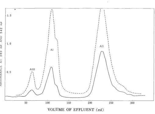
Fig. 1 shows three peaks, which were designated AIII, Al, and All in the elution order. Their yields were 4.8%, 23.8%, and 71.4%, respectively. The recovery was 87%. MANWELL28) also obtained almost the same results.
2) Isolation of a and p polypeptide chains
The yield of the a polypeptide chain was 44% and that of the Q polypeptide chain, 56%. The recovery was about 60%.
3) Determination of the N-Terminal Structure of the a Polypeptide Chain by the DNP Method.
The N-terminal analysis of the a polypeptide chain resulted in 0.53
mole of DNP-Val, 1.73 mole of DNP-Val-Leu, and none of DNP-Val-
His, indicating that the a polypeptide chain had been isolated almost
in purity as previously reported by MATSUDA et al.31)
VOLUME OF EFFLUENT (ml) Fig. 1. Column chromatogram of adult chicken hemoglobin.
CM-Cellulose column : 2 x 30 cm. Absorbance at 542 mu ... Absorbance at 280 mu.
TUBE NUMBER
Fig. 2. Separation of globin from All Component into a and (3 subunits. Column:
IRC-50 (l'ype II), 3.3 x 45 cm.
4) Determination of Cysteine Residues as Cysteic Acid in the -, Poly peptide Chain.
Estimation of cysteic acid resulted in 1.40 mole per one molec-
ule of the a polypeptide chain.
5) Determination of Tryptophan Residues in the a Polypeptide Chain.
Estimation of tryptophan resulted in 0.14 mole per one molecule of the (i polypeptide chain.
6) Column Chromatography, Paper chromatogrophy, and Amino Acid Corn- position of the Soluble Tryptic Peptides from the a Polypeptide Chain.
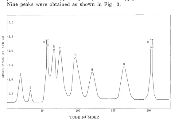
Nine peaks were obtained as shown in Fig. 3.
TUBE NUMBER
Fig. 3. Separation of the tryptic peptides from the a polypeptide chain of All component of adult chicken hemoglobin.
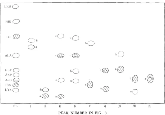
Each peak was submitted to paper chromatography. The result was given in Fig. 4, where 17 main spots were observed. Four of these spots were positive to the SAKAGUEHI reaction ; seven spots pos- itive to the PAULY reaction ; four spots positive to the a-nitrsonaphtol reaction and none positive to the EHRLICH reaction. Table I gives the
amino acid composition of these spots. (i TII-a and a T111-a, a T111-c and a TIV-c were identical in the amino acid composition, respectively.
7) Sequence Analyses of the Soluble Tryptic Peptides from the a,. Polypeptide
Chain.
Amino acid sequence in each tryptic peptide was analyzed by using the DNP method and the PTC method together.
(1) a TI-a (Rf Leu 0.59)
The N-terminal residue was known to be leucine by the DNP met-
PEAK NUMBER IN FIG. 3
Fig. 4. Paper chromatogram of the tryptic peptides from the a polypeptide chain of All Component of adult chicken hemoglobin.
Table I Amino acid compositions of the tryptic peptides from the a polypeptide chain of All Component of
adult chicken homoglobin
la IIa IIb IIlb I IIIc IIId IVa IVd Va Vb via VIb VIIa VIIIb IXa
Lys 1.03 1.00 1.02 1.02 1.01 1.08 '0.98 1.07 1.06 1.12 1.92 0.98 0.93
His 1.02 2.03 0.94 1.88 1.08 1.'02 1.06
Arg 0.90 1.09
Cys
Asp 2.08 2.26 1.12 2.17 1.18 1.09 2.88 2.81
Thr 2.03 1.02 0.93 0.90 0.90 0.94
Ser 0.86 0.88 0.71 1.83 1.74 1.82
Glu 1.03 1.07 1.01 4.01
Pro 0.71 0.96 0.95
Gly 2.03 1.21 1.08 1.10 0.97 0.98 1.96
Ala 1.16 1.10 0.97 1.21 4.93 4.84 3.00
Val 0.92 0.92 1.98 0.63 1.15 0.91
Met + i
Ileu 1.15 0.95 0.91 2.96 2.93 0.94
Leu 1.10 0.78 1.90 0.98 1.14 2.08 2.02 1.05
Tyr 0.91 0.72 0.76
Phe 0.81 0.94 0.88 1.65
hod. The sequence is Leu-Arg. D indicates the DNP method hereafter.
D (2) a TII-a (Rf Leu 0.07)
The following result was obtained by the PTC method.
Lys His Gly
composition 1.03 1.02 2.03
stage 1 1.00 0.92 1.08
2 1.00 0.30 1.00
The squence is, therefore, Gly-His-Gly-Lys. P indicates the PTC method hereafter. --> - )
P P
(3) a TII-b (RfLeu 0.18)
It was confirmed to be a single lysine by the descending paper chromatography and amino acid analyzer.
(4) a TIII-b (Rf Leu 0.24)
The following result was obtained by the PTC method.
Lys Asp Val
composition 1.02 2.08 0.92
stage 1 n . d . 1.17 0.83
2 n.d. 0.66 1.00
n . d . indicates "not determined", hereafter. The sequence was there- fore determined to be Asp-Asp-Val-Lys.
P P
Distinction between acidic amino acids and their amides was hereafter performed by paper electrophoresis.
(5) a TIII-c (Rf Leu 0.50)
The N-terminal amino acid turned out to be tyrosine by the DNP method. The sequence was Tyr-Arg.
D (6) a TIII-d (Rf Leu 0.71)
Analysis by the PTC method resulted as follow;
Lys Thr Pro Gly MetO2 Ileu Phe
Composition 1.02 2.03 0.71 1.21 + 1.15 1.81
stage 1 n . d . 2.18 0.86 0.88 - 1.22 1.83
2 n.d. 2.10 0.93 0.86 -- 1.03 0.84
3 n.d. 2.09 1.08 0.91 - 0.07 0.91
4 n.d. 2.08 0.97 0.02 - 0.00 0.91
5 n.d. 2.08 0.93 0.00 - 0.00 0.12
6 n.d. 2.00 0.23 0.00 - 0.00 0.00
The sequence was, therefore, Met-Phe-Ileu-Gly-Phe-Pro-Thr-Thr-Lys.
P P P P P P (7) aTIV-a (Rf Leu 0.22)
The following result was obtained by the PTC method.
Lys Asp Ser Ala Val Leu
composition 1.01 2.26 0.86 1.16 0.92 0.78
stage 1 n.d. 2.04 0.86 1.18 0.00 0.78
2 n.d. 2.12 0.84 1.04 0.00 0.00
3 n.d. 1.85 0.08 1.07 0.00 0.00
4 n . d . 1.02 0.00 0.98 0.00 0.0-0
5 n.d. 1.00 0.010 0.00 0.00 0.00
The sequence was determined to be Val-Leu-Ser-Asn-Ala-Asp-Lys P P P P P (8) a TIV-d (Rf Leu 0.68)
Analysis by the PTC method gave the following result.
Lys Thr Gly Ileu Phe
composition 1.08 1.02 1.08 0.95 0.94
stage 1 n . d . 1.07 0.00 0.98 0'.95
2 n . d . 0.92 0.00 0.00 1.08
3 n . d . 1.00 0.00 0.00 0.12
The sequence was therefore determined to be Gly-Ileu-Phe-Thr-Lys.
P P P (9) aTV-a (Rf Leu 0.20)
The following result was obtained by the PTC method.
Lys His Asp Ser Ala Leu
composition '0.98 2.03 1.12 0.88 1.10 1.90
stage 1 n.d. n.d. 1.21 0.81 1.11 0.90
2 n.d. n.d. 1.21 0.18 1.07 0.73
3 n.d. n.d. 0.35 0.05 1.00 0.78
4 n.d. n.d. 0.00 0.00 1.00 0.18
5 n.d. n.d. 0.00 0.00 1.00 0.-00
6 n.d. n.d. 0.-00 0.00 0.28 0.00
The sequence was therefore Leu-Ser-Asp-Leu-His-Ala-His-Lys.
> > > >
P P P P P P (10) aTV-6 (Rf Leu 0.61)
It was analyzed by the PTC method as follows.
Lys Asp Pro Val Phe
composition 1.07 2.17 0.96 1.98 0.88
stage 1 n . d . 2.05 0.86 1.06 1.00
2 n.d. 1.24 0.79 0.95 1.01
3 n.d. 1.14 0.16 0.97 0.88
4 n.d. 1.15 0.00 0.13 0.85
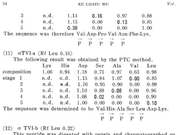
5 n.d. 0.38 0.00 0.00 1.00
The sequence was therefore Vai-Asp-Pro-Val-Asn-Phe-Lys.
P P P P P (11) aTVI-a (Rf Leu 0.16)
The following result was obtained by the PTC method.
Lys His Asp Ser Ala Val Leu
composition 1.06 0.94 1.18 0.71 0.97 0.63 0.98
stage 1 n.d. n.d. 1.15 0.84 1.07 0.00 0.85
2 n.d. n.d. 1.16 0.95 0.90 '0.00 0.98
3 n.d. n.d. 1.10 0.88 0.08 0.00 0.96
4 n.d. n.d. 1.08 0.02 0.00 0.00 0.90
5 n.d. n.d. 1.00 0.00 0.00 0.0.0 0.10
The sequence was determined to be Val-His-Ala-Ser-Leu-Asp-Lys.
P P P P P (12) a TVI-b (Rf Leu 0.32)
This peptide was digested with pepsin and chromatographed on a column (0.9 x 60cm) of Dowex 1X2. As shown in Fig. 5, two peaks were obtained.
TUBE NUMBER
Fig. 5. Isolation of the peptic peptides from the tryptic peptide, a TVI-b.
Column : Dowex 1 X2, 0.9 x 60 cm.
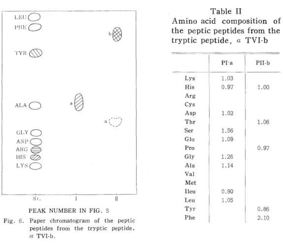
The effluent at each peak was dried under reduced pressure and puri- fied by paper chromatography. Two main spots were observed in Fig. 6.
PEAK NUMBER IN FIG. 5
Fig. 6. Paper chromatogram of the peptic peptides from the tryptic peptide,
a TVI-b.
Table II
Amino acid composition of the peptic peptides from the tryptic peptide, a TVI-b
PI-a PII-b
Lys 1.03
His 0.97 1.00
Arg
Cys
Asp 1.02
Thr 1.06
Ser 1.56
Glu 1.09
Pro 0.97
Gly 1.26
Ala 1.14
Val
Met
Ileu 0.90
Leu 1.05
Tyr 0.86
Phe 2.10
The amino acid composition of these peptide fragments is given in Table II.
(a) aTVI-b, P-I-a (Rf Leu 0.51)
Lys His Asp Ser Glu Gly Ala Ileu Leu composition 1.03 0.97 1.02 1.56 1.09 1.26 1.14 0.90 1.05 stage 1 n.d. n.d. 0.00 1.69 1.08 1.15 1.18 0.89 0.99 2 n. d. n. d. 0.00 1.67 1.12 1.15 1.21 0.85 0.12
3 n. d. n.d. 0.00 1.07 0.96 1.06 1.07 1.09 0.00
4 n.d. n. d. 0.00 1.10 0.99 1.04 1.01 0.90 0.00
5 n. d. n. d. 0.00 1.12 1.08 0.10 1.01 0.81 0.00
6 n. d. n.d. 0.00 0.10 1.11 0.00 1.03 0.85 0.00
7 n.d. n. d. 0.00 0.00 1.11 0.00 0.14 0.89 0.00
8 n. d. n. d. 0.00 0.00 0.23 0.00 0.00 1.0'0 0.00 The sequence was therefore Asp-Leu-Ser-His-Gly-Ser-Ala-Gln-Ileu-Lys,
--). .) -) ->
P P P P P P P P
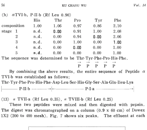
(b) aTVI-b, P-II-b (Rf Leu 0.90)
His Thr Pro Tyr Phe
composition 1.00 1.06 0.97 0.86 2.10
stage 1 n.d. 0.00 0.91 1.00 2.08
2 n.d. 0.010 0.94 0.00 2.06
3 n.d. 0.00 1.00 0.00 1.00
4 n.d. 0.00 0.00 0.00 1.00
5 n.d. 0.00 0.00 0.00 1.00
The sequence was determined to be Thr-Tyr-Phe-Pro-His-Phe.
P P P P P
By combining the above results, the entire sequence of Peptide a TVI-b was established as follows;
Thr-Tyr-Phe-Pro-His-Phe-Asp-Leu-Ser-His-Gly-Ser-Ala-Gln-Ileu-Lys
P-II-b -- F-- P-I-a -H
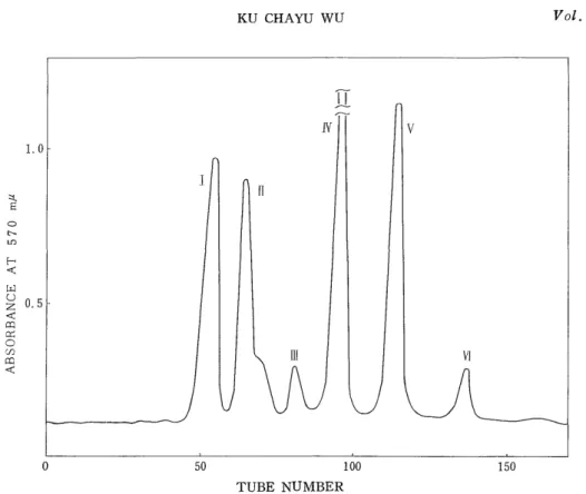
(13) a TVII-a (Rf Leu 0.35), a TVIII-b (Rf Leu 0.25)
These two peptides were mixed and then digested with pepsin.
The digest was chromatographed on a column (0.9 x 60 cm) of Dowex 1X2 (200 to 400 mesh). Fig. 7 shows six peaks. The effluent at each
TUBE NUMBER
Fig. 7. Isolation of the peptic peptides from the tryptic peptide, a TVII-a.
Column: Dowex 1 X2, 0.9 x 60 cm.
PEAK NUMBER IN FIG. 7
Fig. 8. Paper chromatogram of the peptic peptides from the tryptic peptide, a TVII-a.
Trble III Amino acid compositions of the peptic peptides from the tryptic peptide, a TVII-a
Ia Ib IIa IIc IIIa IVa Va VIa VIb
Lys 1.01 1.02 1.10 1
His 0.90 1.00 1.10 1.00
Arg Cys
Asp 1.83 1.02 1.00 2.01 3.10 0.90
Thr 0.96
Ser 1.31 1.92
Glu 1.15 0.86 1.02 0.88
Pro
Gly 0.92 1.03
Ala 0.97 1.98 1.96 2.13 1.06 1.09 1.08 2.08 1.08
Val 1.10 0.86
Met
Ileu 1.04 0.91 1.04 0.8.0 1.81 1.93
Leu 0.79 0.90 1.04 1.01
Tyr
Phe i
peak was dried under reduced pressure and purified by paper chromato- graphy. Nine main spots were observed in Fig. 8.
The amino acid composition of these peptide fragments is given in Table III .
(a) aTVII-a, P-I-a (Rf Leu 0.45)
This fragment was analyzed by the PTC method, suggesting that the sequence was Ser-Gly-Ala-Leu-Ser-Lys.
P P P P
Lys Ser Gly Ala Leu
composition 1.-01 1.31 0.92 0.97 0.79
stage 1 n.d. 0.08 1.02 1.16 0.98
2 n.d. 1.04 0.00 1.07 0.89
3 n.d. 1.09 0.00 0.10 0.91
4 n.d. 1.00 0.00 0'.00 0.15
(b) aTVII-a, P-I-b (Rf Leu 0.83)
Analysis by the PTC method gave the following result, suggesting the sequence was Lys-Val-Ala-Leu-Ala.
P P P P
Lys Ala Val Leu
composition 1.02 1.98 1.10 0.90
stage 1 n.d. 2.01 1.05 0.92
2 n.d. 1.88 0.00 1.12
3 n.d. 0.90 0.00 1.10
4 n.d. 1.00 0.00 0.10
(c) a TVII-a, P-III-a (Rf Leu 0.22)
The following result was obtained by the PTC method.
Asp Ala Ileu
composition 1.02 1.06 0.91
stage 1 0.21 1.12 0.88
2 0.10 0.20 1.00
The sequence was therefore Asn-Ala-Ileu.
-4 -4
P P (d) a TVII-a, P-IV-a (Rf Leu 0.43)
Analysis by the PTC method resulted as follows, suggesting the sequence was Ileu-Glu-His-Ala-Asp.
-4 -4 -4 --~
P P P P
His Asp Glu Ala Ileu
Composition 1.00 1.00 0.86 1.09 1.04
Stage 1 n.d. 1.02 0.84 1.18 0.16
2 n.d. 0.80 0.12 1.20 0.00
3 n.d. 0.97 0.00 1.03 0.00
4 n.d. 1.00 0.00 0.18 0.00
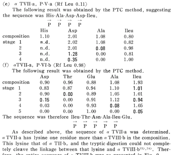
(e) a TVII-a, P-V-a (Rf Leu 0.11)
The following result was obtained by the PTC method, suggesting the sequence was His -Ala-Asp-Asp-Ileu .
P P P P
His Asp Ala Ileu
composition 1.10 2.01 1.08 0.80
stage 1 n.d. 2.02 1.08 0.82
2 n.d. 2.01 0.08 0.98
3 n.d. 1.28 0.010 0.81
4 n.d. 0.35 0.00 1.00
(f) aTVII-a, P-VI-b (Rf Leu 0'.98)
The followiug result was obtained by the PTC method.
Asp Thr Glu Ala Ileu
composition 0.90 0.96 -0.88 1.08 1.93
stage 1 0.83 0.87 0.94 1.10 1.01
2 0.90 0.00 0.89 1.05 1.101
3 0.15 0.00 0.91 1.12 0.94
4 0.03 0.00 0.93 0.08 1.05
5 0.00 0.00 1.00 0.00 0.05
The sequence was therefore Ileu-Thr-Asn-Ala-Ileu-Glu.
P P P P P
As described above, the sequence of a TVII-a was determined.
a TVII-a has lysine one residue more than a TVIII-b in the composition.
This lysine that of a TII-b, and the tryptic digestion could not comple- tely cleave the linkage between that lysine and a TVIII-b15,,24> Ther- fore, the entire sequence of a TVIII-b was as presented in Fig. 9.
i
Fig. 9. Amino acid sequence in the tryptic peptide, a TVIII-b.
(14) aTIX-a (Rf Leu 0.23)
This peptide was further digested with pepsin, and then the digest
was subjected to Dowex 1X2 column chromatography. As shown in
TUBE NUMBER
Fig. 10. Isolation of the peptic peptides from the tryptic peptide, aTIX-a.
Column: Dowex 1 x 2, 0.9 x 60cm.
PEAK NUMBDER IN FIG 10
Fig. 11. Paper chromatogram of the peptic peptides front the tryptic peptide, aTIX-a.
Fig. 10, six peak were obtained. The effluent at each peak was dried under reduced pressure and then purified by paper chromatography.
Five main spots were detected.
The amino acid composition of these peptide fragments is summarized in Table IV.
Table IV Amino acid compositions of the peptic peptides from the tryptic peptide, a TIX-a
Ib IIa IVa Vb VC
Lys
His 0.99
Arg 0.98 0.95
Cys Asp
Thr 0.84 0.88
Ser
Glu 1.03 1.09 1.81 1.15 1.05
Pro
Gly 1.08 1.13 1.13
Ala 2.11 1.12 1.05
Val Met
Ileu 0.96
Leu 1.09 1.05
Tyr 0.71 0.75
Phe
(a) a TIX-a, P-II-a (Rf Leu 0.59)
The following result was obtained by the PTC method, suggesting the sequence was Thr-Leu-Glu-Arg.
P P
Arg Thr Glu Leu
composition 0.95 0.84 1.09 1.05
stage 1 n.d. 0.00 1.04 0.96
2 n.d. 0.00 1.00 0.06
(b) a TIX-a, P-IV-a (Rf Leu 0.43)
This fragment was analyzed by the PTC method as follows.
His Glu Gly Ala Ileu
composition 0.99 1.81 1.08 2.11 0.96
stage 1 n.d. 2.05 1.04 1.90 0.00
2 n.d. 2.00 0.94 1.05 0.00
3 n.d. 2.01 0.26 0.96 0.00
4 n.d. 1.90 0.00 1.03 0.00
5 n.d. 2.00 0.00 0.00. 0.00
The sequence was determined to be Ileu-Ala-Gly-His-Ala-Glu-Glu, P P P P P (c) a TIX-a, P-V-b (Rf L eu 0.68)
Analysis by the PTC method gave the following result, suggesting the sequence was Tyr-Gly-Ala-Glu-Thr.
> >
P P P P
Thr Glu Gly Ala Tyr
composition 0.88 1.15 1.13 1.12 0.71
stage 1 0.82 1.08 1.05 1.04 0.00
2 0.83 1.11 0.00 1.05 0.00
3 0.92 1.08 0.00 0.00 0.00
4 1.00 0.08 0.00 0.00 0.00
By combining the above results from the peptide fragments toge- ther, the entire sequence of a TIX-a was determined as shown in Fig.
12.
Fig. 12. Amino acid sequence in the tryptic peptide, aTIX-a
8) Determination of the N-Terminal Residue of the Lpsoluble Tryptic Peptide by the DNP Method.
The N-terminal residue of the insoluble tryptic peptide was known to be leucine by the DNP method.
9) Column Chromatography, Paper Chrcrnatogi afhy, and Amino Acid Composition of Peptic Peptides from the Insoluble Tryptic Peptide.
Five peaks are observed in Fig. 13. The effluent at each peak was dried under reduced pressure and purified by paper chromatography.
As shown in Fig. 14, eight main spots were obtained. The amino acid composition of these spots is given in Table V.
10) Amino Acid Sequence of the Peptic Peptides from the Insoluble Tryptic Peptide
(1) P-I-b (Rf Leu 0.36)
TUBE NUMBER
Fig. 13. Isolation of the peptic peptides from the insoluble tryptic peptide
PEAK NUMBER IN FIG. 13
Fig. 14. Paper chromatogram of the peptic peptides from the insoluble tryptic peptide.
Table V Amino acid compositions of the peptic peptides from the insoluble tryptic peptide
lb Ha IIIa IIIb IIIc IIId I Va Va
Lys 0.88 1.01 1.01
His 1.12 1.03
Arg
Cys + +
Asp 1.01
Thr 0.96 0.96 0.83
Ser 0.96
Glu 0.89 1.08
Pro 0.97 1.05
Gly 1.05 1.06 1.17
Ala 2.03 1.04 2.03 1.16
Val 0.98 0.97 1.98 2.25 1
.96 Met
lieu
Leu 1.07 1.03 1.04 1.01 1.85 1.94 1.97
Tyr
Phe 0.90 0.89
Analysis by the PTC method resulted as follows.
Lys His Pro Ala Val Leu
composition 0.88 1.12 0.97 2.03 0.98 1.07 stage 1 0.98 1.02 0.95 2.08 0.99 0.08
2 0.96 1.03 1.07 1.01 0.92 0.00
3 0.98 1.03 0.18 1.00 0.93 0.0'0
4 0.10 0.98 0.00 1.00 0.92 0.00
5 0.00 0.99 0.00 1.00 0.10 0.00
6 0.00 0.15 0.00 1.00 0.010 0.00
The sequence was therefore determined to be Leu-Ala-Pro-Lys-Val-His-
Ala. P P P P P P
(2) P-II-a (Rf Leu 0.32)
The following result was obtained by the PTC method, :suggesting the sequence was Thr-Ala-Lys.
P P
Lys Thr Ala
composition 1.01 0.96 1.04
stage 1 n.d. 0.12 1.00
2 n.d. 0.00 0.15
(3) P-III-a (Rf Leu 0.37)
This peptide fragment was analyzed by the PTC method as follows.
His Glu Pro Ala Val Leu
composition 1.03 0.89 1.05 2.03 0.97 1.03
stage 1 n. d. 0.91 1.04 2.04 0.05 1.02
2 n.d. 0.92 1.05 1.03 0.00 1.01
3 n.d. 0.88 1.05 1.03 0.00 0.85
4 n.d. 0.95 0.91 1.02 0.00 0.10
5 n.d. 0.94 0.11 1.06 0.00 0.00
6 n.d. 1.00 0.00 0.16 0.00 0.00
The sequence was therefore determined to be Val-Ala-His-Leu-Pro-Ala-
--~- - --± - * --> ---).
Glu. P P P P P P
(4) P-III-b (Rf Leu 0.78)
The N-terminal amino acid was known to be serine by the DNP method, suggesting the sequence was Ser-Leu.
D (5) P-III-c (Rf Leu 0.86)
Analysis by the PTC method resulted as follows.
Thr Gly Val Leu
composition 0.96 1.05 1.98 1.01
stage 1 0.98 1.04 1.01 1.02
2 0.96 0.13 1.02 1.02
3 0.08 0.00 0.98 1.01
4 0.00 0.00 0.15 1.00
The sequence was therfore determined to be Val-Gly-Thr-Val-Leu P P P P (6) P-III-d (Rf Leu 1.00)
The following result was obtained by the PTC method.
Val Leu
composition 2.25 1.85
stage 1 2.03 0.97
2 1.06 0.94
3 0.18 1.00
The sequence was therefore Leu-Val-Val-Leu.
P P P (7) P-IV-a (Rf Leu 0.92)
Analysis by the PTC method resulted as follows.
Cys Glu Gly Leu Phe
composition + 1.08 1.06 1.94 0.90
stage 1 + 1.09 0.97 0.99 0.93
2 + 1.10 1.01 0.11 0.89
3 + 1.04 0.10 0.010 0.96
4 + 0.28 0.00 0.00 1.00
5 - 0-.00 0.00 '0.00 1.00
The sequence was therefore Leu-Leu-Gly-Gln-Cys-Phe.
> > >
P P P P P (8) P-V-a (Rf Leu 0.75)
Lys Cys Asp Thr Gly Ala Val Leu Phe composition 1.01 + 1.01 0.83 1.17 1.16 1.96 1.97 0.89 stage 1 n . d . + 0.05 0.90 1.15 1.10 1.98 1.98 0.91 2 n.d. + 0.00 0.89 1.16 1.09 1.99 2.01 0.92
3 n.d. + 0.00 0.91 1.13 1.09 1.98 2.00 0.08
4 n.d. + 0.00 0.91 1.10 1.11 1.97 1.01 0.00
5 n.d. - 0.00 0.88 1.08 1.06 1.98 1.02 0.00
6 n.d. - 0.00 0.90 1.06 0.10 1.98 1.02 0.00
7 n.d. - 0.00 0.91 1.05 0.00 1.03 1.01 0.00 The above result obtained by the PTC method suggests the sequence was Asp-Lys-Phe-Leu-Cys-Ala-Val-(Gly, Thr, Val, Leu).
P P P P P P P
Since Peptide P-III-c corresponds to the C-terminal portion of this fragment, the sequence of P-V-a was determined to be Asp-Lys-Phe- Leu-Cys-Ala-Val-Gly-Thr-Val-Leu.
By combining the above results together, the whole sequence of the insoluble tryptic peptide was determined as shown in Fig. 15.
Leu-Leu-GIy-GIn-Cys-Phe- Leu-Val-VaI-Leu-Val-Ala-His- Leu-Pro-Ala- Glu-Leu-Ala-Pro
PIV-a PIII-d P111-a
-~
Lys-Val-His-Ala-Ser-Leu-Asp-Lys-Phe-Leu-Cys-Ala-Val-Gly-Thr-Val-Leu-Thr-Ala-Lys
- PI-b P111-b P111-c PII-a
PV- a