下顎側方偏位における咀嚼運動パターンと大臼歯の歯軸傾斜の関連性について
日本大学大学院松戸歯学研究科歯学専攻
鈴木 雄士
(指導:葛西 一貴 教授)
1
[ABSTRACT]
【
Introduction
】A lateral deviation of the mandible leads to aesthetic problems and impaired mastication because it can result in a molar crossbite. Previous studies on participants with lateral deviations of the mandible have identified characteristic patterns of masticatory movement (e.g., reversed and crossover chewing patterns) that indicate that the masticatory movement exhibited by these individuals is asymmetric. In addition, a previous study reported that chewing movements after orthognathic surgery were similar to those in individuals with normal occlusion. Other reports indicated that chewing patterns are not changed before and after orthognathic surgery. Thus, no consistent findings have been obtained on how chewing movements are improved with orthognathic surgery. Therefore, the aim of the this study was to determine the change in chewing pattern after improvement in occlusion with orthognathic surgery and the relationship of improvement status in chewing pattern after treatment with relapse.
The present study was conducted in the following two parts;.in the study 1, to investigate the relationship between molar occlusion and chewing patterns in individuals with laterally deviated mandibles and to, and in the study 2, to investigate the relationship between changes of masticatory movement and changes of inclination of first molars was investigated in individuals after one year retention.
【
Materials and Methods
】The six degrees of freedom jaw-tracking system (Gnatho-Hexagraph III, GC, Tokyo Japan)was used
to measure the masticatory movement. The chewing pattern was classified as normal(N Type),
reversed(R Type), or crossover(C Type). And a three-dimensional digital scanner was used to obtain
3D data of Dental models, and to measure the axial inclination of the maxillary and mandibular first
molars.The study 1: 23 patients with mandibular deviation from the midline (4 mm or more) and
skeletal class I (0°≤ANB≤4°), were divided into two groups: normal bite (NB group: mean age:
2
19.5±4.5 years) and crossbite (CB group: mean age: 25.1±5.1 years). The percentage of chewing patterns on the affected side and the unaffected side were calculated. The chewing patterns were found to be normally distributed in each group. Student’s t-test was used to test for the significant differences between the groups for the width of the maxillary and mandibular dentition, palatal width, and axial inclination of the maxillary and mandibular first molars on the affected side and the unaffected side.
In the study 2: 21 participants (mean age: 23.2±5.4 years) with a menton deviation ≥ 4 mm from the midline. Changes in the masticatory movement patterns were measured from before treatment (T1) to after treatment (T2) and from T2 to after 1 year or more retention (T3). The changes in the masticatory movement patterns were analyzed using McNemar’s test. The differences in the axial inclination of the maxillary and mandibular first molars and width of the dentition between cases involving normal, crossover, and reversed chewing patterns were analyzed using Kruskal–Wallis and Mann–Whitney U tests.
【
Results
】1) In normal occlusion on the affected side, chowing pattern showed normal type, on the contrary, in crossbite on the affected side, chowing pattern showed crossover type and reverse type chowing patterns.
2) The width of the maxillary dentition and palatal width were significantly larger in normal bite group than those in crossbite group. Also the difference maxillary dentition and mandibular dentition was significantly larger in normal bite group than that in crossbite group.
3) In the inclination of mandibular molar axis on the affected side, normal bite group showed more lingually inclination than that crossbite group. There was no significant difference on unaffected side between two groups.
4) Six of the 15 participants with reversed chewing patterns on the affected side at T1 changed their
chewing patterns to normal chewing patterns by T2. There was a significant difference between T1
and T2 in the masticatory movement patterns on the affected sides. On the contrary, two of the 5
participants with reversed chewing patterns on the unaffected side at T1 changed their chewing
3
patterns to normal chewing patterns by T2. There were no significant differences in the patterns between T1 and T2 on the unaffected sides.
5) On the affected side between T2 and T3 , the changes in the inclination of the teeth and dental arch widths were significantly greater for patients with reversed chewing patterns compared to those with normal or crossover chewing patterns. Reversed chewing patterns showed a tendency to relapse.
【
Conclusion
】A normal chewing pattern may lead to lingual inclination of the mandibular molars on the affected
side, and it may be a factor that prevents the onset of crossbite. After orthognathic surgery, in the
reverse type chewing pattern, the change of the tooth axis was large and a tendency of relapse was
observed. These results suggested that the masticatory movement pattern might be associated with
the stability of occlusion after orthognathic surgery.
4
[
緒 言]
顔面非対称を呈する顎変形症には下顎側方偏位を有する者が
25%
存在する1)。下顎側 方偏位は上顎咬合平面の傾斜,下顎枝の長さや下顎骨体および下顎頭形態の左右差,臼 歯の鋏状咬合やcrossbite
などの問題を有している2~4)。下顎側方偏位に伴い偏位側臼歯部に
crossbite
がみられる症例が多いものの,crossbite
がみられない症例もみうけられる。Crossbite
は遺伝的および環境的な要因によって発症するが環境的要因の一つとして片咀嚼があり,咀嚼運動が関与している可能性があると考えられる5,6)。
Crossbite
における咀 嚼運動について,成人のcrossbite
では咀嚼運動パターンが逆サイクルになるという報告7~10)や,小児期の
crossbite
では咀嚼運動パターンが逆サイクルを示していたが,crossbite
改善後に咀嚼運動パターンが正常になったとの報告11)がみられる。また外科的矯正治療 においては,治療後に咀嚼運動が正常咬合者の咀嚼運動に類似していたとの報告12,13)が あるが,一方,術前後での咀嚼運動パターンが変化しない14)との報告もある。このよう に外科的矯正治療によって咀嚼運動がどのように改善するかについて,一定の知見が得 られていないのが現状である。治療が長期に及ぶ矯正歯科治療後に咬合が改善すること で咀嚼機能が改善する15~17)と言われているものの,短期間で咬合が顕著に変わる外科的 矯正治療では不明な点が多い。さらに,術前に大臼歯の歯軸を適正に改善した後に骨切 り手術を行うが,外科的矯正治療後の咀嚼運動パターンの変化が大臼歯歯軸へどの程度 影響しているか検討した報告は見られない。
そこで,外科的矯正治療による咬合改善後に咀嚼運動パターンがどのように変化し,
また,治療後の咀嚼運動パターンの変化が歯列の後戻りとどのように関連しているか明 らかにするため,研究
1
として,下顎側方偏位における大臼歯crossbite
の有無と咀嚼運 動および大臼歯歯軸傾斜の関連性について調査し,研究2
では外科的矯正治療前後と保 定開始1
年後の咀嚼運動パターンの変化と大臼歯歯軸傾斜の関係について検討した。5
【資料および方法】
1 .
被験者 研究1
被験者は,本学付属病院矯正歯科を受診した顎変形症患者
23
名である。選定条件は,正面・側面セファロによる計測結果から鶏冠と前鼻棘を結ぶ直線を正貌の正中線
(VRL)
か らMenton
が4mm
以上偏位を認めた下顎側方偏位2, 3),Skeletal Class I (0°
≦ANB
≦4)
とし た。前頭断において上下顎第一大臼歯が偏位側で正常咬合しているものをNB
群(14
名:男性
2
名,女性12
名,平均年齢19.5
歳±4.5)
,偏位側でcrossbite
を呈するものをCB
群(9
名:男性1
名,女性8
名,平均年齢25.1
歳±5.1)
とし,2
群に分類した。研究
2
被験者は,本学付属病院矯正歯科を受診した顎変形症患者のうち治療を終了し,保定開 始
1
年以上経過しているもの21
名(
男性5
名,女性16
名 平均年齢23.2
歳±5.4)
,であ る。選定条件として,術前正面・側面セファロによる計測結果から研究1
と同様にMenton
が4mm
以上偏位を認めた下顎側方偏位のうち2, 3),Skeletal Class I
およびIII (-2°
≦
ANB
≦4°)
を選択した。また,外科手術を執刀した術者は同一人物で行った症例とし た。研究
1
および2
の除外条件を下記に示す。① 顎関節の疼痛,開口障害などの顎関節症状が認められる。
② 歯科矯正治療の既往歴がある。
③ 臼歯部に補綴装置
(
ブリッジ,義歯およびインプラント)
を認める場合やカリエス,1
歯以上の欠損が認められる場合がある。④ 先天的奇形や症候群,外傷などの既往がある。
⑤ 機能性交叉咬合を認める。
なお,本研究は,日本大学松戸歯学部倫理委員会にて研究倫理の承認を得ている。
(
承認番号:研究1: EC16-14-042-1
,研究2: EC16-15-004-2)
6
2 .
方法1)
頭部エックス線規格写真研究
1, 2
において正面および側面頭部エックス線規格写真を通法に従い撮影し,分析方法は,
Steiner analysis
に準じて行った18~21)。なお,計測点,基準線ならびに計測項目はFigure 1
とFigure 2
に示した。2)
咀嚼運動の記録・分類咀嚼運動の計測は研究
1, 2
ともに3
次元6
自由度顎運動測定装置(
ナソヘキサグラフIII
,((
株)GC,
東京)
を用いた。被験者の下顎前歯部にクラッチを装着し,頭部を固定せず リラックスした状態でフランクフルト平面が水平になるように椅子に座らせた後,ヘッ ドフレームおよびフェイスボウを装着した。基準平面である両側外耳孔上縁および左側 眼窩下縁の3
点よりなるフランクフルト平面と両側下顎頭,下顎左右第一大臼歯近心頬 側咬頭と下顎左右中切歯を計測点とした22)。被験者には自由咀嚼を行わせて十分に軟性 ガムを軟化させた後,被験者に左右片側ずつ咬頭嵌合位からの軟性ガム咀嚼を30
秒間行 わせ,その咀嚼運動を記録した。なお,被験食品には軟性ガム(100%
キシリトールガ ム,(
株)
オーラルケア,東京:以下,軟性ガム) 1
粒(1.5g)
を使用した。咀嚼運動の解析 は,下顎切歯点における主咀嚼側の咀嚼開始第5
ストロークから第14
ストロークまでの 計10
ストロークを対象として9, 23),咀嚼運動経路形態について顎運動測定器付属の解析 ソフトを用いて咀嚼運動を解析した。 研究1, 2
において咀嚼運動パターンは,Normal
Type (N Type
:中心咬合位から咀嚼側あるいは非咀嚼側に向かって開口し,その後中心咬合位へ閉口するもの
)
,Cross Type (C Type
:開閉口路が交叉するもの)
,およびReverse Type (R Type
:開閉口路がNormal Type
と逆のもの)
の3
タイプに分類した24,25)(Figure 3)
。研究
2
では初診時(T1)
,矯正装置除去時(T2)
,保定開始1
年以上経過時(T3)
にて咀嚼運 動の計測を行い,各計測時期における咀嚼運動パターンの分類を行った。偏位側・非偏 位側におけるT1-T2
,T2-T3
での咀嚼運動パターンの変化については,N Type
とC Type
および
R Type
の変化を検定した。7
3)
口腔模型計測計測は,
Eguchi
ら26)の方法に基づき,計測項目をFigure 4
とFigure 5
に示した。研究1
では,矯正治療開始前の口腔模型にて,3D-
デジタルスキャナー(
マエストロ3D
オルソ システム,(
株)
安永社)
を用いて口腔模型を3D
データにし,得られた3D
データを3D
解析ソフト
BODY-RUGLE (Medic(
株))
を使用し,上顎歯列幅径(U6-6CW)
,下顎歯列幅径(L6-6CW)
,上顎第一大臼歯口蓋幅径(U6GW)
と上下顎第一大臼歯歯軸傾斜角を計測した26~29)
(Figure 4
,Figure 5)
。またT1
での偏位側・非偏位側において第一大臼歯頬舌的被蓋を確認し,正常に咬合しているものを
normal bite
,交叉咬合しているものをcrossbite
と した。研究
2
は研究1
と同様に上顎歯列幅径(U6-6CW)
,下顎歯列幅径(L6-6CW)
,上顎第一大 臼歯口蓋幅径(U6GW)
と上下顎第一大臼歯歯軸傾斜角を計測した27~30)。さらに,計測項目 ごとにT2
からT3
への変化量を算出し,各咀嚼運動パターンにて比較した。研究
1,2
ともに上顎基準平面は,切歯乳頭頂および左右側第二小臼歯と第一大臼歯の歯 間乳頭頂の3点を通る平面とした。なお,左右側第二小臼歯と第一大臼歯歯間乳頭頂を 通る直線をX
軸,X
軸と直交する直線をY
軸,基準平面上でX
軸とY
軸の交点と直交 する直線をZ
軸とした。下顎基準平面は,下顎左右側中切歯間乳頭頂,下顎左右側第二 乳臼歯(第二小臼歯)と第一大臼歯の歯冠乳頭頂の3
点を通る平面上とした。計測部位 は,第一大臼歯の頬側面の溝から咬合面の頬側溝への移行点と舌側面の溝から咬合面の 舌面溝への移行点2
点を通る直線がZ
軸となす角度を歯軸傾斜角とし偏位側大臼歯歯軸 傾斜角,非偏位側大臼歯歯軸傾斜角とした。研究
2
において計測項目ごとにT2-T3
における上下顎偏位側・非偏位側第一大臼歯歯 軸傾斜角から各咀嚼運動パターンでの変化量を算出し比較した。また,T2-T3
における 偏位側同一咀嚼運動パターンでの歯列幅径変化量(U6-6CW
,L6-6CW
,U6GW)
を絶対値と して算出した。変化量は計測項目ごとにT3
からT2
を引いた数値を変化量とした。8
4)
統計方法研究
1
では,偏位側,非偏位側の咀嚼運動パターンをType
別に発現頻度(
%)
を算出し た。各群は,正規分布を認め2
群間における上下顎歯列幅径,上顎第一大臼歯口蓋幅 径,上下偏位側・非偏位側第一大臼歯歯軸傾斜角についてStudent t-test
による有意差検 定を行った。研究
2
では,T1-T2
,T2-T3
の咀嚼運動パターンの変化と変化割合(%)
を算出した(Figure 6(a)
,(b))
。T1-T2
,T2-T3
の咀嚼運動パターンの変化は,McNemar’s test
による有 意差検定を行った。N Type
,C Type
,R Type
の3
群間でのT2-T3
における同一咀嚼タイ プによる偏位側上下顎大臼歯歯軸変化量,上顎第一大臼歯口蓋幅径変化量,上下顎歯列 幅径変化量についてBonferroni
によるKruskal–Wallis
とMann–Whitney U tests
の有意差検 定を行った。統計分析ソフトはIBM
○RSPSS
○RStatistics 21(SPSS-IBM
社,MD
,米国)
を用 い,有意水準は5%
とした。9
【結 果】
研究
1
1)
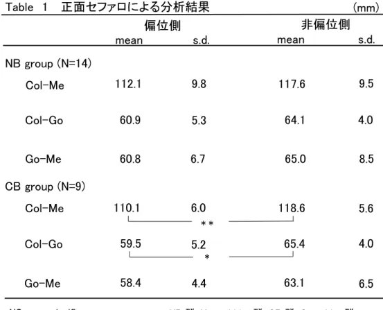
正面セファロによる計測結果(Table 1)
正面セファロにより
Me
偏位量は,NB
群で7.3mm±2.2mm
,CB
群では8.9mm±4.2mm
であり差は認められなかった。また,Occlusal plane angle
も同様に差は認められなかった。
NB
群のCol-Me, Col-Go, Go-Me
において偏位側より非偏位側の方が値は大きいものの有意差はみられなかった。一方,
CB
群では,非偏位側のCol-Me, Col-Go
が偏位側よ り有意に大きかった。2)
咀嚼運動パターンの結果(Table 2)
NB
群の偏位側ではN Type
が100%
,非偏位側ではN Type
が92.8%
,R Type
が7.2%
で
1
症例のみ認められた。C Type
は偏位側,非偏位側ともに0%
であった。一方,CB
群 の偏位側ではN Type
が0%
,R Type
は55.6%
,C Type
は44.4%
であり,非偏位側ではN Type
が55.6%
,R Type
は33.3%
,C Type
は11.1%
であった。3)
上下顎歯列幅径と上顎第一大臼歯口蓋幅径および上下顎第一大臼歯歯軸傾斜角の比較(Table 3)
上顎歯列幅径
(U6-6CW)
および上顎第一大臼歯口蓋幅径(U6GW)
はNB
群がCB
群より 大きい値を示し,上下顎歯列幅径較差(U6-L6CW)
ではNB
群がCB
群より有意に大きか った。下顎歯列幅径では差は認められなかった。偏位側上顎大臼歯歯軸傾斜角において は差が認められなかったが,非偏位側上顎大臼歯歯軸傾斜角ではCB
群はNB
群に比べ 有意に小さい値を示した。偏位側下顎大臼歯歯軸傾斜角においては,NB
群はCB
群に 比べ有意に小さい値を示したが,非偏位側下顎大臼歯歯軸傾斜角では差は認められなか った。10
研究
2
1)
正面セファロによる計測結果正面セファロにより
Me
偏位量は7.8mm±3.1mm
,Occlusal plane angle
は2.4mm±1.4mm
であった。2) T1-T2
,T2-T3
における咀嚼運動パターンの変化について(Figure 6(a))
偏位側
T1-T2
において,[C Type
およびR Type]
は[N Type]
へ変化した比率は15
症例中6
症例であり,9
症例は[C Type
およびR Type]
のままであった。Mcnemar
検定の結果,偏位側
T1-T2
における変化は有意であった(p = 0.03)
。非偏位側T1-T2
では[C Type
およ びR Type]
から[N Type]
へ変化した比率は5
症例中2
症例であり,3
症例は[C Type
および
R Type]
のままで有意な変化を認めなかった。また偏位側・非偏位側ともにT2-T3
においての咀嚼運動パターンの変化はごく少数であり有意な変化を認めなかった。
3)
偏位側・非偏位側における咀嚼運動パターンの変化(Figure 6(b))
T1-T2
およびT2-T3
における咀嚼運動パターンの変化とその割合(%)
を(Fig.6)
に示す。これらの咀嚼運動パターンの変化を
Type
別に検討した結果,偏位側のT1
における咀嚼 運動パターンは,21
名中6
名(28.5%)
にN Type
を認め,C Type
は3
名(14.2%)
,R Type
は12
名(57.1%)
認めた。そのうち,crossbite
を認めたのはN Type 2
名,C Type 1
名,R Type 7
名であった。T2
における咀嚼運動パターンは,N Type
が12
名(57.1%)
,C Type
が3
名(14.2%)
,R Type
が5
名(28.5%)
であった。また,T3
ではN Type
は12
名(57.1%)
,C Type
は4
名(19.0%)
,R Type
は5
名(23.8%)
認めた。偏位側
T1-T2
における変化では,N Type
において全く変化を認めなかったが,C Type
では
3
名中2
名(
⑤,
⑱)
がR Type
となり,1
名は変化しなかった。R Type
では,12
名中6
名(
❷,
④,
⑦,
⑨,
⑩,
⓱)
がN Type
となり,2
名(
⓭,
⓰)
がC Type
,4
名が変化せずにR Type
のままであった。偏位側T2-T3
における変化では,N Type
とC Type
は変化を認めな かったが,R Type
は6
名中5
名がR Type
のままであり,1
名(
⑱)
がC Type
へ変化した。非偏位側において
T1
は21
名中16
名(76.1%)
がN Type
を認め,C Type
は2
名(9.5%)
,R
Type
は3
名(14.2%)
認めた。T2
ではN Type
は15
名(71.4%)
,C Type
は3
名(14.2%)
,R
11
Type
は3
名(14.2%)
認めた。また,T3
ではN Type
は17
名(80.9%)
,C Type
は2
名(9.5%)
,R Type
は2
名(9.5%)
認めた。非偏位側T1-T2
における変動ではN Type
では16
名中13
名,C Type
では2
名中2
名が変化しなかったが,R Type
は2
名(
④,
⑥)
がN Type
へ変化 し,1
名は変化しなかった。非偏位側T2-T3
における変動では,100% N Type
のままであ り,C Type
では3
名中2
名がそのままであり,1
名(
⑫)
がN Type
へ変化した。R Type
は3
名中2
名が変化せずにR Type
のままであり,1
名(
⓴)
がN Type
となった。以上のことから,偏位側
T1-T2
における変化は,C Type
およびR Type
がN Type
へと 有意な変化を認めたが非偏位側T1-T2
および偏位側・非偏位側T1-T2
,T2-T3
は有意な変 化はなく咀嚼運動パターンは変化しないことを認めた。4) T2-T3
における大臼歯歯軸変化量(Figure 7(a)
,(b))
N Type
,C Type
,R Type
の3
群での有意差検定を行った結果,偏位側では上下顎ともに大臼歯歯軸変化量は
R Type
がN Type
およびC Type
より有意に大きい値を認め,非偏 位側では上下顎ともに大臼歯歯軸変化量に有意差を認めなかった。5) T2-T3
における歯列幅径変化量(Figure 8)
N Type
,C Type
,R Type
の3
群での有意差検定を行った結果,U6-6CW
,L6-6CW
ならびに
U6GW
においてR Type
がN Type
およびC Type
より有意に大きい値を示した。12
【考 察】
1.
大臼歯咬合と咀嚼運動の関連性について(
研究1)
不正咬合における咀嚼運動パターンについて,
YANO
ら6),Proeschel
ら 31)の報告は全体の約
80%
以上がN Type
,C Type
ならびにR Type
の3
つの咀嚼運動パターンであり,詳細に検討した
Nie
ら9)は8
パターンに分類している。本研究では咀嚼運動パターンの特徴をよ り明瞭に分類するために代表的なN Type
,C Type
,R Type
の咀嚼運動パターンに分類し た。N Type
は大臼歯正常被蓋の偏位側,非偏位側において認められることが多く,研究1
において
NB
群の咀嚼運動パターンでは,ほぼN Type
を認めた。Jemt
ら32)は,骨格性,歯性 ともに正常咬合での咀嚼運動パターンは,規則的で安定しているとし,3
つの咀嚼運動パタ ーンの中でN Type
は最も安定していると考えられる。C Type
は咀嚼運動中に開閉口路が交叉するものである。本研究1
の結果からCB
群のみに
C Type
を認めた。過去に咀嚼運動中に開閉口路が交叉するものは側方運動干渉時や顎関節症患者に認められる 33,34)と報告がある。また,下顎側方偏位は顎関節症を生じやすいと いった報告3)もある。本研究の被験者は開口障害や疼痛などを認めない下顎側方偏位である が,下顎側方偏位であるため開口障害や疼痛など顎関節症状がなくとも側方運動干渉時に 顎運動として
C Type
が認められた可能性があると考えられる。R Type
は開閉口路がN Type
と逆のものを示し,臼歯crossbite
になりやすい下顎側方偏位や下顎前突でよく認められる。本研究
1
の結果より,CB
群の咀嚼運動パターンは偏位側に おいてN Type
を認めず,R Type(55.6%,)
,C Type(44.4%)
認めた。また非偏位側はN Type(55.6%)
も認めるがR Type(33.3%)
,C Type(11.1%)
であった。中南ら6)は,臼歯部crossbite
の症例において
R Type
が高率で出現する傾向にあるとし,Tomonari
ら7)は臼歯部被蓋と咀嚼運動との関係について,
crossbite
の咀嚼運動パターンとして開口路と閉口路が逆転していると述べ ている。また大臼歯crossbite
を認めないケースでも前歯部や小臼歯部にもcrossbite
があることで
R Type
を生じるとの報告7)がある。このことから,臼歯部crossbite
により偏位側咀嚼時に臼歯部による咬頭干渉が生じることで咀嚼運動路が制限され,
N Type
と比較して咀13
嚼経路幅が狭い
Re Type
とC Type
が出現したと考えられた。上下顎大臼歯歯軸傾斜角では,非偏位側上顎大臼歯歯軸傾斜角において
CB
群はNB
群よ り有意に小さい値を認め舌側傾斜がみられ,上下顎歯列幅径においてNB
群はCB
群より上 顎歯列幅径,上顎第一大臼歯口蓋幅径,上下顎歯列幅径較差が有意に大きい値となった。Figure 9
に示すように,上顎大臼歯歯軸傾斜角の変化に付随してNB
群とCB
群の上顎歯列幅径,上顎第一大臼歯口蓋幅径に差が生じたと考えられる。また,偏位側下顎大臼歯歯軸傾 斜角において
NB
群はCB
群より有意に小さい値を認めたことからCB
群に比較してNB
群 の偏位側下顎大臼歯は舌側傾斜をしていることがわかった。また,顎骨の偏位に対する歯牙補償機構 35)の観点から,顔面非対称者における口腔内圧 力と下顎第一大臼歯の傾斜との間に有意な正の相関があり,下顎歯列の非対称および歯牙 補償機構に関連していること 36)や歯牙補償機構は顎骨の前後的不調和に生じるだけではな く,水平的不調和に対しても生じ 35),被蓋を維持することに関わっている 37,38)と考えられ ている。
2)
外科的矯正治療後の咀嚼運動と大臼歯歯軸傾斜について(
研究2)
外科的矯正治療後咀嚼運動パターンの変化において,偏位側
T1-T2
では有意な咀嚼運動 パターンの変化を認めたが,非偏位側では変化は認められなかった。これにより,T2-T3
すなわち保定期間において咀嚼運動パターンは変化しないことが明らかになった。咬合の改善による咀嚼運動パターンの変化において,小児期の
crossbite
を改善した後,逆サイクルが改善した報告10,39) や
Skeletal Class
Ⅲにおける外科的矯正治療前後ではR Type
が術後減少したとの報告がある40)。このことからT1-T2
にかけて咀嚼運動パターン に変化が生じる原因として骨切り手術終了後,偏位側・非偏位側ともに臼歯被蓋関係は正 常になることから,咬合接触の変化に伴い咬合干渉などが軽減することで咀嚼運動パター ンが改善したと考えられる。一方,非偏位側
R Type
のT1-T2
では33%
において変化を認めず,同様に非偏位側C
Type
でもT1-T2
にかけて変化はみられなかった。既報告によれば咀嚼運動パターンは,橋14
と延髄に位置する
centaral pattern generator(CPG)
により生成され41),成人の場合ではCPG
が確立されてから変化することが少ないと報告42)されており,動物においても咀嚼サイク ルの形状は大部分が不変のままであると報告されている43,44)。従って,T1-T2
にて咬合接 触の変化が生じても,CPG
が術前の状態のまま維持されており,咀嚼運動パターンの変化 は認められなかった可能性がある。T2-T3
大臼歯歯軸変化量において,N Type
,C Type
,R Type
の偏位側上下顎大臼歯歯軸変化量を比較した結果,
R Type
が有意な大きい値を認めた。また。上下顎歯列幅径変化量 および上顎第一大臼歯口蓋幅径においてもN Type
,C Type
と比較しR Type
は有意な大き い値を認めた。従って,保定期間においてR Type
は歯軸の傾斜の変化に伴い歯列幅が変 化しており,術前の状態に戻るいわゆる後戻り傾向が示唆された。R Type
はN Type
,C Type
と異なり咬合接触時に加わる咬合力の方向が逆であり,咬合の安定が得られにくいことが考えられる。このように咀嚼運動と歯軸傾斜の変化は密接に関 係していた。外科的矯正治療による咀嚼運動パターンの変化は偏位側のみ認められ、
R Type
から
N Type
へと変化し,非偏位側の変化は認められなかった。また保定後の咀嚼運動パターンの変化は偏位側・非偏位側ともに変化しないことが明らかになり,外科的矯正治療後に おいても咀嚼運動パターンが
R Type
の状態であると後戻り傾向が示唆された。15
【結 論】
下顎側方偏位における大臼歯
crossbite
の有無と咀嚼運動および大臼歯歯軸傾斜の関連 性ならびに外科的矯正治療後における咀嚼運動パターンの変化と大臼歯歯軸傾斜の関係 ついて検討した結果,以下の結論を得た。1)
下顎側方偏位において大臼歯が正常咬合の場合,咀嚼運動パターンはN Type
であっ た。一方,大臼歯がcrossbite
の場合では咀嚼運動パターンは,C Type
およびR Type
であった。2)
上顎歯列幅径および上顎第一大臼歯口蓋幅径はNB
群がCB
群より有意に大きい値を 示し,上下顎歯列幅径較差もNB
群がCB
群より有意に大きかった。3)
偏位側下顎大臼歯歯軸傾斜角は,NB
群はCB
群に比べ有意に小さく舌側傾斜していた が,非偏位側下顎大臼歯歯軸傾斜角では差は認められなかった。4) T1-T2
の偏位側において,C Type
およびR Type
からN Type
へ変化したものは15
症 例中6
症例であり変化は有意(p = 0.03)
であったが,非偏位側では5
症例中2
症例で有 意な変化を認めなかった。また偏位側・非偏位側ともにT2-T3
において咀嚼運動パタ ーンの有意な変化を認めなかった。5) T2-T3
の大臼歯歯軸変化量において,R Type
の偏位側上下顎大臼歯歯軸変化量はN Type
および
C Type
より有意に大きく,また歯列幅径変化量も同様に有意に大きい値を認め,R Type
は後戻り傾向が示唆された。以上のことから,下顎側方偏位において
N Type
咀嚼運動は偏位側下顎大臼歯を舌側傾斜させ
crossbite
の発症を防ぐ要因となること。また外科的矯正治療後において,R Type
咀嚼運動では大臼歯歯軸変化量が大きく後戻り傾向がみられ,下顎側方偏位における外科 的矯正治療後の咬合の安定性に咀嚼運動が関与していることが示唆された。
16
【文 献】
1. Severt TR
,Proffit WR
:The Prevalence of Facial Asymmetry in the Dentofacial Deformities Population at the University of North Carolina
,Int J Adult OrthodOrthognath Surg,12:171-176,1997.
2. Uesugi S
,Yonemitsu I
,Kokai S
,et al
:Features in Subjects with the Frontal Occlusal Plane Inclined Toward the Contralateral Side of the Mandibular Deviation
,Am J OrthodDentofacial Orthop,149:46-54,2016.
3.
山田恭平,大森浩子,桐本博章ほか:下顎側方偏位を伴う顎変形症患者における顎 関節雑音と下顎頭形態との関連,Orthod Waves Jpn Ed,75:1-7,2016.4. Takeshita N
,Ishidad M
,Watanabe H
,et al
:Improvement of Asymmetric Stomatognathic Functions, Unilateral Crossbite, and Facial Esthetics in a Patient with Skeletal Class III Malocclusion and Mandibular Asymmetry
,Treated With Orthognathic Surgery
,Am JOrthod Dentofacial Orthop,144:441-454,2013.
5.
佐藤貞雄,高木建雄,後藤幹子ほか:下顎骨の側方偏位を伴う骨格性交叉咬合の成立 機序に関する考察,神奈川歯学,25,93-98,1990.6.
矢野圭介,久保田雅人,篠原親ほか:顎の非対称を伴う骨格性下顎前突症における 下顎頭長軸角と習慣性咀嚼との関連性について,顎変形誌,102:110-116,2000.7.
中南国史,西尾公一,宮内修平ほか:臼歯部交叉咬合が顎口腔機能に及ぼす影響に 関する研究,顎機能,6:87-96,1988.8. Tomonari H
,Ikemori T
,Kubota T
,et al
:First Molar Cross-bite is More Closely Associated with a Reverse Chewing Cycle Than Anterior or Pre-molar Cross-bite During Mastication
,J Oral Rehabil,41:890-896,2014.9. Nie Q
,Kanno Z
,Xu T
,et al
:Clinical Study of Frontal Chewing Patterns in Various Crossbite Malocclusions
,Am J Orthod Dentofacial Orthop,138:323-329,2010.10. Riloa B
,Silvaa JL
,Moraa MJ
,et al
:Unilateral Posterior Crossbite and Mastication
,Archives of Oral Biology,52:474-478,2007.
17
11. Kwak YY
,Jang I
,Choi DS
,et al
:Functional Evaluation of Orthopedic and Orthodontic Treatment in a Patient with Unilateral Posterior Crossbite and Facial Asymmetry
,Korean JOrthod,44:143-153,2014.
12. Wang D
,Fu H
,Zeng R
,et al
:Changes of Mandibular Movement Tracings after the Correction of Mandibular Protrusion by Bilateral Sagittal Split Ramus Osteotomy
,J OralMaxillofac Surg,67:2238-2244,2009.
13. Ko EWC
,Huang CS
,Lo LJ
,et al
:Longitudinal Observation of Mandibular Motion Pattern in Patients with Skeletal Class III Malocclusion Subsequent to Orthognathic Surgery
,J Oral Maxillofac Surg,70:e158-e168,2012.14.
橋本志織,菅原準二,友寄裕子ほか:下顎非対称を伴う骨格性下顎前突症の外科的 矯正治療後における顎顔面形態および咀嚼運動経路の評価,東北矯歯誌,12:3-12,2004.
15. Nagai I
,Tanaka N
,Noguchi M
,et al
:Changes in Occlusal State of Patients with Mandibular Prognathism after Orthognathic Surgery : A Pilot Study
,Br J Oral Maxillofac Surg,39:429-433,2001.
16. Ohkura K
,Harada K
,Morishima S
,et al
:Changes in Bite Force and Occlusal Contact Area after Orthognathic Surgery for Correction of Mandibular Prognathism
,Oral Surg Oral MedOral Pathol,91:141-145,2001.
17. Iwase M
,Ohashi M
,Tachibana H
,et al
:Bite Force, Occlusal Contact Area and Masticatory Efficiency Before and After Orthognathic Surgical Correction of Mandibular Prognathism
,JOral Maxillofac Surg,35:1102-1107,2006.
18. Proffit WR
; 高田健治,北井則行,山城隆(訳):新版プロフィトの現代歯科矯正学,
175-177
,クインテッセンス出版,東京,2004
;Contemporary orthodontics
,3nd
ed
,Mosby
,Canada
,2000.
18
19. Damstra J
,Fourie Z
,Ren Y
:Evaluation and Comparison of Postero-Anterior Cephalograms and Cone-Beam Computed Tomography Images for the Detection of Mandibular Asymmetry
,Eur J Orthod,35:45-50,2013.20. Janson G
,Lima KJRS
,Woodside DG
,et al
:Class II Subdivision Malocclusion Types and Evaluation of Their Asymmetries
,Am J Orthod Dentofacial Orthop,131:57-65, 2007.
21. Masuoka N
,Muramatsu A
,Ariji Y
,et al
:Discriminative Thresholds of Cephalometric Indexes in the Subjective Evaluation of Facial Asymmetry
,Am J Orthod DentofacialOrthop,131:609-613,2007.
22. Hayashi R
,Kawamura A
,Kasai K
:Relationship Between Masticatory Function, Dental Arch Width, and Bucco-Lingual Inclination of the First Molars
,Orthod Waves,65:120-126,2006.
23. Shiga H
,Kobayashi Y
,Arakawa I
,et al
:Relationship Between Pattern of Masticatory Path and State of Lateral Occlusal Contact
,J Oral Rehabil,36:250-256,2009.24.
秋山仁志,志賀博,小林義典:咀嚼運動の機能的分析-
正常咬合者における前頭面 でみた切歯点の経路のパターン,補綴誌,35:609-621,1991.25. Proschel P
,Hofmann M
:Frontal Chewing Patterns of the Incisor Point and Their Dependence on Resistance of Food and Type of Occlusion
,J Prosthet Dent,59:617-624,1988.26. Eguchi S
,Townsend GC
,Togy H
,et al
:Genetic and Environmental Contributions to Variation in the Inclination of Human Mandibular Molar
,Orthod Waves,63:95-100,2004.
27. Oliveira NL
,Silveira ACD
,Kusnoto B
,et al
:Three-Dimensional Assessment of the
Maxilla: A Comparison of 2 Kinds of Palatal Expanders
,Am J Orthod DentofacialOrthop,126:354-362,2004.
19
28. Veli I
,Yuksel B
,Uysal T
,et al
:Longitudinal Evaluation of Dental Arch Asymmetry in Class II Subdivision Malocclusion with 3-Dimensional Digital Models
,Am J OrthodDentofacial Orthop,145:763-770,2014.
29.
岡野 美紀:第一大臼歯の頬舌的歯軸傾斜と歯列幅の成長パターンについて,日本人 と南太平洋諸国の2
集団との比較,Orthod Waves Jpn Ed,65:112-121,2006.30. Langberg BJ
,Arai K
,Miner RM
:Transverse Skeletal and Dental Asymmetry in Adults with Unilateral Posterior Crossbite
,Am J Orthod Dentofacial Orthop,127:6-16,2005.
31. Proeschel PA
:Chewing Patterns in Subjets with Normal Occlusion and with Malocclusions
,Semin Orthod,12:138-149,2006.32. Jemt T
,Hedegard B
:Reproducibility of Chewing Rhythm and of Mandibular Displacements During Chewing
,J Oral Rehabil,9:531-537,1982.33.
伊藤陽介,志賀博,児玉秀夫ほか:実験的咬合干渉が咀嚼運動に及ぼす影響,第6
報前頭面における干渉付与前後の咀嚼パターンの変化,顎機能,7:165-170,1989.34. Kuwahara T
,Miyauchi S
,Maruyama T
:Characteristics of Condylar Movements During Mastication in Stomatognathic Dysfunction
,Int J Prosthodont,Nov/Dec 3, 555-566,1990.
35.
阿部朗子,玉置幸雄,尾崎正雄ほか:顎態模型の三次元形状計測による下顎側方偏 位症例における歯系の補償の検討,Orthod Waves Jpn Ed,70:97-110,2011.36. Takada J
,Ono T
,Miyamato J
,et al
:Association Between Intraoral Pressure and Molar Position and Inclination in Subject with Facial Asymmetry
,Eur J Orthod,33:243-249, 2010.
37. Ishikawa H
,Nakamura S
,Iwasaki H
,et al
:Dentoalveolar Compensation Related to Variations in Sagittal Jaw Relationships
,Angle Orthod,69:534-538,1999.38. Ishikawa H
,Nakamura S
,Iwasaki H
,et al. Dentoalveolar Compensation in Negative
Overjet Cases
,Angle Orthod,70:145-148,2000.20
39. Piancinoa MG
,Fallab D
,Merloc A
,et al
:Effects of Therapy on Masseter Activity and Chewing Kinematics in Patients with Unilateral Posterior Crossbite
,Arch of Oral Biol,67:61-67,2016.
40. Piancino MG
,Frongia G
,Dalessandri D
,et al
:Reverse Cycle Chewing Before and After Orthodontic-Surgical Correction in Class III Patients
,Oral Surg Oral Med Oral Pathol,115:328-331,2013.
41. Lund JP
,Kolta A
:Generation of the Central Masticatory Pattern and its Modification by Sensory Feedback
,Dysphagia,21:167-174,2006.42. Gibbs CH
,Wickwire NA
,Jacobson AP
,et al
:Comparison of Typical Chewing Patterns in Normal Children and Adults
,JADA,105:33-42,1982.43. Byrd KE
,Luschei ES
:Cerebellar Ablation and Mastication in the Guinea Pig (Cavia Porcellus)
,Brain Res,197:577-581,1980.
44. Huang X
,Zhang G
,Herring SW
:Alterations of Muscle Activities and Jaw Movements after
Blocking Individual Jaw-Closing Muscles in the Miniature Pig
,Arch Oral Biol,38:291-297,1993.
21
【図および表】
22
Table 1
正面セファロによる分析結果(mm)
Go-Me
NS : no significant
*: p < 0.05
**: p < 0.01
Col-Me
Go-Me Col-Go NB group (N=14)
CB group (N=9) Col-Me Col-Go
58.4 4.4 63.1 6.5
NB群:Normal bite群, CB群:Crossbite群 Col-Me:ColとMeの距離
Col-Go:ColとGoの距離 Go-Me:GoとMeの距離
mean s.d.
112.1 60.9
9.8 5.3
60.8 6.7
偏位側
110.1 59.5
6.0 5.2
mean s.d.
117.6 64.1
9.5 4.0
65.0 8.5
非偏位側
118.6 65.4
5.6 4.0
* *
*
R Type C Type N Type
7.2 0 100
Normal bite
群 (N=14)92.8
0 0
(14/14) (13/14)
(0/14) (1/14) (0/14) (0/14)
偏位側 非偏位側(%) Crossbite
群 (N=9)0 55.6
55.6 33.3
44.4 11.1
(0/9) (5/9)
(5/9) (3/9)
(4/9) (1/9)
偏位側 非偏位側
N Type: Normal Type NB群:Normal bite群, CB群:Crossbite群 C Type: Cross Type
R Type: Reverse Type
Table 2
咀嚼運動パターンの結果23
Table 3
上下顎第一大臼歯歯列幅径と上顎第一大臼歯口蓋幅
径および上下顎第一大臼歯歯軸傾斜角の比較
*: p < 0.05
**: p < 0.01
53.3 3.5
mean s.d.
45.8 3.0
7.5 2.8
36.9 2.9
96.0 4.8
90.8 6.1
64.6 5.0
74.1 8.1
50.0 3.7
46.1 2.6
3.9 2.8
33.6 3.3
mean s.d.
98.6 4.0
86.0 2.9
71.0 5.4
70.8 6.5
(U6-6CW)
(U6-L6CW) (U6GW)
角度(degree)
(L6-6CW)
距離計測(mm)NS : no significant
偏位側上顎大臼歯歯軸傾斜角 非偏位側上顎大臼歯歯軸傾斜角
偏位側下顎大臼歯歯軸傾斜角 非偏位側下顎大臼歯歯軸傾斜角 上顎歯列幅径
上顎第一大臼歯 口蓋幅径
下顎歯列幅径
上下顎歯列幅径較差
Normal bite
群 (N=14)Crossbite
群 (N=9)*
* *
* *
*
*
)LJXUHṇ㠃ࢭࣇࣟࡢィ Ⅼ࠾ࡼࡧᇶ‽⥺
/2/DWHUR2UELWDOH5LJKWVLGH /2̓/DWHUR2UELWDOH/HIWVLGH
$16DQWHULRUQDVDOVSLQH *2̓㠀೫ഃ *RQLRQ
*2೫ഃ *RQLRQ0H0HQWRQ
02೫ഃ ILUVWPRODU 0R̓㠀೫ഃ ILUVWPRODU
1&㭜ෙ㢕㒊ࡢ᭱⊃✽㒊95/1& ࢆ㏻ࡾỈᖹᇶ‽⥺┤⾜ࡍࡿ┤⥺
;D[LVỈᖹᇶ‽⥺/2/2̓ 0H ೫㔞95/ 0H ࡢ㊥㞳 2FFOXVDOSO0R0R̓ ;D[LV ࡢ࡞ࡍゅᗘ
)LJXUHṇ㠃ࢭࣇࣟࡢィ 㡯┠
&RO*R&RO *2 ࡢ㊥㞳&RO̓*R̓&RO̓ *2̓ࡢ㊥㞳
*20H*2 0H ࡢ㊥㞳*2̓0H*2̓ 0H ࡢ㊥㞳
&RO0H&RO 0H ࡢ㊥㞳&RO̓0H&RO̓ 0H ࡢ㊥㞳
)LJXUHᄮ㐠ືࣃࢱ࣮ࣥࡢศ㢮
17\SH୰ᚰညྜࡽᄮഃ࠶ࡿ࠸ࡣ㠀ᄮഃྥࡗ࡚㛤ཱྀࡋ㸪 ࡑࡢᚋ୰ᚰညྜ㛢ཱྀࡍࡿ㐠ືࣃࢱ࣮ࣥ
&7\SH㛤㛢ཱྀ㊰ࡀཫࡍࡿ㐠ືࣃࢱ࣮ࣥ
57\SH㛤㛢ཱྀ㊰ࡀ 1RUPDO7\SH㏫ࡢ㐠ືࣃࢱ࣮ࣥ
&HQWULFRFFOXVLRQ&2
)LJXUHୖୗ㢡ṑิ࠾ࡅࡿィ 㡯┠
ୖ㢡ᕥྑ➨୍⮻ṑ㏆ᚰ㢋ഃည㢌㡬㛫㊥㞳8&:
ୖ㢡ᕥྑ➨୍⮻ṑ⯉㠃⁁┤ୗࡢṑ㢁㒊᭱῝Ⅼ㛫㊥㞳8*:
ୗ㢡ᕥྑ➨୍⮻ṑ㏆ᚰ㢋ഃည㢌㡬㛫㊥㞳/&:
)LJXUHཱྀ⭍ᶍᆺࡢィ 㡯┠
➨୍⮻ṑṑ㍈ഴᩳゅࡣୖୗ㢡➨୍⮻ṑࡢ㢋⯉ഃ㠃ࡢ
⁁ࡽညྜ㠃ࡢ㢋㠃⁁ࡢ⛣⾜Ⅼ⯉ഃ㠃ࡢ⁁ࡽညྜ㠃 ࡢ⯉㠃⁁ࡢ⛣⾜Ⅼࡢ Ⅼࢆ㏻ࡿ┤⥺ࡀ = ㍈࡞ࡍゅᗘࠋ
)LJXUHD77㸪77 ࠾ࡅࡿᄮ㐠ືࣃࢱ࣮ࣥࡢኚ
7ึデ7▹ṇ⨨㝖ཤ7ಖᐃ㛤ጞ ᖺ௨ୖ⤒㐣
۵ึデQRUPDOELWH ەึデFURVVELWH
)LJXUHEྛ FDVH ࡢ೫ഃ࣭㠀೫ഃ࠾ࡅࡿᄮࢱࣉࡢኚྜ㸦㸧
7ึデ7▹ṇ⨨㝖ཤ7ಖᐃ㛤ጞ ᖺ௨ୖ⤒㐣
۵ึデQRUPDOELWH ەึデFURVVELWH
)LJXUHD ೫ഃ࣭➨୍⮻ṑṑ㍈ഴᩳゅࡢ 77 ࡢኚ㔞
೫ഃ࡛ࡣୖୗ㢡ࡶ⮻ṑṑ㍈ኚ㔞ࡣ 5 7\SH ࡀ 1 7\SH ࠾ࡼࡧ & 7\SH ࡼࡾ
᭷ពࡁ࠸್ࢆㄆࡵࡓࠋ
)LJXUHE 㠀೫ഃ࣭➨୍⮻ṑṑ㍈ഴᩳゅࡢ 77 ࡢኚ㔞
㠀೫ഃ࡛ࡣୖୗ㢡ࡶ⮻ṑṑ㍈ኚ㔞᭷ពᕪࢆㄆࡵ࡞ࡗࡓࠋ
)LJXUH ୖୗ㢡ṑิᖜᚄኚ㔞ࡢ 77 ࡢኚ㔞
8&:㸪8*: ࡞ࡽࡧ /&: ࠾࠸࡚ 57\SH ࡀ 17\SH ࠾ࡼࡧ &7\SH
ࡼࡾ᭷ពࡁ࠸್ࢆ♧ࡋࡓࠋ
)LJXUH ⮻ṑṑ㍈ഴᩳ
೫ഃୗ㢡⮻ṑ⯉ഃഴᩳ㸪㠀೫ഃୖ㢡⮻ṑ⯉ഃഴᩳࢆㄆࡵࡓࠋ