Functional analysis of DCAF7 and ERCC1‑XPF interaction identified by
affinity‑purification and mass spectrometry
著者 川原 弘明
著者別表示 Kawara Hiroaki journal or
publication title
博士論文本文Full 学位授与番号 13301甲第4986号
学位名 博士(創薬科学)
学位授与年月日 2019‑09‑26
URL http://hdl.handle.net/2297/00059280
doi: https://doi.org/10.1016/j.bbrc.2019.08.147
Creative Commons : 表示 ‑ 非営利 ‑ 改変禁止 http://creativecommons.org/licenses/by‑nc‑nd/3.0/deed.ja
1
Chapter 1 Identification of interacting factors with ERCC1-XPF
1-1. Introduction
DNA damage response
Our genomic DNA is constantly exposed to various kinds of DNA damaging
factors, which can be exogenous and endogenous, such as ultraviolet (UV) light,
ionizing radiation, reactive oxygen species, and replication stresses (Friedberg et al.,
2006). If such DNA damage are not properly processed, they will cause mutations,
cellular senescence, death, and oncogenesis. To prevent from developing those
harmful consequences and maintain DNA sequence integrity, our cells have multiple
DNA damage response pathways; mismatch repair (MMR), nucleotide excision
repair (NER), base excision repair (BER), interstrand crosslink repair (ICL),
homologous recombination (HR), non-homologous end-joining (NHEJ) and so on
(Bouwman et al., 2012). Analysis of the detailed mechanisms of DNA damage
response would provide us with promising ways for prevention and treatment of
many diseases, including cancer.
2
UV-induced photolesions and nucleotide excision repair
Nucleotide excision repair (NER) is a major mechanism for repairing dimeric
pyrimidine photolesions by ultraviolet light or bulky chemical base adducts. The
major UV photoproducts are the cyclobutane pyrimidine dimers (CPD) and (6-4)
photoproducts (6-4PP) (Friedberg et al., 2006; Schärer, 2013). The biochemical
mechanism of NER has been well characterized, which includes damage recognition,
dual incisions, release of excised oligonucleotides, repair synthesis to fill the gap and
ligation (Hu et al., 2017; Adebali et al., 2017a; Adebali et al., 2017b; Li et al., 2018)
(Fig. 2).
Deficiency in the NER is related to a rare hereditary disease, xeroderma
pigmentosum (XP), which is classified into seven XP complementation groups, A (XP-
A) through G (XP-G) (Friedberg et al., 2006; Cleaver et al., 2009). All seven
responsible genes were already identified and are known to code proteins essential
or sub-essential for NER reaction. XP patients are characterized by
photosensitivity, pigment changes and a predisposition to skin cancer on areas
exposed to sun light, which are potentially caused by their inability to repair UV-
induced DNA lesions.
3 ERCC1-XPF heterodimer essential for NER
ERCC1-XPF is one of the indispensable NER factors, which is involved in not only
NER, but also other DNA repair systems, including ICL repair (De Silva et al., 2000;
Kuraoka et al., 2000; De Silva et al, 2002; Niedernhofer et al., 2004; Fisher et al.,
2008; Klein et al., 2014), HR (Sargent et al., 1997; Adair et al., 2000; Ahmad et al.,
2008; Al-Minawi et al., 2008; Bennardo et al., 2008). The ERCC1-XPF complex is a
structure specific endonuclease, which makes a nick at the junction of double-
stranded to single-stranded DNA transition with a polarity of 5’ to 3’ direction
(Matsunaga et al., 1996; Sijbers et al., 1996; de Laat et al., 1998a). As a result, DNA
substrates for this endonuclease contain a special structure such as a splayed arm,
stem loop, bubble, or 3’ flap (Fig. 2B). ERCC1 and XPF interact with each other via
their C-terminal helix-hairpin-helix (HhH) regions (de Laat et al., 1998b) (Fig. 2A).
XPF protein contains a catalytic domain, although the structural support of ERCC1
is required for its endonuclease activity (Fig. 1A and B) (Enzlin and Scharer 2002;
Tripsianes et al., 2005).
Pathology associated with ERCC1-XPF malfunction
In humans, germline mutations in both alleles of XPF/ERCC4 gene causes not only
4
XP complementation group F (shortly XP-F), but also other severer genetic disorders,
XFE progeroid syndrome, Fanconi anemia (FA), Cockayne syndrome (CS) and a
combined form of XP/CS (Gregg et al., 2011; Manandhar et al., 2015). In contrast,
no ERCC1 mutations have been identified in XP patients so far. It was recently
reported that one of the patients suffered from cerebro-oculo-facio-skeletal (COFS)
syndrome harbors mutations in ERCC1 gene (Kashiyama et al., 2013). This patient
manifested severe developmental failure, microcephaly and skeletal abnormalities.
In mouse models, two ERCC1 knockout strains were independently generated by
two groups (McWhir et al., 1993; Weeda et al., 1997), and showed severe growth
failure and nuclear abnormality and died at approximately 3 weeks before weaning.
The mice lacking ERCC1 also developed various symptoms including progressive
neurodegeneration, rapid turnover of hematopoietic cells, musculo-skeletal and
nervous system defects, and impaired liver function (Núñez et al., 2000;
Niedernhofer et al., 2006). Taken together, it is clear that the normal function of
ERCC1-XPF is important for not only cancer prevention but also normal
development as well as the suppression of neurodegeneration.
5 Cellular levels of ERCC1-XPF
The heterodimer formation of ERCC1 and XPF is also known to be also necessary
for their stability (Biggerstaff et al., 1993; Reardon et al., 1993; van Vuuren et al.,
1993). XP-F patients carry a missense mutation in at least one allele of
XPF/ERCC4 genes (Gregg et al., 2011; Manandhar et al., 2015) and show only
marginal or undetectable level of XPF protein, whereas ERCC1 protein level is also
markedly reduced in various XP-F cell strains (Yagi et al., 1997). Similarly, ERCC1-
mutated cells showed low XPF level (Jaspers et al., 2007) and ERCC1 knockdown
caused the significant reduction of XPF protein level without affecting mRNA level
(Arora et al., 2010). It has been also shown that ERCC1 can fold only in the
presence of XPF in vitro, suggesting that XPF acts as a scaffold for the proper folding
of ERCC1 (Tripsianes et al., 2005; Choi et al., 2005). In addition, the cellular level
of ERCC1-XPF is well known to influence the efficacy of platinum-based cancer
chemotherapy. However, the detailed mechanism of how the cellular levels of
ERCC1 and XPF is regulated still remains elusive.
6 Objective of this study
In this study, I set a goal to identify novel interacting factors with ERCC1-XPF and
uncover their functions to obtain the information regarding regulatory mechanisms
of ERCC1-XPF. At Chapter 1, I have tried to identify novel interacting factors with
ERCC1-XPF using Flp-In T-REx 293 cells conditionally expressing FLAG- and
6xHis-tagged ERCC1 or XPF and tandem purification followed by mass spectrometry.
I obtained 699 candidates from the fractions and found 14 kinds of E3 ligases and 5
kinds of deubiquitinases included. After immunoprecipitation experiments and
subsequent siRNA-mediated knockdown experiments, I found that DCAF7 and
USP11 are novel interacting proteins with ERCC1-XPF and their knockdown affects
cellular protein levels of ERCC1-XPF. At Chapter 2, I have focused on the
functional analysis of DCAF7 and ERCC1-XPF interaction and found that DCAF7 is
a novel regulator for maintaining the cellular level of ERCC1-XPF and NER activity.
7
1-2. Results
Overexpression of double-tagged ERCC1 or XPF and their tandem affinity
purification.
To explore a novel interacting protein(s) of ERCC1-XPF, I employed affinity
purification and mass spectrometry using Flp-In T-REx 293 cells conditionally
expressing FLAG- and 6xHis-tagged ERCC1 or XPF upon the treatment of
tetracycline or its derivatives. The cell lines were established by Dr. Joyce T.
Reardon, and were kindly provided by Dr. Aziz Sancar, University of North Carolina
at Chapel Hill, USA. The two cell lines and their parental cells (negative control)
were treated with tetracycline for 48 h and each proteins were visualized by
immunoblotting (Fig. 3A). I could detect overexpressed double-tagged ERCC1 or
XPF, which are much higher levels compared to the endogenous ERCC1 or XPF.
Furthermore, the endogenous ERCC1 was downregulated and almost depleted due
to the overexpression of FLAG-ERCC1-6xHis. Next, I optimized tandem
purification of those bait proteins by first Ni-NTA agarose and subsequent anti-
FLAG antibody resin, and found the condition under which FLAG-ERCC1-6xHis can
be efficiently isolated (Fig. 3B). I validated the interaction between overexpressed
FLAG-ERCC1-6xHis and endogenous XPF or ectopic FLAG-XPF-6xHis and
endogenous ERCC1 using co-immunoprecipitation experiments (Fig. 3C). Finally,
8
I visualized all proteins in each sample by silver staining to check how abundant
non-specific proteins are included (Fig. 3D). After the first Ni-NTA purification,
there are still many bands detected in the sample from parental cells, indicating that
the fractions contains non-specific proteins (Lane 2). After the subsequent anti-
FLAG antibody resin purification, however, most of non-specific bands were
eliminated and only a few bands were visible (Lane 3). Additionally, a strong signal
was observed at the estimated molecular size 103 kDa in FLAG-ERCC1-6xHis
fraction (Lane 7) or at the estimated band size 36 kDa in FLAG-XPF-6xHis fraction,
suggesting the detection of endogenous XPF or ERCC1 co-purified, respectively.
More importantly, in both tandem-purified fractions, there were many other bands
which were not seen in the fraction of parental cells (Lane 3 vs 7). I concluded that
the fractions after the tandem purification would be applicable to mass spectrometry
analysis.
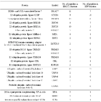
List of ERCC1- or XPF-interacting factors identified by mass spectrometry analysis
I submitted the purified fractions to the UNC Michael Hooker Proteomics
Center, University of North Carolina at Chapel Hill (USA) for mass spectrometry.
In total, polypeptides from 699 proteins were detected, which include several
9
proteins previously reported to interact with ERCC1-XPF, XPA, MSH2 and SLX4
(Park and Sancar, 1994; Lan et al., 2004; Muñoz et al., 2009), indicating that the
purification and mass spectrometry were properly conducted for our purposes. On
the other hand, RPA (a known XPF interactor) and USP45 (a known ERCC1
interactor) (de Laat et al., 1998; Perez-Oliva et al., 2015) were not detected. The
lists include many proteins related to the ubiquitination/deubiquitination system,
DNA replication, molecular chaperons, heat shock proteins, ribosomal proteins and
so on (data not shown). In particular, I was interested in the proteins related to the
ubiquitination/deubiquitination system, since they are possibly implicated in the
regulation of ERCC1-XPF stability. In this category, 14 kinds of E3 ubiquitin
ligases and 5 kinds of deubiquitinases were included (Table 1).
Validation of each interaction by transient co-expression and co-
immunoprecipitation.
Mass spectrometry is very powerful and sensitive, but often includes false-
positive proteins. I next carried out co-immunoprecipitation experiments by
overexpressing epitope-tagged candidate interactors along with tagged or non-
tagged ERCC1 in HEK293T cells to ascertain their associations as a second
10
screening. Among the listed proteins related to the ubiquitination/deubiquitination
system, I could obtain or make expression constructs of 3xMyc-STUB1, 3xMyc-
KCTD13, DCAF7-Myc, GFP-RNF220, 3xMyc-FBXO22, USP11-Myc, USP30-Myc,
USP46-Myc, V5-TRIM28, 3xMyc-RNF40, 3xMyc-RNF2, MID1-Myc, GFP-CBL,
FLAG-TRIM25, and FLAG-USP7. Those proteins were co-overexpressed with
3xFLAG-ERCC1 or non-tagged ERCC1 and co-immuprecipitated by various epitope-
tag antibodies. As a result, 3xMyc-STUB1, 3xMyc-KCTD13, DCAF7-Myc, GFP-
RNF220, 3xMyc-FBXO22, USP11-Myc, USP30-Myc and USP46-Myc were co-
precipitated with ERCC1, while V5-TRIM28, 3xMyc-RNF40, 3xMyc-RNF2, MID1-
Myc, GFP-CBL, FLAG-TRIM25 and FLAG-USP7 were not (Fig. 4).
Depletion of DCAF7 or USP11 affects the cellular levels of ERCC1-XPF.
Among the eight proteins that were verified to interact with ERCC1-XPF by
immunoprecipitation experiments, I tested the impacts of siRNA-mediated
knockdown of six interactors, DCAF7, STUB1, KCTD13, USP11, USP30 and USP46,
on ERCC1-XPF protein level. As a result, I found that the knockdown of DCAF7 or
USP11 in U2OS cells affects cellular level of ERCC1-XPF (Fig. 6), while the
knockdown of other four interactors did not. In case of DCAF7 knockdown, the
11
cellular levels of both ERCC1 and XPF were markedly diminished. On the other
hand, USP11 knockdown moderately reduced only ERCC1 level. These results
suggest that DCAF7 and USP11 may play a regulatory role in controlling ERCC1-
XPF level.
12 1-3. Discussion
In DNA maintenance systems, ERCC1-XPF is one of the most important factors
which functions in not only NER but also other several DNA damage responses.
ERCC1 and XPF need to form heterodimer to stabilize each other (Yagi et al., 1997;
Jaspers et al., 2007; Tripsianes et al., 2005; Choi et al., 2005; Sanjeevani et al., 2010).
ERCC1 was reported to interact with USP45 deubiquitinase, which appears to
facilitate ERCC1 recruitment to DNA lesions rather than ERCC1 stability (Perez-
Oliva et al., 2014). No other proteins involved in ubiquitination-mediated
regulation of ERCC1-XPF were currently known.
For further understanding the function and regulation of ERCC1-XPF complex,
I thought it is essential to identify novel interacting factors. In this study, I
conducted tandem purification of double-tagged ERCC1 or XPF and mass
spectrometry analysis, leading me to identify 699 candidate proteins. In particular,
I have focused on 14 kinds of E3 ligases and five kinds of deubiquitinases included
in the candidates. Verification process by co-immunoprecipitation experiments
indicated that STUB1, KCTD13, DCAF7, RNF220, FBXO22, USP11, USP30 and
USP46 appear truly positive as ERCC1-XPF interactors tested so far (Fig, 4).
STUB1 is known to interact with heat shock proteins, HSP70 and HSP90, and
13
ubiquitinate misfolded protein for their proteolytic degradation (Ballinger at al.,
1999; Connel et al., 2001). STUB1 also ubiquitinates DNA Pol involved in base
excision repair (BER) and induces its degradation to regulate BER activity (Parsons
et al., 2008). So STUB1 might also be involved in NER activity regulation by
ubiquitinating ERCC1-XPF. KCDT13 is induced by TNF- and interacts with
PCNA and DNA Pol to activate DNA Pol (He et al., 2001). DCAF7, also known
as WDR68 or HAN11, controls the cellular signaling as a scaffold protein or an
adaptor protein and also functions as a substrate receptor of Cul4-DDB1 E3 ligase
complex (see the Introduction section of Chapter 2). USP11 was reported to be
deubiquitinase and stabilize p53 promoting DNA damage responses (Ke et al., 2014).
Knockdown experiments by siRNAs in selected six interactors revealed that
DCAF7 and USP11 affect the steady state level of ERCC1-XPF (Fig. 6). Specially,
the impact of DCAF7 was more prominent and ERCC1-XPF level was reduced to
~20%. I decided to focus on DCAF7 and at Chapter 2, the functional analysis of the
interaction between DCAF7 and ERCC1-XPF was extensively analyzed.
14
Chapter 2 Functional analysis of one of the interacting proteins, DCAF7
2-1. Introduction
Background of DCAF7, a novel interacting protein with ERCC1-XPF
DDB1- and CUL4-associated factor 7 (DCAF7, also known as WDR68 or
HAN11) is a WD40 domain which was discovered by a research of a kinase DYRK1A
(dual-specificity tyrosine-phosphorylation regulated protein kinase/dual-specificity
YAK1-related protein kinase) (Skurat and Dietrich, 2004). DCAF7 has five WD40
repeats. The N- and C-terminal have no manifest sequence similarity with the
WD40 repeats. Because most of the structurally specified WD40 proteins contain
seven WD40 repeats and form seven-bladed-propeller structures (Stirnimann et al.,
2010), it is speculated that DCAF7 has as least five -propeller blades (Fig. 5A).
Miyata et al., constructed a structural model of mammalian DCAF7 by analyzing its
amino acid sequence (Miyata et al., 2014). DCAF7 is ecclesial for the proliferation
and survival of mammalian cells (Ritterhoff et al., 2010; Miyata et al., 2011).
DCAF7 can function as a substrate receptor of Cullin4-DDB1 complex and
ubiquitinates target proteins, such as DNA ligase I and regulate their cellular levels
(Peng et al., 2016). Since DCAF7 doesn’t have nuclear localization signal, DCAF7
15
need to interact with DYRK1A which is a family of mammalian protein kinases
(Miyata et al., 2011). DCAF7 is required for normal cellular levels of DYRK1A and
DYRK1B (Yousefelahiyeh et al., 2018). DCAF7 works as a scaffold and mediates
the interaction between kinases, DYRK1A, HIPK2 and MEKK (Glenewinkel et al.,
2016; Ritterhoff et al., 2010). DCAF7 and DYRK1A form a complex and
phosphorylates the C-terminal region of RNA polymesase II and promote myogenesis
(Yu et al., 2019).
Molecular chaperon TRiC
In cells, newly synthesized proteins need to be properly folded. Molecular
chaperones assist folding of many proteins. The chaperonin-containing T-complex
(TRiC) is an ATP-dependent type II (group II) chaperon, prevents misfolding and
aggregation during folding of many proteins (Hartle et al., 2002; Spiess et al., 2004;
Horwich et al., 2007). TRiC consists of eight subunits (TCP1 to TCP1) (Fig. 5B)
(Dunn et al., 2001). TRiC mainly recognizes exposed hydrophobic residues
(Horwich et al., 2007), but charged animo acid residues can also be recognized (Spiess
et al., 2006). It is reported that TRiC binds to DCAF7 and promotes its folding and
nuclear localization (Miyata et al., 2014).
16 Functional analysis of DCAF7
Here at Chapter 2, I conducted several functional analysis to determine the
mechanisms of DCAF7 against ERCC1-XPF. By co-immunoprecipitation analysis,
I found out that DCAF7 interacts with XPF, not ERCC1. I further tried to
determine the interacting region by partial XPF expression vectors. I speculate
that DCAF7 interacts with XPF via not only one site but also multiple sites. The
stabilization of ERCC1-XPF by DCAF7 was not depend on Cullin4-DDB1 complex.
The reduction of ERCC1-XPF by depleted DCAF7 was not fully dependent on 26S
proteasome, but some newly synthesized ERCC1 was partly dependent.
Furthermore, I identified the molecular chaperon, TRiC as another regulator of the
cellular levels of ERCC1-XPF. Importantly, DCAF7-depleted cells showed less
efficient removal of UV-induced (6-4) photoproducts from the genome, indicating
DCAF7 as a novel regulator of cellular NER.
17 2-2. Results
DCAF7 binds to ERCC1-XPF heterodimer through the association with XPF
I first tried to confirm the interaction between DCAF7 and ERCC1 or XPF by
co-immunoprecipitation experiments with HEK293T cells transiently expressing
epitope-tagged DCAF7 and ERCC1 (Figs. 7A) or XPF (Figs. 7B and C). In all types
of combination, I could detect the co-immunoprecipitation between DCAF7 and
ERCC1 or XPF, suggesting that DCAF7 indeed associates with ERCC1-XPF
heterodimer in cells.
I next asked which subunit of the heterodimer interacts with DCAF7. For this
purpose, instead of HEK293T cells, I used XP2YO(SV) cells from XP-F patient, in
which mutant XPF protein is undetectable and the protein level of ERCC1 is
marginal due to its destabilization (Matsumura et al., 1998). Interestingly, similar
immunoprecipitation experiments exhibited co-precipitated DCAF7 and XPF (Figs.
8A and B), but not DCAF7 and ERCC1 (Figs. 8C and D). However, co-transfection
of an increasing amount of XPF plasmid enabled us to detect ERCC1 coprecipitated
with FLAG-DCAF7 by anti-FLAG antibody resin (Fig. 8E). These results strongly
suggest that DCAF7 directly associates with XPF but indirectly with ERCC1.
18 XPF interacts with DCAF7 via multiple sites.
XPF consists of Helicase-like domain, nuclease domain and Helix-hairpin-helix
motif, class 2. To identify which part of XPF is responsible for the DCAF7 binding,
I made deletion mutants of Myc-tagged XPF (Fig. 9A) and examined their ability to
interact with FLAG-tagged DCAF7 by co-immunoprecipitation analysis in
XP2YO(SV) cells. Unexpectedly, both XPF (1-515)-Myc and XPF (516-916)-Myc
which are not partially overlapped were co-immunoprecipitated with FLAG-DCAF7
(Fig. 9B and 9C). I assumed that the central part of XPF associates with DCAF7.
Shorter region of C-terminus, XPF (719-916)-Myc was also co-immunoprecipitated
in XP2YO(SV) (data not shown). Shorter region of N-terminus XPF (1-457)-Myc
couldn’t be overexpressed in XP2YO(SV) cells, but could be in HEK293T cells and
also co-immunoprecipitated with FLAG-DCAF7 (data not shown). For these results,
it is speculated that DCAF7 interact with XPF via multiple sites. DCAF7 might
interact with their target proteins via several regions. I couldn’t achieve any valuable
functional insights from partial XPF and DCAF7 interaction analysis.
Depletion of DCAF7 reduces the cellular level of ERCC1-XPF.
To examine a functional role of DCAF7 and ERCC1-XPF interaction, U2OS
19
cells were transfected with DCAF7 siRNA (#1) and monitored for the cellular levels
of XPF and ERCC1, in addition to DCAF7. Under the condition that DCAF7 was
gradually decreased in a time-dependent manner, the protein levels of XPF and
ERCC1 were also downregulated with similar kinetics (Fig. 10A). The DCAF7
depletion-induced ERCC1-XPF downregulation was reproducible (Fig. 10C),
whereas the cellular levels of other NER factors (DDB1, DDB2 and XPB) were not
affected by DCAF7 depletion (Fig. 10B). Moreover, I had a similar observation
using HeLa cells with two different DCAF7 siRNAs, #1 and #2, targeting distinct
regions of DCAF7 mRNA (Fig. 11A), indicating that this phenomenon is neither cell
line-specific nor due to off-target effects.
The molecular chaperon TRiC, which plays an important role in proper folding
of many proteins (Spiess et al., 2004; Horwich et al., 2007), has been reported to
interact with DCAF7 and promote its folding and nuclear localization (Miyata et al.,
2014). I examined the impact of depletion of TCP1, one of eight subunits of TRiC,
on the protein level of ERCC1-XPF. As shown in Fig. 10D, transfection of specific
siRNA (#1) caused efficient TCP1knockdown, but no change in DCAF7 protein
level as reported previously (Miyata et al., 2014). The cellular level of ERCC1-XPF
was also downregulated as expected, although no striking impacts on DDB2 and XPA
20
levels. Again, this phenomenon was reproducible (Fig. 10E) and was observed
with another TCP1siRNA sequence (#2) as well (Fig. 11B). Taken together, these
results suggest that DCAF7 is required for maintaining the normal cellular levels of
ERCC1-XPF.
DCAF7-mediated regulation of ERCC1-XPF level is independent of Cul4-DDB1 E3
ligase and 26S proteasome.
DCAF7 is known to form a complex with Cul4-DDB1 and to function as a
substrate receptor that determines the substrate specificity of the E3 ligase complex.
I wished to know whether the depletion of DDB1 also causes the downregulation of
ERCC1-XPF. However, transfection of DDB1 siRNA affected neither the basal
levels of ERCC1-XPF (Fig. 12A) nor the downregulation of ERCC1-XPF upon DCAF7
depletion (Fig. 12B), indicating that DDB1 and probably also Cul4 are unlikely to
participate in this mechanism.
I further asked whether the downregulation of ERCC1-XPF by DCAF7
depletion is dependent on 26S proteasome. After transfection with DCAF7 siRNA,
a proteasome inhibitor MG-132 was added to cell culture at 12 h and treated for 12
h before cell lysis. Immunoblot analysis showed that MG132 partially suppresses
21
the downregulation of ERCC1, while XPF level is unchanged (Fig. 12C). Non-
ATPase regulatory subunit 14 (PSMD14), one of proteasome subunits, possesses a
deubiquitinase activity and is essential for the proteolytic function of proteasome
(Verma et al., 2002). Partial knockdown of PSMD14 by siRNA elevated basal levels
of p53 and also DCAF7, XPF and ERCC1. Importantly, cotreatment of DCAF7
siRNA again caused the downregulation of ERCC1 and XPF (but not p53) (Fig. 12D),
suggesting that the accumulation of ERCC1 and XPF by PSMD14 knockdown and
their downregulation by DCAF7 knockdown are mutually independent.
Taken together, I suggest that the ubiquitin-proteasome system is not mainly
implicated in DCAF7-mediated regulation of ERCC1-XPF level, although some
fraction of ERCC1 regulation might be proteasome-dependent.
Depletion of DCAF7 causes the suppression of NER activity.
ERCC1-XPF is an indispensable factor for the dual incision reaction of NER.
Given that DCAF7 depletion reduces the protein level of ERCC1-XPF, I wished to
know the impact of DCAF7 depletion on cellular NER activity. To this end, I
transfected HeLa cells with DCAF7 siRNA (#1) and measured the repair kinetics of
6-4PP induced by UV-C irradiation. DCAF7-depleted cells showed a significantly
22
slower rate of 6-4PP removal, compared to the undepleted control cells (Fig. 13A). I
also performed the same experiment with Flp-In T-REx 293/FLAG-XPF-3xHis cells
to check whether inducible expression of XPF can rescue the delay of 6-4PP repair.
As seen in Fig. 13B, DCAF7 knockdown again suppressed 6-4PP repair, although
less significantly compared with DCAF7-depleted HeLa cells, probably due to low
knockdown efficiency in this cell line. Importantly, the treatment of doxycycline
elevated the cellular level of exogenous FLAG-XPF-6xHis and even endogenous
ERCC1, and restored the full 6-4PP repair ability. Taken together, these results
suggest that DCAF7 plays an important role in maintaining normal NER activity by
stabilizing ERCC1-XPF endonuclease.
DYRK1A kinase activity inhibitor, Harmine doesn’t affect the cellular levels of
ERCC1-XPF
There are several reports that DCAF7 acts as a scaffold of kinases and it is
necessary for kinase pathways. Kinase DYRK1A interacts with DCAF7 and it is
essential for the nuclear localization of DCAF7 (Miyata et al., 2011). DCAF7 is also
required for normal cellular levels of DYRK1A and DYRK1B (Mina et al., 2018).
The complex of DYRK1A and DCAF7 phosphorylates the C-terminal region of RNA
23
polymerase II and promote myogenesis (Dan et al., 2019). DCAF7 needs to serve a
scaffold for kinases, HIPK2 and MEKK1 and their phosphorylation pathways
(Stefanie et al., 2010). I speculate that DCAF7 acts as a scaffold protein and
mediate the interaction between DYRK1A and XPF, and DYRK1A phosphorylates
XPF to stabilize ERCC1-XPF. I used DYRK1A kinase activity inhibitor Harmine
and analyzed its effects against ERCC1 and XPF levels (Figure 14). Even though
the marker of kinase inhibition, Cyclin D1 levels were elevated (Chen et al., 2013),
cellular levels of ERCC1 and XPF were not strikingly changed. DYRK1A kinase
activity might not be involved in the mechanism of stabilization of ERCC1-XPF.
24 2-3. Discussion
In this study, I identified DCAF7 (also known as WDR68 or HAN11) as a novel
interacting protein of ERCC1-XPF by a proteomic approach. Co-
immunoprecipitation experiments confirmed their interaction and suggested a direct
association of DCAF7 with XPF but not ERCC1, leading us to speculate an indirect
interaction with ERCC1 probably via XPF.
Like most of WD40 proteins with seven WD40 repeats, DCAF7 forms a seven-
bladed -propeller structure, although it contains only five WD40 repeats (Miyata et
al., 2014). One of known functions of DCAF7 is to control the cellular signaling as
a scaffold protein of multiprotein complex or an adaptor protein recruiting
downstream proteins by facilitating protein-protein interactions. DCAF7 binds to
DYRK1A and DYRK1B belonging to the dual specificity tyrosine phosphorylation-
regulated protein kinase (DYRK) family (Skurat and Dietrich, 2004; Nissen et al.,
2006; Mazmanian et al., 2010; Miyata et al., 2011). DCAF7 also binds to MEKK1
and HIPK2, facilitating complex formation and signal transduction between the two
protein kinases (Ritterhoff et al., 2010). Conversely, DYRK1A appears to contribute
to the nuclear localization of DCAF7 through the physical interaction, independent
of its kinase activity (Miyata et al., 2011).
25
Another known function of DCAF7 is as a substrate receptor of Cul4-DDB1 E3
ligase complex. The Cul4-DDB1 complex is a member of Cullin-RING finger E3
ligase family, which ubiquitinates target proteins (substrates) and mainly induces
their proteasomal degradation. A large number of substrate receptors, DCAFs,
have been identified (Lee and Zhou, 2007) and play an important role in determining
the substrate specificity of the complex. DCAF7 was found to interact with DNA
ligase I involved in DNA replication by joining Okazaki fragments, and to target it
for ubiquitination by Cul4-DDB1 E3 ligase upon inhibition of cell proliferation (Peng
et al., 2016).
Here, I found that DCAF7 depletion reduced the steady state level of ERCC1-
XPF (Fig. 6) and hence the cellular NER activity (Fig. 13). The mechanism how
DCAF7 stabilizes ERCC1-XPF is currently unknown, but several possible models
can be proposed. First of all, the Cullin4-DDB1 complex is unlikely to participate
in this mechanism, since knockdown of DDB1 influenced neither the basal level of
ERCC1-XPF nor the downregulation of ERCC1-XPF upon DCAF7 depletion (Figs.
12A and B). On the other hand, Serine 256 of DCAF7 was recently reported to be a
potential reactive site center of deubiquitination based on enhanced actively-based
probes (ABPs) (Hewings et al., 2018). It is possible to speculate that the potential
26
deubiquitinase activity of DCAF7 might protect ERCC1-XPF from proteasomal
degradation. However, it is currently unclear whether DCAF7 indeed possesses
deubiquitinase activity.
A more plausible model would be that DCAF7 may function as an adopter
protein to stabilize ERCC1-XPF. In fact, it was recently reported that DCAF7 is
required for normal levels of DYRK1A and DYRK1B at a posttranslational level in a
ubiquitin-proteasome-independent manner, although its detailed mechanism
remains elusive (Yousefelahiyeh et al., 2018). In the context of this model, the
molecular chaperone TRiC (TCP1 ring complex), also known as CCT (chaperonin
containing TCP1), is possibly involved in DCAF7-mediated ERCC1-XPF
stabilization. Nishida’s group reported that TRiC binds to DCAF7 and promotes its
folding, DYRK1A binding and nuclear accumulation (Miyata et al., 2014). Similarly,
it has been also reported that TRiC binds to CSA, another DCAF protein with WD40
repeats, ensures its proper folding and complex formation with Cul4-DDB1 (Pines et
al., 2018). It should be noted that, in our mass spectrometry analysis of ERCC1- or
XPF-bound fraction, all of eight components of TRiC were detected (Table 2). I also
obtained the result that TCP1 knockdown causes the downregulation of ERCC1-XPF
level, as seen in DCAF7 knockdown (Figs. 10D and E), suggesting the possible
27
implication of TRiC in the DCAF7-mediated stabilization of ERCC1-XPF.
Another potential coplayer with DCAF7 may be protein kinase(s) that interacts
with DCAF7. DCAF7 was reported to recruit DYRK1A and HIPK2 to the
adenovirus E1A protein and mediate their interaction for phosphorylation
(Glenewinkel et al., 2016). More recently, DCAF7 was found to stabilize and tether
DYRK1A to RNA polymerase II, which promotes the hyperphosphorylation of its C-
terminal domain (CTD) by DYRK1A and hence transcription of myogenic genes (Yu
et al., 2019). In this model, some kinase(s) is possibly recruited to ERCC1-XPF
complex or XPF before hetero-dimerization and facilitate the phosphorylation of XPF
and/or ERCC1, thereby enhancing ERCC1-XPF stability. I need to test the
possibility that protein kinases such as DYRK1A/B, HIPK2 and MEKK1 are also
involved in the DCAF7-mediated ERCC1-XPF stabilization in near future.
In this study, I demonstrated that DCAF7, a novel interacting protein of
ERCC1-XPF, plays some role in maintaining the cellular levels of ERCC1-XPF and
nucleotide excision repair. As already mentioned, DCAF7 also functions in the
negative regulation of the cellular level of DNA ligase I in quiescent cells, collectively
suggesting that DCAF7 is one of key players controlling DNA metabolism. The
cellular level of ERCC1-XPF is well known to influence the efficacy of platinum-
28
based cancer chemotherapy. This study provides a new insight into the regulatory
mechanism of the cellular levels of ERCC1-XPF heterodimers and its application to
anticancer therapy.
29 Materials and Methods
Cell lines and culture conditions
Parental Flp-In™ T-REx™ 293 (Invitrogen) and its derivative cell lines conditionally
expressing FLAG-ERCC1-6xHis or FLAG-XPF-6xHis upon tetracycline or
doxycycline treatment were grown in Dulbecco’s modified Eagle medium (DMEM)
(Gibco) supplemented with 10% fetal bovine serum (Sigma) and 10 g/ml Penicillin-
Streptomycin (Gibco). U2OS, HeLa and HEK293T cells were grown in DMEM
(Wako) with 10% fetal bovine serum (Sigma) and 50 g/ml Gentamicin (Nacalai
tesque). All cell lines were cultured at 37oC in a humidified 5% CO2 atmosphere.
Silver stain
The gel of SDS-PAGE is fixed by incubating with fix solution (50% MeOH, 12% Acetic
Acid, 0.0185% HCHO) for one hour. The gel was washed with 50% ethanol for three
times for 20 min each. The gel was pre-treated by incubating with pre-treat solution
(0.02% sodium thiosulfate) for one minute, washed with water three times for 20
seconds each. The gel was impregnated by incubating with impregnate solution
(12mM silver nitrate, 0.02775% HCHO) for 20 min, and washed twice by water for
20 seconds each. The gel was developed by incubated with develop solution (0.02%
30
sodium thiosulfate, 12% w/v NaCO3) until bands of proteins become visible level,
washed with water. The reaction was stopped by incubating with distain solution
(12% acetic acid, 60% methanol, 1.2% glycerol).
Identification of ERCC1-XPF-interacting proteins by mass spectrometry
Flp-InTM T-RExTM 293 cell line (control) and Flp-InTM T-RExTM 293/FLAG-ERCC1-
6xHis or FLAG-XPF-6xHis cell lines were incubated with 1 g/ml tetracycline for 48
h to induce double-tagged bait proteins. Cell lysates were prepared in the lysis
buffer (50 mM Tris-HCl, pH 7.4, 150 mM NaCl, 1 mM EDTA, 1% Triton X-100)
containing EDTA-free protease inhibitor cocktail (Roche), supplemented with
imidazole (final conc. 10 mM) (Sigma) and incubated with Ni-NTA agarose (Qiagen)
for 1 h at 4oC. After washing with the lysis buffer containing 10 mM imidazole,
bound fractions were eluted with the lysis buffer containing 200 mM imidazole. The
eluates were incubated with anti-FLAG antibody agarose (Sigma) for 12 h at 4oC.
The proteins bound to agarose were washed three times and eluted by incubating
with the lysis buffer containing 150 g/ml 3xFLAG peptide (Sigma) for 1 h at 4oC.
The eluates were concentrated by chloroform-methanol extraction, dissolved in SDS
sample buffer and fractionated by NuPAGE 10% Bis-Tris Protein Gels (Invitrogen).
31
The mass spectrometry analysis was conducted in the UNC Michael Hooker
Proteomics Center.
Construction of expression vectors
A plasmid clone containing human DCAF7 cDNA was obtained from the Riken DNA
bank (#HGX001752, Clone name: IRAK004G08). DCAF7 cDNA was subcloned into
the pEF6/Myc-His vector (Thermo Fisher Scientific) using PCR with the primers, 5'-
GTGGAATTCATGTCCCTGCACGGCAAACG (sense) and 5'-
CGAGCGGCCGCCCACTCTGAGTATCTCCAGGC (antisense), or the pEF6-2xFLAG
vector (Kind gift from Dr. Yamashita) by PCR with the primes, 5'-
ATACATATGTCCCTGCACGGCAAACGGAA (sense) and 5'-
CGAGCGGCCGCCTACACTCTGAGTATCTCCA (antisense). MID1 was also
obtained from Riken DNA bank (Catalog#: HGX046310, Clone #: IRAK115M22) and
Myc-tagged by subcloned into pEF6-Myc-His B. cDNAs of STUB1 (912, 11052 E08);
KCTD13 (990, 11032 F11); RNF2 (1011, 11033 C01); FBXO22 (1212, 31006 F02) were
obtained from Lineberger Tissue Culture Facility, hORFeome V5.1. and were
inserted into the empty vector, pcDNA3-3Myc-DEST, which was kindly provided by
Dr. Yue Xiong and 3xMyc-tagged by Gateway™ LR Clonase™ Enzyme mix (Thermo
32
Fisher Scientific) according to the manufacturer’s instruction. TRIM25 (#12449),
USP7 (#46751), USP11 (#22566), USP30 (#22578), USP46 (#22584) were obtained
from Addgene. cDNAs of USP11, USP30, and USP46 are Myc-tagged by subcloned
into pEF6-Myc-His B. GFP-RNF220 (pcDNA5-FRT/TO-GFP RNF220, DU Number:
DU33230), GFP-CBL (pcDNA5-FRT/TO-GFP CBL, DU Number: DU31284) were
obtained from MRC Protein Phosphorylation and Ubiquitylation Unit.
Immunoprecipitation
HEK 293T cells were transfected with various combinations of plasmids, pEF6/Myc-
His, pEF6/DCAF7-Myc-His, pEF6/XPF-Myc-His, pEF6-2xFLAG, pEF6-2xFLAG-
DCAF7, pEF6/3xFLAG-ERCC1, pcDNA3.1(+)/ERCC1 and pcDNA3.1(+)/XPF, and
incubated for 48 h. Cell lysates were prepared with the NP-40 lysis buffer (50 mM
Tris-Cl, pH 7.4, 150 mM NaCl, 1% Nonidet P-40) supplemented with EDTA-free
protease inhibitor cocktail (Roche or Nacalai tesque), and incubated with anti-FLAG
antibody resin (Wako) or anti-Myc antibody resin (Wako) for 1 h. After washing
with the NP-40 lysis buffer three times, bound proteins were eluted by boiling with
SDS sample buffer and analyzed by western blotting.
33 Antibodies and siRNA
The antibodies used in this study were as follows: anti-DCAF7 (EPR8712, Abcam),
anti-ERCC1 (GTX129282, GeneTex), anti-TCP1 (sc-53454, Santa Cruz
Biotechnology), anti-PSMD14 (A303-857A-T, Bethyl), anti-USP11 (A301-613A-T,
Bethyl), anti-FLAG (PM020, MBL), anti-Myc (562, MBL). I also used home-made
monoclonal antibodies against XPF (clone 19-16) and DDB1 (clone 43233-3-1)
(Cosmo bio).
The siRNAs were obtained from Sigma-Aldrich: DCAF7 #1, 5’-
GGAACAAGCAGGACCCUAAdTdT; DCAF7 #2, 5’-
GGUGUUAGGGCGAGUGAAUdTdT. TCP1 #1, 5’-
GACCAAAUUAGACAGAGAdTdT; TCP1#2, 5’-
GCAAGGAAGCAGUGCGUUAdTdT; PSMD14, 5’-
GGCAUUAAUUCAUGGACUAdTdT. USP11, 5’-
ACCGATTCTATTGGCCTAGTAdTdT. DDB1 siRNA was obtained from Cosmo Bio,
5’-GAAUUCAAUAAGGGCCUUdTdT. I also used siRNA Universal Negative
Control #1 (Sigma) as control siRNA. U2OS or HeLa cells were transfected with
siRNA using LipofectamineTM RNAiMAX Transfection Reagent (Thermo Fisher
Scientific) according to the manufacturer’s instruction and incubated normally for
34 72 h.
Enzyme-linked immunosorbent assay (ELISA)
Cells grown in 35-mm dish were irradiated with 10 J/m2 of UV-C from germicidal
lamps (Toshiba, GL-10) and incubated for various periods. Genomic DNAs were
isolated with the Geno Plus Genomic DNA Extraction Miniprep system (Viogene),
and the amount of 6-4PP was measured by using 64M-5 antibody as described
previously (Mori et al., 1991).
35
ACKNOWLEDGEMENTS
The first half of this study was carried out by the support of Tobitate! (Leap for
Tomorrow) Study Abroad Initiative. Thanks to the scholarship, I could achieve a
precious opportunity to visit the University of North Carolina at Chapel Hill to study.
I’d like to thank all people who are working for the scholarship, including Ministry
of Education, Culture, Sports, Science and Technology, and supporting companies.
I would like to express my deepest gratitude to Dr. Aziz Sancar for giving me a great
opportunity to study at his laboratory and a lot of supports. His comments and
suggestions were innumerably valuable. I was also helped by members of Dr.
Sancar’s laboratory, Dr. Laura Lindsey-Boltz, Dr. Christpher Selby, Dr. Michael
Kemp, Dr. Yi-Ying Chiou, Dr. Jinchuan Hu, Dr. Yangyan Yang, Dr. Wentao Li, Dr.
Sheera Adar, Dr. Fazile Canturk, Dr. Muhammet Karaman, Dr. Wentao Li, Dr.
Gulnihal Kulaksiz. I employed the Flp-In T-REx 293 cells which were constructed by
Dr. Joyce Reardon. To conduct mass spectrometry analysis, Laura Herring gave me
a lot of advices. I got a support from Dr. Yue Xiong’s laboratory for proteomics
analysis of uniquitination and Dr. Naoya Sasaki directed me how to proceed the
research.
Also, I would like to thank Professor Tsukasa Matsunaga for his constant devoting
36
attitude towards me, not only for my research but also my health, future careers,
and financial conditions. I also thank Dr. Mitsuo Wakasugi for appropriate guides,
comments for proceeding my research, and improvement of my journal. Dr. Katsumi
Yamashita kindly gave me a plasmid and a lot of supportive advices. Daily I
communicate and share a lot of data, tips, and opinions with Ryo Akahori. He gave
me a plasmid he made. I strongly hope that he will continue this research and
determine further molecular mechanisms of cellular level and localization of the
heterodimer, ERCC1-XPF. Additionally, I’ve been supported by all of other members
of the Human Molecular Genetics laboratory. My family gave me a valuable
opportunity to study at the lab, and gave me a lot of supports. Many people helped
me with my mental conditions. I’d like to thank all of those people who have been
supporting me.
This research is based in part upon work conducted using the UNC Michael Hooker
Proteomics Center, which is supported in part by the NIH-NCI Grant No. CA016086
to the Lineberger Comprehensive Cancer Center.
37
References
Adair, G. M., Rolig, R. L., Moore-Faver, D., Zabelshansky, M., Wilson, J. H., Nairn, R. S.
(2000) Role of ERCC1 in removal of long non-homologous tails during targeted homologous recombination. The EMBO J. 19, 5552–5561.
Adebali, O., Chiou, Y. Y., Hu, J., Sancar, A., Selby CP. (2017a) Genome-wide transcription-coupled repair in Escherichia coli is mediated by the Mfd translocase. Proc Natl Acad Sci U S A. 114, E2116–E2125.
Adebali. O., Sancar, A., Selby, C. P. (2017b) Mfd translocase is necessary and sufficient for transcription-coupled repair in Escherichia coli. J Biol Chem. 292, 18386-18391.
Ahmad, A., Robinson, A. R., Duensing, A., van Drunen, E., Beverloo, H. B., Weisberg, D.
B., Hasty, P., Hoeijmakers, J. H., Niedernhofer, L. J. (2008) ERCC1-XPF endonuclease facilitates DNA double-strand break repair. Mol Cell Biol. 28, 5082–5092.
Al-Minawi, A. Z., Saleh-Gohari, N., Helleday, T. (2008) The ERCC1/XPF endonuclease is required for efficient single-strand annealing and gene conversion in mammalian cells.
Nucleic Acids Res 36, 1–9.
Araújo, S. J., Tirode, F., Coin, F., Pospiech, H., Syväoja, J. E., Stucki, M., Hübscher, U., Egly, J. M., Wood, R. D. (2000) Nucleotide excision repair of DNA with recombinant human proteins: definition of the minimal set of factors, active forms of TFIIH, and modulation by CAK. Genes Dev. 14, 349–359.
Arora, S., Kothandapani, A., Tillison, K., Kalman-Maltese, V., Patrick. S. M. (2010) Downregulation of XPF-ERCC1 enhances cisplatin efficacy in cancer cells. DNA Repair 9, 745–753.
Ballinger, C. A., Connell, P., Wu, Y., Hu, Z., Thompson, L. J., Yin, L. Y., Patterson, C.
(1999) Identification of CHIP, a novel tetratricopeptide repeat-containing protein that interacts with heat shock proteins and negatively regulates chaperone functions. Mol Cell Biol. 19, 4535–4545.
38
Bennardo, N., Cheng, A., Huang, N., Stark, J. M. (2008) Alternative-NHEJ is a mechanistically distinct pathway of mammalian chromosome break repair. PLoS genetics 4, e1000110.
Biggerstaff, M., Szymkowski, D. E., Wood, R. D. (1993) Co-correction of the ERCC1, ERCC4 and xeroderma pigmentosum group F DNA repair defects in vitro. The EMBO J.
12, 3685–3692.
Bouwman, P, Jonkers, J. (2012) The effects of deregulated DNA damage signalling on cancer chemotherapy response and resistance. Nat Rev Cancer. 12, 587–598.
Chen, J. Y., Lin, J. R., Tsai, F. C., Meyer, T. (2014) Dosage of Dyrk1a shifts cells within a p21-cyclin D1 signaling map to control the decision to enter the cell cycle. Mol Cell. 52, 87–100.
Choi, Y. J., Ryu, K. S., Ko, Y. M., Chae, Y. K., Pelton, J. G., Wemmer, D. E., Choi, B. S.
(2005) Biophysical characterization of the interaction domains and mapping of the contact residues in the XPF-ERCC1 complex. J Biol Chem. 280, 28644–28652.
Cleaver, J. E., Lam, E. T., Revet, I. (2009) Disorders of nucleotide excision repair: the genetic and molecular basis of heterogeneity. Nat Rev Genet. 10, 756–768.
Connell, P., Ballinger, C. A., Jiang, J., Wu, Y., Thompson, L. J., Höhfeld, J., Patterson, C.
(2001) The co-chaperone CHIP regulates protein triage decisions mediated by heat-shock proteins. Nat Cell Biol. 3, 93–96.
de Laat W. L., Sijbers, A. M., Odijk, H., Jaspers, N. G., Hoeijmakers, J. H. (1998b) Mapping of interaction domains between human repair proteins ERCC1 and XPF.
Nucleic Acids Res. 26, 4146–4152.
de Laat, W. L., Appeldoorn, E., Sugasawa, K., Weterings, E., Jaspers, N. G., Hoeijmakers, J. H. (1998) DNA-binding polarity of human replication protein A positions nucleases in nucleotide excision repair. Genes Dev. 12, 2598–609.
de Laat, W.L., Appeldoorn, E., Jaspers, N. G., Hoeijmakers, J. H. (1998a) DNA structural elements required for ERCC1-XPF endonuclease activity. J Biol Chem. 273, 7835–7842.
39
De Silva, I. U., McHugh, P. J., Clingen, P. H., Hartley, J. A. (2000) Defining the roles of nucleotide excision repair and recombination in the repair of DNA interstrand cross- links in mammalian cells. Mol Cell Biol 20, 7980–7990.
De Silva, I. U., McHugh, P. J., Clingen, P. H., Hartley, J. A. (2002) Defects in interstrand cross-link uncoupling do not account for the extreme sensitivity of ERCC1 and XPF cells to cisplatin. Nucleic Acids Res. 30, 3848–3856.
Dunn, A. Y., Melville, M. W., Frydman, J. (2001) Review: cellular substrates of the eukaryotic chaperonin TRiC/CCT. J Struct Biol. 135, 176–184.
Enzlin, J. H., Schärer, O. D. (2002) The active site of the DNA repair endonuclease XPF- ERCC1 forms a highly conserved nuclease motif. EMBO J. 21, 2045–2053.
Fisher, L. A., Bessho, M., Bessho, T. (2008) Processing of a psoralen DNA interstrand cross-link by XPF-ERCC1 complex in vitro. J Biol Chem. 283, 1275–1281.
Friedberg, E. C., Aguilera, A., Gellert, M., Hanawalt, P. C., Hays, J. B., Lehmann, A.R., Lindahl, T., Lowndes, N., Sarasin, A., Wood, R. D. (2006) DNA repair: from molecular mechanism to human disease. DNA Repair 5, 986–996.
Friedberg, E. C., Walker, G. C., Siede, W., Wood, R. D., Schultz, R. A. Ellenberger, T.
(2006) DNA Repair and Mutagenesis. 2nd Edition, ASM Press, Washington DC.
Glenewinkel, F., Cohen, M. J., King, C. R., Kaspar, S., Bamberg-Lemper, S., Mymryk, J.
S., Becker, W. (2016) The adaptor protein DCAF7 mediates the interaction of the adenovirus E1A oncoprotein with the protein kinases DYRK1A and HIPK2. Sci Rep. 6, 28241.
Gregg, S. Q., Robinson, A. R., and Niedernhofer, L. J. (2011) Physiological consequences of defects in ERCC1-XPF DNA repair endonuclease. DNA Repair 10, 781–791.
Hartl, F. U., Hayer-Hartl, M. (2002) Molecular chaperones in the cytosol: from nascent chain to folded protein. Science 295, 1852–1858.
He, H., Tan, C. K., Downey, K. M., So, A. G. (2001) A tumor necrosis factor alpha- and interleukin 6-inducible protein that interacts with the small subunit of DNA polymerase
40
delta and proliferating cell nuclear antigen. Proc Natl Acad Sci U S A. 98, 11979–11984.
Hewings, D. S., Heideker, J., Ma, T. P., AhYoung, A. P., El Oualid, F., Amore, A., Costakes, G. T., Kirchhofer, D., Brasher, B., Pillow, T., Popovych, N., Maurer, T., Schwerdtfeger, C., Forrest, W. F., Yu, K., Flygare, J., Bogyo, M., Wertz, I. E., (2018) Reactive-site-centric chemoproteomics identifies a distinct class of deubiquitinase enzymes. Nat Commun. 9, 1162.
Horwich, A. L., Fenton, W. A., Chapman, E., Farr, G. W. (2007) Two families of chaperonin: physiology and mechanism. Annu Rev Cell Dev Biol. 23, 115–145.
Horwich, A. L., Fenton, W. A., Chapman, E., Farr, G. W. (2007) Two families of chaperonin: physiology and mechanism. Annu. Rev. Cell Dev. Biol. 23, 115–145.
Hu, J., Selby, C. P., Adar, S., Adebali, O., Sancar, A. (2017) Molecular mechanisms and genomic maps of DNA excision repair in Escherichia coli and humans. J Biol Chem. 292, 15588–15597.
Jaspers, N. G., Raams, A., Silengo, M. C., Wijgers, N., Niedernhofer, L. J., Robinson, A.
R., Giglia-Mari, G., Hoogstraten, D., Kleijer, W. J., Hoeijmakers, J. H., Vermeulen, W.
(2007) First reported patient with human ERCC1 deficiency has cerebro-oculo-facio- skeletal syndrome with a mild defect in nucleotide excision repair and severe developmental failure. Am J Hum Genet. 80, 457–466.
Kashiyama, K., Nakazawa, Y., Pilz, D. T., Guo, C., Shimada, M., Sasaki, K., Fawcett, H., Wing, J. F., Lewin, S. O., Carr, L., Li, T. S., Yoshiura, K., Utani, A., Hirano, A., Yamashita, S., Greenblatt, D., Nardo, T., Stefanini, M., McGibbon, D., Sarkany, R., Fassihi, H., Takahashi, Y., Nagayama, Y., Mitsutake, N., Lehmann, A. R., Ogi, T. (2013) Malfunction of nuclease ERCC1-XPF results in diverse clinical manifestations and causes Cockayne syndrome, xeroderma pigmentosum, and Fanconi anemia. Am J Hum Genet. 92, 807–19.
Ke, J. Y., Dai, C. J., Wu, W. L., Gao, J. H., Xia, A. J., Liu, G. P., Lv, K. S., Wu, C. L. (2014) USP11 regulates p53 stability by deubiquitinating p53. J Zhejiang Univ Sci B. 15, 1032–
1038.
Klein, Douwel, D., Boonen, R. A., Long, D. T., Szypowska, A. A., Raschle, M., Walter, J.C., Knipscheer, P. (2014) XPF-ERCC1 acts in Unhooking DNA interstrand crosslinks in cooperation with FANCD2 and FANCP/SLX4. Mol Cell 54, 460–471.
41
Kuraoka, I., Kobertz, W. R., Ariza, R. R., Biggerstaff, M., Essigmann, J. M., Wood, R. D.
(2000) Repair of an interstrand DNA cross-link initiated by ERCC1-XPF repair/recombination nuclease. J Biol Chem. 275, 26632–26636.
Lan, L., Hayashi, T., Rabeya, R. M., Nakajima, S., Kanno, Si, Takao, M., Matsunaga, T., Yoshino, M., Ichikawa, M., Riele, Ht, Tsuchiya, S., Tanaka, K., Yasui, A. (2004) Functional and physical interactions between ERCC1 and MSH2 complexes for resistance to cis-diamminedichloroplatinum(II) in mammalian cells. DNA Repair 3, 135–
143.
Lee, J., Zhou, P. (2007) DCAFs, the missing link of the CUL4-DDB1 ubiquitin ligase. Mol Cell. 26, 775–780.
Li, W., Adebali, O., Yang, Y., Selby, C. P., Sancar, A. (2018) Single-nucleotide resolution dynamic repair maps of UV damage in Saccharomyces cerevisiae genome. Proc Natl Acad Sci U S A. 115, E3408–E3415.
Manandhar, M., Boulware, K. S., Wood, R. D. (2015) The ERCC1 and ERCC4 (XPF) genes and gene products. Gene 569, 153–161.
Matsumura, Y., Nishigori, C., Yagi, T., Imamura, S., Takebe, H. (1998) Characterization of molecular defects in xeroderma pigmentosum group F in relation to its clinically mild symptoms. Hum Mol Genet. 7, 969–974.
Matsunaga, T., Park, C. H., Bessho, T., Mu, D., Sancar, A. (1996) Replication protein A confers structure-specific endonuclease activities to the XPF-ERCC1 and XPG subunits of human DNA repair excision nuclease. J Biol Chem. 271, 11047–11050.
Mazmanian G, Kovshilovsky M, Yen D, Mohanty A, Mohanty S, Nee A, Nissen RM.
(2010) The zebrafish dyrk1b gene is important for endoderm formation. Genesis. 48, 20–
30.
McWhir, J., Selfridge, J., Harrison, D. J., Squires, S., Melton, D. W. (1993) Mice with DNA repair gene (ERCC-1) deficiency have elevated levels of p53, liver nuclear abnormalities and die before weaning. Nat Genet. 5, 217–224.
42
Miyata, Y., Nishida, E. (2011) DYRK1A binds to an evolutionarily conserved WD40- repeat protein WDR68 and induces its nuclear translocation. Biochim Biophys Acta.
1813, 1728–1739.
Miyata, Y., Shibata, T., Aoshima, M., Tsubata, T., Nishida, E. (2014) The molecular chaperone TRiC/CCT binds to the Trp-Asp 40 (WD40) repeat protein WDR68 and promotes its folding, protein kinase DYRK1A binding, and nuclear accumulation. J Biol Chem. 289, 33320–33332.
Muñoz, I. M., Hain, K., Déclais, A. C., Gardiner, M., Toh, G. W., Sanchez-Pulido, L., Heuckmann, J. M., Toth, R., Macartney, T., Eppink, B., Kanaar, R., Ponting, C. P., Lilley, D. M., Rouse, J. (2009) Coordination of structure-specific nucleases by human SLX4/BTBD12 is required for DNA repair. Mol Cell. 35, 116–127.
Muñoz, P., Blanco, R., Flores, J. M., Blasco, M. A. (2005) XPF nuclease-dependent telomere loss and increased DNA damage in mice overexpressing TRF2 result in premature aging and cancer. Nat Genet. 37, 1063–1071.
Niedernhofer, L. J., Garinis, G. A., Raams, A., Lalai, A. S., Robinson, A. R., Appeldoorn, E., Odijk, H., Oostendorp, R., Ahmad, A., van Leeuwen, W., Theil, A. F., Vermeulen, W., van der Horst, G. T., Meinecke, P., Kleijer, W. J., Vijg, J., Jaspers, N. G., Hoeijmakers, J.
H. (2006) A new progeroid syndrome reveals that genotoxic stress suppresses the somatotroph axis. Nature 444, 1038–1043.
Niedernhofer, L. J., Odijk, H., Budzowska, M., van Drunen, E., Maas, A., Theil, A. F., de Wit, J., Jaspers, N. G., Beverloo, H. B., Hoeijmakers, J. H., Kanaar, R. (2004) The structure-specific endonuclease Ercc1-Xpf is required to resolve DNA interstrand cross- link-induced double-strand breaks. Mol Cell Biol. 24, 5776–5787.
Nissen, R. M., Amsterdam, A., Hopkins, N. (2006) A zebrafish screen for craniofacial mutants identifies wdr68 as a highly conserved gene required for endothelin-1 expression. BMC Dev Biol. 7, 28.
Núñez, F., Chipchase, M. D., Clarke, A. R., Melton, D. W. (2000) Nucleotide excision repair gene (ERCC1) deficiency causes G2 arrest in hepatocytes and a reduction in liver binucleation: the role of p53 and p21. FASEB J. 14, 1073–1082.
43
Park, C. H., Sancar, A. (1994) Formation of a ternary complex by human XPA, ERCC1, and ERCC4 (XPF) excision repair proteins. Proc Natl Acad Sci U S A. 91, 5017–5021.
Parsons, J. L., Tait, P. S., Finch, D., Dianova, I. I., Allinson, S. L., Dianov, G. L. (2008) CHIP-mediated degradation and DNA damage-dependent stabilization regulate base excision repair proteins. Mol Cell. 29, 477–487.
Peng, Z., Liao, Z., Matsumoto, Y., Yang, A., Tomkinson, A. E. (2016) Human DNA Ligase I Interacts with and Is Targeted for Degradation by the DCAF7 Specificity Factor of the Cul4-DDB1 Ubiquitin Ligase Complex. J Biol Chem. 291, 21893–21902.
Perez-Oliva, A. B., Lachaud, C., Szyniarowski, P., Muñoz, I., Macartney, T., Hickson, I., Rouse, J., Alessi, D. R. (2015) deubiquitylase controls ERCC1-XPF endonuclease- mediated DNA damage responses. EMBO J. 34, 326–343.
Pines, A., Dijk, M., Makowski, M., Meulenbroek, E. M., Vrouwe, M. G., van der Weegen, Y., Baltissen, M., French, P. J., van Royen, M. E., Luijsterburg, M. S., Mullenders, L. H., Vermeulen, M., Vermeulen, W., Pannu, N. S., van Attikum, H. (2018) TRiC controls transcription resumption after UV damage by regulating Cockayne syndrome protein A.
Nat Commun. 9, 1040.
Reardon, J. T., Thompson, L. H., Sancar, A. (1993) Excision repair in man and the molecular basis of xeroderma pigmentosum syndrome. Cold Spring Harb Symp Quant Biol. 58, 605–617.
Ritterhoff, S., Farah, C. M., Grabitzki, J., Lochnit, G., Skurat, A. V., Schmitz, M. L. (2010) The WD40-repeat protein Han11 functions as a scaffold protein to control HIPK2 and MEKK1 kinase functions. EMBO J. 29, 3750–3761.
Sargent, R. G., Rolig, R. L., Kilburn, A. E., Adair, G. M., Wilson, J. H., Nairn, R. S. (1997) Recombination-dependent deletion formation in mammalian cells deficient in the nucleotide excision repair gene ERCC1. Proc Natl Acad Sci U S A. 94, 13122–13127.
Schärer, O. D. (2013) Nucleotide excision repair in eukaryotes. Cold Spring Harb Perspect Biol. 5, a012609.
Sijbers, A. M., de Laat, W. L., Ariza, R. R., Biggerstaff, M., Wei, Y. F., Moggs, J. G., Carter, K. C., Shell, B. K., Evans, E, de Jong M. C., Rademakers, S., de Rooij, J., Jaspers, N. G.,
44
Hoeijmakers, J. H., Wood, R. D. (1996) Xeroderma pigmentosum group F caused by a defect in a structure-specific DNA repair endonuclease. Cell. 86, 811–822.
Skurat, A. V., Dietrich, A.D. (2004) Phosphorylation of Ser640 in muscle glycogen synthase by DYRK family protein kinases. J Biol Chem. 279, 2490–2498.
Spiess, C., Meyer, A. S., Reissmann, S., Frydman, J. (2004) Mechanism of the eukaryotic chaperonin: protein folding in the chamber of secrets. Trends Cell Biol. 14, 598–604.
Spiess, C., Miller, E. J., McClellan, A. J., Frydman, J. (2006) Identification of the TRiC/CCT substrate binding sites uncovers the function of subunit diversity in eukaryotic chaperonins. Mol Cell. 24, 25—37.
Stirnimann, C. U., Petsalaki, E., Russell, R. B., Müller, C. W. (2010) WD40 proteins propel cellular networks. Trends Biochem Sci. 35, 565–574.
Tripsianes, K., Folkers, G., Ab, E., Das, D., Odijk, H., Jaspers, N. G., Hoeijmakers, J. H., Kaptein, R., Boelens, R. (2005) The structure of the human ERCC1/XPF interaction domains reveals a complementary role for the two proteins in nucleotide excision repair.
Structure 13, 1849–1858.
van Vuuren, A. J., Appeldoorn, E., Odijk, H., Yasui, A., Jaspers, N. G., Bootsma, D., Hoeijmakers, J. H. (1993) Evidence for a repair enzyme complex involving ERCC1 and complementing activities of ERCC4, ERCC11 and xeroderma pigmentosum group F.
EMBO J. 12, 3693–3701.
Verma, R., Aravind, L., Oania, R., McDonald, W. H., Yates, J. R. 3rd ., Koonin, E. V., Deshaies, R. J. (2002) Role of Rpn11 metalloprotease in deubiquitination and degradation by the 26S proteasome. Science. 298, 611–615.
Wan, B., Yin, J., Horvath, K., Sarkar, J., Chen, Y., Wu, J., Wan, K., Lu, J., Gu, P., Yu, E.
Y., Lue, N. F., Chang, S., Liu, Y., Lei, M. (2013) SLX4 assembles a telomere maintenance toolkit by bridging multiple endonucleases with telomeres. Cell Rep. 4, 861–869.
Weeda, G., Donker, I., de Wit, J., Morreau, H., Janssens, R., Vissers, C. J., Nigg, A., van Steeg, H., Bootsma, D., Hoeijmakers, J. H. (1997) Disruption of mouse ERCC1 results in a novel repair syndrome with growth failure, nuclear abnormalities and senescence.
45 Curr Biol. 7, 427–439.
Wu, Y., Mitchell, T. R., Zhu, X. D. (2008) Human XPF controls TRF2 and telomere length maintenance through distinctive mechanisms. Mech Ageing 129, 602–610.
Yagi, T., Wood, R. D., Takebe, H. (1997) A low content of ERCC1 and a 120 kDa protein is a frequent feature of group F xeroderma pigmentosum fibroblast cells. Mutagenesis.
12, 41–44.
Yousefelahiyeh, M., Xu, J., Alvarado, E., Yu, Y., Salven, D., Nissen, R. M. (2018) DCAF7/WDR68 is required for normal levels of DYRK1A and DYRK1B. PLoS One 13, e0207779.
Yu, D., Cattoglio, C., Xue, Y., Zhou, Q. (2019) A complex between DYRK1A and DCAF7 phosphorylates the C-terminal domain of RNA polymerase II to promote myogenesis.
Nucleic Acids Res. 47, 4462–4475.
Zhu, X. D., Niedernhofer, L., Kuster, B., Mann, M., Hoeijmakers, J. H., de Lange, T.
(2003) ERCC1/XPF removes the 3' overhang from uncapped telomeres and represses formation of telomeric DNA-containing double minute chromosomes. Mol Cell. 12, 1489–
1498.
46
Table
Table 1. Proteomic analysis data on E3 ligases and deubiquitinases.
The numbers of peptides from 14 kinds of E3 ligases and 5 kinds of deubiquitinases detected in mass spectrometry are listed. Data on XPA, MSH2 and SLX4, well-known interactors of ERCC1-XPF was also shown.
Table 2. Proteomic analysis data on TRiC/CCT components
All eight components of TRiC/CCT were detected in mass spectrometry and the numbers of peptides from each component are listed.
47
Figure
Figure 1. Structure and functions of ERCC1-XPF
(A) Schematic representation of various domains and known interaction regions of XPF and ERCC1.
(B) Schematic representation of the structures of DNA substrates for ERCC1-XPF endonuclease.
48
Figure. 2 Outline of Nucleotide excision repair core reaction
49
Figure. 3 Overexpression of ERCC1 or XPF in Flp-In TREx 293 cells and tandem affinity purification.
(A) Parental Flp-In™ T-REx™ 293 and its derivative cell lines conditionally expressing FLAG-ERCC1- 6xHis or FLAG-XPF-6xHis were treated with or without 1 g/mL tetracycline for 48 h and lysed. Each cell lysate was analyzed by western blotting with the indicated antibodies. (B) Cell lysate from tetracycline-treated Flp-In T-REx 293 /FLAG-ERCC1-6xHis cells was incubated with Ni-NTA agarose and subsequently with anti-FLAG antibody resin. Bound proteins were eluted with 3xFLAG peptides five times and remaining proteins bound to the resin were eluted by boiling with SDS sample buffer.
Aliquots of samples from each step were separated on SDS-PAGE and analyzed by western blotting with anti-FLAG antibody. (C) Cell lysates from the three cell lines were processed for tandem purification as described in (B), and the lysates (Input) and the precipitates (Pellet) were analyzed by western blotting with the indicated antibodies. (D) Each fraction of tandem purification steps was separated on SDS-PAGE and visualized by silver staining.
1 2 3 4 5 6 7 8 9 10 11 12
50
Figure.4 Co-immunoprecipitation experiments after transient expression of various candidate interactors and ERCC1.
An epitope-tagged version of various candidate interactors and FLAG-tagged or non-tagged ERCC1 were transiently co-expressed in HEK293T cells for 48 h. Cell lysates were precipitated by anti-FLAG or anti-Myc antibody resin and analyzed by western blotting with the indicated antibodies.
51 Figure. 5 Structures of DCAF7 and TRiC/CCT
(A) Domain structure of DCAF7/WDR68/HAN11. (B) Ring structure of TRiC/CCT complex
Figure. 6 DCAF7 depletion causes reduction of cellular levels of ERCC1 and XPF, while USP11 depletion diminishes only ERCC1 level.
U2OS cells were transfected with control siRNA, DCAF7 siRNA (#1) or USP11 siRNA and incubated for 72 h. Cell lysates were prepared and analyzed by western blotting with the indicated antibodies.
52 Figure 7. ERCC1-XPF binds to DCAF7
(A and B) DCAF7-Myc and/or FLAG-ERCC1 were ectopically expressed in HEK293T cells and cell lysates were immunoprecipitated by anti-FLAG antibody resin (A) or anti-Myc antibody resin (B). (C and D) FLAG-DCAF7 and/or XPF-Myc were expressed in HEK293T cells and precipitated by either anti-Myc (C) or anti-FLAG antibody resin (D). The precipitates were analyzed by western blotting with the indicated antibodies.
Figure 8. DCAF7 binds to XPF but not ERCC1
(A and B) DCAF7-Myc and/or FLAG-ERCC1 were ectopically expressed in XP2YO(SV) cells and cell lysates were immunoprecipitated by anti-FLAG antibody resin (A) or anti-Myc antibody resin (B). (C and D) FLAG-DCAF7 and/or Myc-tagged XPF (C) or non-tagged XPF (D) were expressed in XP2YO(SV) cells and precipitated by either anti-Myc (C) or anti-FLAG (D) antibody resin. (E) The same amounts of Non-tagged ERCC1 and FLAG-DCAF7 expression plasmids (0.65 g each) were co-transfected with an increasing amount (0.65-5.2 g) of XPF-Myc expression plasmid into XP2YO(SV) cells. The total amount of DNA was adjusted by empty vector pEF6/Myc-His. After 48 h, cell lysates were prepared and immunoprecipitated by anti-FLAG antibody resin. Each sample was analyzed by western blotting with the indicated antibodies.
53
Figure 9. DCAF7 interacts with both N-terminal or C-terminal half of XPF.
(A) A schematic illustration of two kinds of XPF deletion mutants. (B and C) FLAG-DCAF7 and XPF(1- 515)-Myc or XPF(516-916)-Myc were ectopically expressed in XP2YO(SV) cells and cell lysates were immunoprecipitated by anti-FLAG antibody resin. The precipitates were analyzed by western blotting with the indicated antibodies.