Table of contents
I.
Introduction ・・・・・・・・・・・・・・・・・・ 3
II.
Material and methods・・・・・・・・・・・・・・・ 6
III.
Results ・・・・・・・・・・・・・・・・・・・・ 15
Loss of systemic PLCδ1 causes granulocytosis 15
PLCδ1-/- mice display local and systemic IL-17 upregulation 22
Epidermal PLCδ1 is sufficient for normal IL-17 levels 25
Epidermal PLCδ1 regulates local and systemic IL-17 levels 30
Epidermal PLCδ1 regulates IL-23 expression in skin 36
cKO skin shares features of human psoriasis 41
Loss of PLCδ1 in keratinocytes exacerbates contact hypersensitivity responses 46
IV.
Discussion・・・・・・・・・・・・・・・・・・・47
V.
References・・・・・・・・・・・・・・・・・・・ 48
2
Abbreviation
IL, interleukin
CHS, contact hypersensitivity
G-CSF, granulocyte colony stimulating factor PLC, phospholipase C
IP3, inositol 1, 4, 5-trisphosphate; cKO, conditional KO DAG, diacylglycerol
PKC, protein kinase C
FACS, fluorescence-activated cell sorting BrdU, 5-bromo-2’-deoxy-uridine
CFU, Colony-forming unit ILNs, inguinal lymph nodes MLNs, mesenteric lymph nodes PMA, phorbol 12-myristate 13-acetate ELISA, enzyme-liked immunosorbent assays
RT-PCR, reverse-transcription polymerase chain reaction GAPDH, glyceraldehyde 3-phosphate dehydrogenase STAT3, Signal transducer and activator of transcription 3 DNFB, dinitrofluorobenzene
CM, conditioned medium IMQ, imiquimod
TCR, T-cell receptor IFN, interferon
I.
Introduction
Interleukin-17 (IL-17) has pleiotropic effects and is important in the pathology of many disease processes, and has emerged as a central player in the mammalian immune system. Although this cytokine exerts a host defense role in many infectious diseases, it promotes inflammatory pathology in autoimmunity and other settings (Iwakura et al., 2008). For example, IL-17 plays important roles in allergic responses, including delayed-type hypersensitivity, contact hypersensitivity (CHS), and allergic airway inflammation (Nakae et al., 2002). IL-17 also plays critical roles in autoimmune diseases such as multiple sclerosis (Goverman, 2009), rheumatoid arthritis (Nakae et al., 2003; Lubberts et al., 2005), psoriasis (Lee et al., 2004), and inflammatory bowel disease (Fujino et al., 2003; Yen et al., 2006). IL-17 is mainly produced by T lymphocytes, under the regulation of IL-23 (Aggarwal et al., 2003; Happel et al., 2003), and in turn, regulates granulopoiesis through induction of granulocyte colony-stimulating factor (G-CSF) (Schwarzenberger et al., 1998; Schwarzenberger et al., 2000; Forlow et al., 2001). Granulocytes are key players in the pathogenesis of several inflammatory diseases. They are an essential component of the acute-phase response and a major contributor to inflammation. The short lifespan of granulocytes requires continuous replacement and robust mechanisms that tightly regulate their numbers in the circulating blood of humans and mice. Increased baseline circulating granulocyte number is a risk factor for all-cause mortality and the progression of chronic diseases such as atherosclerosis and chronic renal failure (Fried et al., 2004; Margolis et al., 2005; Ruggiero et al., 2007). In addition, granulocytosis is often a feature of several autoimmune diseases, such as rheumatoid arthritis. IL-17-producing T lymphocytes play an important role in granulocyte homeostasis. Phagocytes are another critical regulator of granulocyte numbers. Phagocytosis of apoptotic granulocytes by macrophages and dendritic cells is a major homeostatic mechanism for the regulation of granulocyte production in vivo through the secretion of IL-23 by these phagocytes (Stark et al., 2005).
4
a
b
Figure 1 Phospholipase C.
(a) Phospholipase C (PLC), a key enzyme in phosphoinositide turnover, catalyzes the hydrolysis
of phosphatidylinositol 4,5-bisphosphate (PIP2), leading to the generation of two second
messengers, namely, diacylglycerol (DAG) and inositol 1,4,5-triphosphate (IP3). DAG stimulates
protein kinase C (PKC) activation and IP3 releases Ca2+ from the intracellular stores.
(b) Domain structure of each type PLC. Catalytic and regulatory domains are shown. PH,
Although the functions of PLC have been extensively studied at the single-cell level, its physiological role in interactions among different cell types in vivo remains largely unknown. One of the PLC isozymes, PLCδ1, was abundantly expressed in the epidermis (Nakamura et al., 2005) and that systemic loss of PLCδ1 resulted in epidermal hyperplasia associated with the infiltration of immune cells (Ichinohe et al., 2007). The epidermis is mainly composed of keratinocytes and is characterized by a polarized pattern of epithelial growth and differentiation, with a single basal layer of proliferating keratinocytes and multiple, overlying differentiated layers (Fig. 2). Epidermal keratinocytes not only act as a barrier to the external environment, but also exert important functions in skin immune responses by secreting a variety of cytokines that initiate local inflammatory responses (Swamy et al., 2010). Indeed, keratinocytes play pivotal roles in the pathogenesis of human inflammatory skin diseases, including psoriasis and atopic dermatitis (Nestle et al., 2009). However, little is known about the ability of keratinocytes to regulate systemic inflammatory responses.
Here I demonstrate that loss of epidermal PLCδ1 results not only in skin inflammation associated with aberrant activation of IL-23/IL-17 axis, but also in systemic inflammation, characterized by increase in serum cytokine levels and systemic granulocytosis.
Figure 2 The structure of epidermis.
The epidermis is mainly composed of keratinocytes and is characterized by a polarized pattern of epithelial growth and differentiation, with a single basal layer of proliferating keratinocytes and
6
II.
Methods
Mice.
PLCδ1-/- mice and PLCδ1 flox/flox mice (RIKEN CDB, CDB0552K) were produced as
described (Nakamura et al., 2003) (http://www.cdb.riken.jp/arg/Methods.html). In brief, a
floxed allele of PLCδ1 was generated by inserting loxP sites upstream of exon 4 and
downstream of exon 5. The resulting mutant mice carrying the floxed allele of PLCδ1 were crossed with B6-Tg (CAG-FLPe)36 mice (RIKEN BRC, RBRC01834) to remove the neomycin-resistant cassette, and then with K14-Cre transgenic mice (Dassule et al., 2000) (#004782, Jackson Laboratory, Bar Harbor, ME) to remove the floxed exons. Foxn1::PLCδ1 transgenic mice (RIKEN CDB, CDB0437T) were developed as per a standard protocol. In brief, murine PLCδ1 was subcloned into a plasmid that contained a 27,970-bp Foxn1 promoter fragment (gift from Dr. T. Boehm) (Bleul, C.C. & Boehm, T., 2005). The construct was linearized and injected into C57BL/6N or BDF1 pronuclei according to standard protocols. Tg/KO mice were generated with two independent transgenic mouse lines. Adult mice or pups were routinely genotyped by PCR. The sequences of genotyping primers were listed in Table 1. Age- and sex-matched littermates were used to minimize any effects of genetic background. All animal studies were approved by the animal experiments review board of Tokyo University of Pharmacy and Life Sciences.
Antibodies.
Antibodies and their dilutions used in this study were listed in Table 2.
Fluorescence-activated cell sorting (FACS) analysis of cells from peripheral blood and tissues.
Fluorophor-conjugated monoclonal antibodies were used in various combinations to stain peripheral blood mononuclear cells, splenocytes, and bone marrow. Red blood cells were depleted with 1× RBC Lysis Buffer (eBioscience, San Diego, CA). For staining, 2–5 × 10⁶ cells were used. FcR was blocked by CD16/32 antibody. After staining, the cells were fixed with 1% paraformaldehyde. Stained and fixed cells were assayed using a FACSCanto flow cytometer (BD, San Diego, CA) and further analysed with FlowJo software (Tree Star, Inc., Ashland, OR).
5-bromo-2’-deoxy-uridine (BrdU) incorporation assay.
Colony-forming unit (CFU) assays.
Colony-forming cell assays were performed using bone marrow cells and MethoCult M3434 (Stem Cell Technologies Inc., Vancouver, British Columbia, Canada). Colonies were counted after 12 days’ incubation in a humidified atmosphere with 5% CO2 and characterized according to their unique morphologies.
Bone marrow transplantation.
Recipient mice were irradiated with 9 Gy whole-body irradiation. Donors were PLCδ1+/- or
PLCδ1-/- (CD45.2+) mice, while recipients were of B6.SJL (CD45.1+) background. A total of
4 × 10⁶ donor bone marrow cells were intravenously injected into each recipient. Peripheral blood, spleen, and bone marrow chimerism were analysed by immunostaining for CD45 congenic marker isoforms in leukocytes 1 month after transplantation.
Intracellular IL-17 staining.
Cells from inguinal lymph nodes (ILNs) and mesenteric lymph nodes (MLNs) were cultured for 4 hr in RPMI-1640 (Invitrogen, San Diego, CA) containing 10% fetal bovine serum (FBS) in the presence of phorbol 12-myristate 13-acetate (PMA) (50 ng/mL; Sigma, St. Louis, MO) and ionomycin (1 µg/mL; Invitrogen). Brefeldin A (10 µg/mL; Sigma) was added for the last 2 hr of incubation. Cells were harvested and stained with antibodies against cell surface antigens. The cells were then subjected to intracellular cytokine staining using the mouse Foxp3 buffer set (BD Pharmingen), according to the manufacturer’s instructions.
Enzyme-linked immunosorbent assays (ELISA).
Serum G-CSF and IL-17 levels were determined using the Quantikine Mouse G-CSF and IL-17 Immunoassay kits (R&D Systems, Minneapolis, MN), respectively, according to the manufacturer’s instructions.
Real-time reverse-transcription polymerase chain reaction (RT-PCR).
Total RNA was isolated using the RNeasy Mini kit (Qiagen, Hilden, Germany), according to the manufacturer’s protocol. Template cDNA was synthesized from total RNA using the QuantiTect Reverse Transcription kit (Qiagen) or the ReverTra Ace qPCR RT kit (Toyobo, Osaka, Japan). Real-time PCR was performed using the THUNDERBIRD SYBR qPCR Mix (Toyobo) in a CFX96 thermocycler (Bio-Rad, München, Germany). The relative amounts of mRNA were normalized to glyceraldehyde 3-phosphate dehydrogenase (GAPDH) mRNA.
Immunofluorescence and immunohistochemistry.
8
was performed with Hoechst 33258 (Invitrogen). Immunofluorescence analysis of mouse PLCδ1 was performed using paraffin sections with TSA, Plus Cyanine 3 System (PerkinElmer). Sections were observed under a BZ-8000 microscope (Keyence, Tokyo, Japan). Immunohistochemistry for phosphorylated signal transducer and activator of transcription 3 (STAT3) was carried out on paraffin sections, according to the manufacturer’s instructions. Immunohistochemical assays for human PLCδ1 were performed using paraffin sections with a Vectastain Elite rabbit ABC kit (Vector Laboratories, Burlingame, CA). Sections were examined under a BX51 microscope (Olympus Co., Ltd., Tokyo, Japan).
Measurement of PLC Activity.
Epidermis was homogenized in 40 mM HEPES-KOH, pH 7.0, 120 mM KCl containing 0.1 % sodium deoxycholate. The PLC activity of these epidermal lysates was assayed by hydrolysis of PI(4,5)P2 in a 50 µl reaction mixture containing 20,000 dpm of [3H]PI(4,5)P2 (PerkinElmer Life Sciences), 40 µM PI(4,5)P2, and 50 µM phosphatidylethanolamine as phospholipids micelles. The micelles were incubated with epidermal lystaes at 37 °C for 5 min, and the reaction was stopped by adding chloroform/methanol (2:1, v/v). Radioactive IP3 was extracted with 1 N HCl, and radioactivity in the upper aqueous phase was measured for 1 min in a liquid scintillation counter (Kouchi et al., 2011).
Hapten-induced CHS.
Mice were sensitized with dinitrofluorobenzene (DNFB) (Sigma) by painting the shaved dorsal skin with 50 µL of 0.5% (w/v) DNFB dissolved in acetone:olive oil (4:1). Five days later, 10 µL of 0.2% (w/v) DNFB was applied to both sides of the right ear. The same volume of acetone:olive oil (4:1) was applied to the left ear as an unchallenged control. Ear swelling was calculated by subtracting the thickness of the left ear from that of the right ear after measurement with a pair of callipers. To detect the role of IL-17 in the elicitation of CHS, mice were sensitized and treated twice intraperitoneally with anti-IL-17 antibody (R&D Systems) (200 µg/mouse) or normal rat IgG (R&D Systems) (200 µg/mouse) on days 4 and 5 after sensitization. Mice were challenged on day 5 and CHS was measured.
Explant culture of epidermal sheet.
Ear or tail skin was removed from adult mice and incubated for 30 min at 37°C in 0.25% trypsin (Invitrogen) to separate the epidermis from the dermis. For stimulation with PMA and ionomycin, epidermal sheets were cultured for 6 hr in RPMI-1640 containing 10% FBS with or without PMA (100 ng/mL) and ionomycin (2.5 µg/mL). For IL-23 neutralization, epidermal sheets were cultured for 24 hrs in RPMI1640 containing 10% FBS with 4 µg of anti-IL-23p19 antibody (R&D Systems) or normal goat IgG (R&D Systems).
G-CSF induction.
RPMI-1640 containing 10% FBS and conditioned medium (CM) was collected. Swiss 3T3 cells were cultured for 24 hr in DMEM containing 10% FBS and skin-draining lymph-node CM. Skin-draining lymph-node CM was preincubated with anti-IL-17 antibody (1 µg/mL) or normal rat IgG for 1 hr before adding to Swiss 3T3 cells.
Intracellular IL-17 staining of epidermal single-cell preparation.
Ear skin was removed from adult mice and incubated for 1 hr at 37°C in 0.5% trypsin to separate the epidermis from the dermis. Single-cell suspensions were prepared from the epidermis by incubation for an additional 15 min with 0.5% trypsin. Leukocyte enrichment was performed by overlaying a single-cell suspension on a Percoll density gradient and centrifuging. Epidermal cell suspensions were then stained with antibodies against CD3, and the cells were subjected to intracellular IL-17 staining using the mouse Foxp3 buffer set (BD Pharmingen), according to the manufacturer’s instructions.
Imiquimod (IMQ) treatment.
Balb/c mice were treated on the shaved back skin or inner side of the right ear with a daily topical dose of 62.5 or 12.5 mg of commercially available IMQ cream (5%) (Beselna Cream; Mochida Pharmaceuticals, Tokyo, Japan) for 6 days, respectively. Left ears were untreated and used as control. Back skin or ears were harvested 24 hr after the last treatment. For the preparation of epidermal samples, ear skin was incubated for 30 min at 37°C in 0.25% trypsin to separate the epidermis from the dermis.
Human subjects.
Patients with psoriasis and healthy volunteers without psoriasis were enrolled. Informed consent was obtained from all participants. The study protocol was approved by the Ethics Committee of Kyoto University and was conducted according to the Declaration of Helsinki Principles. Skin biopsies were analysed by immunohistochemistry.
Keratinocyte culture.
Epidermis was separated from dermis with 0.25% trypsin and single cell suspension was prepared from isolated epidemis. Then, isolated keratinocytes were cultured with LoCa medium (Lichti et al., 2008). Keratinocytes were harvested 72 hr after plating. Differentiation of keratinocytes was induced by addition of 1 mM CaCl2 to cultures for 48 hr before harvest.
Phagocytosis analysis.
Peripheral blood samples were collected from adult mice via the heart immediately following euthanasia and mixed with heparin. Phagocytosis analysis was performed with PHAGOTEST (Orpegen Pharma, Heidelberg, Germany), according to the manufacturer’s instructions.
Respiratory burst.
10
FACSCanto flow cytometer (BD) (von Vietinghoff, S. & Ley, K., 2009).
Antibiotic treatment.
Antibiotic treatment was performed by adding 100 µg/mL geneticin (Sigma) and 10 µg/mL polymyxin B sulphate (Sigma) to the drinking water for 8 weeks after birth (Ishigami, et al., 2009).
Measurement of serum cytokine concentrations.
Serum was collected from control (n = 4) and PLCδ1-/- (n = 4) mice and pooled. Serum
concentrations of IL-3, keratinocyte-derived chemokine (KC), macrophage inflammatory protein 2 (MIP-2) and granulocyte macrophage colony-stimulating factor (GM-CSF) were determined using the Proteome Profiler™ Array (R&D Systems), according to the manufacturer’s instructions.
Isolation of γδ-T-cell receptor (TCR)+ Vγ3+ cells from epidermal cell suspensions.
Ear skin was removed from adult mice and incubated for 1 hr at 37°C in 0.5% trypsin to separate the epidermis from the dermis. Single-cell suspensions were prepared from the epidermis. Epidermal cell suspensions were cultured for 14 hr in RPMI-1640 containing 10% FBS to restore expression of cell surface antigens, and γδTCR+ cells were separated by magnetic cell sorting. The purity of these suspensions was confirmed by FACS analysis with anti-γδTCR and anti-Vγ3 antibodies, with over 90% of isolated cells being γδTCR+ Vγ3+ cells.
Depletion of Langerhans cells from epidermal cell suspensions.
Table 2 Antibodies used in this study
Antigen (Format) Application Source Dilution
Gr-1 (FITC) FACS BD Pharmingen 1:200
CD11b (PE) FACS BD Pharmingen 1:200
B220 (PE) FACS BD Pharmingen 1:200
TER-119 (FITC) FACS BD Pharmingen 1:200
c-Kit (PE-Cy7) FACS BD Pharmingen 1:200
CD3e (PE-Cy7) FACS BD Pharmingen 1:200
γδ-TCR (PE) FACS BD Pharmingen 1:200
Vγ3 (FITC) FACS BD Pharmingen 1:200
IL-17A (APC) FACS BD Pharmingen 1:200
CD16/CD32 FACS BD Pharmingen 1:100
CD45.1 (APC) FACS BD Pharmingen 1:200
CD31 (PE) IF BD Pharmingen 1:50 F4/80 IF AbD Serotec (Kidlington, UK) 1:50 CD3 IF Chemicon International (Temecula, CA) 1:50
PLCβ2 WB Santa Cruz Biotechnology
(Santa Cruz, CA)
1:200
PLCβ3 WB Santa Cruz
Biotechnology
1:200
STAT3 WB Cell Signaling Technology Inc.
(Danvers, MA)
1:1000 pSTAT3 WB, IHC Cell Signaling Technology Inc. 1:2000 (WB)
1:400 (IHC) pPKC Substrate WB Cell Signaling Technology Inc. 1:1000
rat IgG (Alexa488) IF Invitrogen 1:2000
rabbit IgG (Alexa568) IF Invitrogen 1:2000
IL-23p19 IF, WB Abcam plc (Cambridge, UK) 1:100 (IF)
1:500 (WB) K1 IF Covance (Emeryville, CA) 1:1000 K5 IF Covance 1:1000 K6 IF Covance 1:1000 Loricrin IF Covance 1:1000 β-actin WB Sigma 1:2000 PLCδ1 IHC Sigma 1:100
14
(Hamburg, Germany)
mouse IgG (HRP) WB DAKO 1:3000
PLCδ1 IF, WB lab-made
rabbit serum
1:1000 (IF) 1:500 (WB) Sca-1 (Microbeads) FACS Miltenyi Biotec (Bergisch
Gladbach, Germany)
1:10
PE (Microbeads) MACS Miltenyi Biotec 1:5
Lineage Cell Detection Cocktail
MACS Miltenyi Biotec 1:10
III.
Results
Loss of systemic PLCδ1 causes granulocytosis
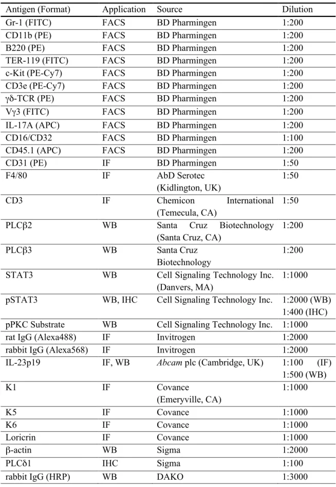
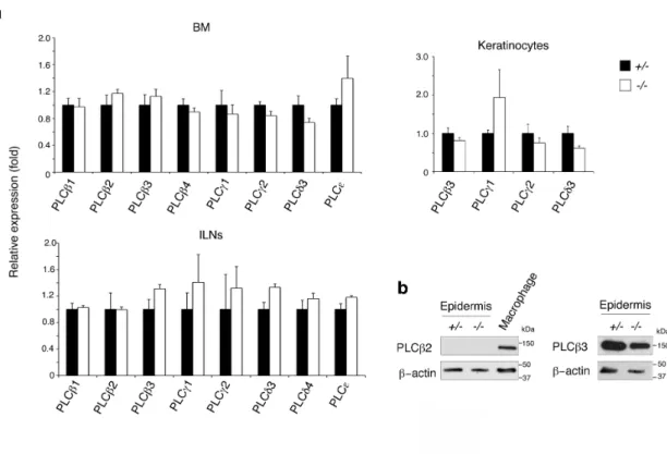
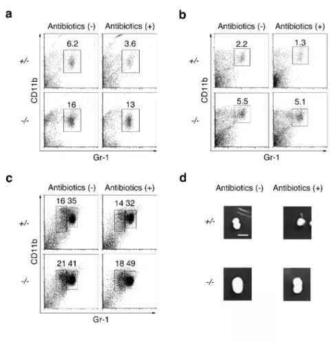
Loss of PLCβ3 in haematopoietic cells alters blood cell counts and populations in a haematopoietic cell intrinsic manner (Xiao et al., 2009). Since PLCβ3 binds to PLCδ1 (Guo et al., 2005) in a manner similar to the binding of PLCβ2, I investigated if the systemic loss of PLCδ1 also affected blood cell counts and populations. Mice lacking the PLCδ1 gene (PLCδ1-/- mice) exhibited a greater than 2-fold increase in peripheral blood leukocytes compared with wild-type mice (mean ± SEM cell numbers, 9.2 ± 0.85 × 10⁶ in wild-type vs 19 ± 1.4 × 10⁶ in PLCδ1-/- mice, n = 6 for wild-type and n = 9 for PLCδ1-/- mice). I also examined the population of CD11b+ Gr-1+ granulocytes. At first, I confirmed that PLCδ1 +/-mice did not exhibit increase in granulocytes (Fig. 3a), and PLCδ1+/- mice were therefore used as a control in subsequent experiments. Granulocytes were markedly increased in the peripheral blood leukocyte in PLCδ1-/- mice, as well as in the spleen and bone marrow (Fig. 3b and Table 3). PLCδ1-/- mice also showed lymphadenopathy of the ILNs and mild splenomegaly (Fig. 4a, b). I confirmed that expression of other PLC isoforms was not affected by loss of PLCδ1 (Fig. 5a, b). Antibiotic treatment of PLCδ1-/- mice had no effect on the
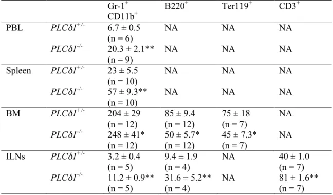
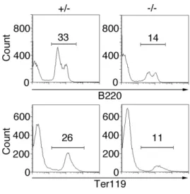
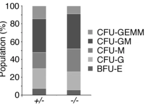
development of granulocytosis and lymphadenopathy (Fig. 6a-d), and PLCδ1-/- granulocytes appeared to be morphologically and functionally normal (Fig. 7a-c), suggesting that the observed granulocytosis was not a secondary effect of bacterial infection or impaired granulocyte function. Enhanced granulopoiesis in the bone marrow is often accompanied by a reduction of B lymphocytes and erythrocytes (Ueda et al., 2005; Walkey et al., 2007; Yang et al., 2007), and reduced numbers of B220+ B lymphocytes and Ter119+ erythrocytes were indeed observed in the bone marrow of PLCδ1-/- mice (Fig. 8 and Table 3). Granulocytes in mouse bone marrow can be categorized into immature and mature subsets (Walkley et al., 2002), both of which were elevated in PLCδ1-/- bone marrow (Fig. 3b), suggesting that the increase in the granulocyte population arose from an immature progenitor population. A BrdU incorporation assay revealed that the proliferative activity of immature granulocytes was higher in PLCδ1-/- than in PLCδ1+/- bone marrow (Fig. 9a), suggesting that the increase in granulocyte number resulted from enhanced proliferation of immature granulocytes. An increased population of myeloid progenitor cells could also increase the granulocyte number in PLCδ1-/- mice. Indeed, the population of myeloid progenitor cells was greater in the bone marrow of PLCδ1-/- than of PLCδ1+/- mice (Fig. 9b), as was the number of myeloid colony-forming units (Fig. 9c), indicating that the loss of PLCδ1 resulted in an increased population of myeloid progenitor cells in the bone marrow, possibly leading to granulocytosis.
16
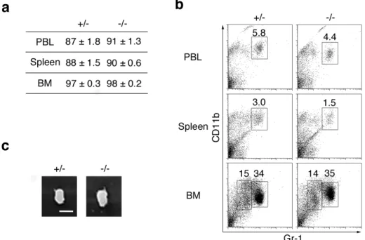
marrow did not develop granulocytosis or lymphadenopathy (Fig. 10a-c), indicating that the loss of PLCδ1 in the haematopoietic system was not responsible for these phenotypes, and that the mechanism of granulocytosis in PLCδ1-/- mice differed from that in mice lacking PLCβ3 (Xiao et al., 2009).
Figure 3 Loss of PLCδ1 resulted in granulocytosis.
(a) Representative FACS profiles of CD11b+ Gr-1+ granulocytes in the peripheral blood leukocytes
(PBL), spleen, and bone marrow (BM) (n = 3 per genotype). PLCδ1+/+
(b) Representative FACS profiles of CD11b+ Gr-1+ granulocytes in peripheral blood leukocytes
(PBL), spleen, and bone marrow (BM) (n = 14). Granulocytes in bone marrow were categorized
into immature (CD11b+ Gr-1low) and mature (CD11b+ Gr-1high) subsets. Mice used in all
Table 3 Absolute numbers of granulocytes, erythrocytes, and B and T lymphocytes in peripheral blood, spleen, bone marrow, and inguinal lymph nodes.
Gr-1+ CD11b+ B220 + Ter119+ CD3+ PBL PLCδ1+/- 6.7 ± 0.5 (n = 6) NA NA NA PLCδ1-/- 20.3 ± 2.1** (n = 9) NA NA NA Spleen PLCδ1+/- 23 ± 5.5 (n = 10) NA NA NA PLCδ1-/- 57 ± 9.3** (n = 10) NA NA NA BM PLCδ1+/- 204 ± 29 (n = 12) 85 ± 9.4 (n = 12) 75 ± 18 (n = 7) NA PLCδ1-/- 248 ± 41* (n = 12) 50 ± 5.7* (n = 12) 45 ± 7.3* (n = 7) NA ILNs PLCδ1+/- 3.2 ± 0.4 (n = 5) 9.4 ± 1.9 (n = 4) NA 40 ± 1.0 (n = 7) PLCδ1-/- 11.2 ± 0.9** (n = 5) 31.6 ± 5.2** (n = 4) NA 81 ± 1.6** (n = 7)
Statistical significance was assessed using a Student's t-test. *P < 0.05, **P < 0.01.
Cell numbers are represented as number × 105. NA; not available. PBL = peripheral blood lymphocytes; BM =
bone marrow; INLs = inguinal lymph nodes. For ILNs, Gr-1+ cells were counted instead of Gr-1+ CD11b+ cells
a
b
Spleen (mg) ILNs (mg) MLNs (mg) ALNs (mg)
PLCδ1+/- 62.2 ±2.6 (n=9) 4.3 ± 0.6 (n=14) 12.6 ± 1.9 (n=7) 6.6 ± 1.6 (n=7) PLCδ1-/- 83.7 ± 6.7** (n=9) 12.8 ± 1.1** (n=14) 13.6 ± 0.9 (n=7) 16.4 ± 1.7** (n=7)
Figure 4 PLCδ1-/- mice showed lymphadenopathy and splenomegaly.
(a) Macroscopic appearance of inguinal lymph nodes (ILNs). Scale bar = 2 mm. (b) Statistical significance was assessed using a Student's t-test. **P < 0.01.
18
Figure 5 Loss of PLCδ1 did not affect expression of PLC isozymes.
(a) Expression of genes encoding PLC isozymes (PLCβ1-4, γ1-2, δ3-4, and ε) in the bone
marrow (BM), ILNs and differentiated keratinocytes of control and PLCδ1-/- mice was determined
by real-time RT-PCR. PLC isozyme genes with mRNA levels below the limits of detection are not
shown in the graphs. Expression of PLCη1-2 and ζ mRNAs is also omitted because of their
restricted tissue distributions. All values are normalized relative to the expression of GAPDH mRNA, with expression in control mice set at 1. Results are expressed as the mean ± SEM (n = 4 for bone marrow and ILNs, n = 3 for keratinocytes) in arbitrary units. (b) Immunoblotting for
PLCβ2 and PLCβ3 in epidermis of control and PLCδ1-/- mice. A macrophage lysate was included
Figure 6 Antibiotic treatment failed to restore normal granulocyte counts in PLCδ1-/- mice.
(a–c) Water containing antibiotics (100 µg/mL geneticin and 10 µg/mL polymyxin B
sulphate) were supplied for 8 weeks after birth. Representative FACS profiles of CD11b+
Gr-1+ granulocytes in the PBL (a), spleen (b), and BM (c) (n = 3). (d) Macroscopic
20
Figure 7 Granulocyte functions in PLCδ1-/- mice are normal.
(a) May-Grünwald Giemsa-stained peripheral blood smears. Scale bar = 10 µm. (b) Phagocytic activity of peripheral blood granulocytes measured with fluorescein isothiocyanate (FITC)-labelled opsonized bacteria at 37°C for 15 min. Representative FACS profiles are displayed (n = 3). (c) Respiratory burst in bone marrow granulocytes. Measurement of respiratory burst activity assessed by dihydrorhodamine-123 with (blue) or without (red) PMA stimulation (1 µg/µL) (n = 3). Mice used in all experiments were 8–12 weeks old. Data presented in (a) are representative of three mice per genotype.
Figure 8 Loss of PLCδ1 decreased B lymphocytes and erythrocytes in bone marrow.
Representative FACS profiles of B220+ and Ter119+ cells in the BM (n = 7–12).
Figure 9 PLCδ1-/- mice had hypperproliferative immature granulocytes and the increased number of myeloid progenitor cells in bone marrow.
(a) BrdU incorporation in immature granulocytes (CD11b+ Gr-1low) in the BM. Data are expressed
as the relative mean fluorescence intensity (MFI) ± standard error of mean (SEM) (MFI of
PLCδ1+/- mice = 1) (n = 5). (b) Populations of myeloid progenitor-rich Lin- Sca-1- c-Kit+ cells in
the BM. Mean ± SEM (n = 8). (c) In vitro colony-forming assay. The numbers of colony-forming
cells per 6 × 10⁴ cells at 12 days after plating are displayed. Mean ± SEM (n = 4). Mice used in all
experiments were 8–12 weeks old. Statistical significance was assessed using a Student's t-test. *P <0.05;**P <0.01.
Figure 10 Haematopoietic loss of PLCδ1 did not result in granulocytosis and lymphadenopathy.
(a) Haematopoietic chimerism analysis. Chimerism of the PBL, spleen, and BM were determined
in transplanted mice by FACS analysis of CD45.1- cells. Mean percentage ± SEM. Six recipients
in each group. Three donor mice per genotype were used. (b) Representative FACS profiles of
CD45.1- CD11b+ Gr-1+ granulocytes in the PBL, spleen, and BM of CD45.1+ congenic recipients
reconstituted with PLCδ1+/- or PLCδ1-/- BM at 4 weeks post-transplantation. (n = 6). Plots shown
are gated on CD45.1- cells. (c) Macroscopic appearance of the ILNs. Scale bar = 2 mm. Mice used
22
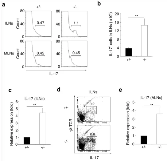
PLCδ1-/- mice display local and systemic IL-17 upregulation.
The granulocyte population was increased and the B220+ B lymphocyte and Ter119+ erythrocyte populations were decreased in the bone marrow of PLCδ1-/- mice (Fig. 3b, Fig. 8,
and Table 3). However, the balance among CFU-
granulocyte/erythroid/macrophage/megakaryocyte (GEMM), -granulocyte/macrophage (GM), -macrophage (M), -granulocyte (G), and burst forming unit- erythroid (BFU-E) was not dramatically disrupted in PLCδ1-/- bone marrow cells cultured in vitro (Fig. 11). I therefore speculated that granulocytosis in PLCδ1-/- mice was induced by humoral factors secreted from peripheral tissues. IL-17 is a critical cytokine for granulopoiesis (Forlow et al., 2001; Schwarzenberger et al., 1998; Schwarzenberger et al., 2000), and I therefore assessed serum IL-17 concentrations. IL-17 levels in the serum of control mice were below the detection limit, but those in PLCδ1-/- mice were detectable (Fig. 12a), indicating that circulating levels of the granulopoietic cytokine IL-17 were increased in PLCδ1-/- mice. The concentrations of other granulopoietic factors were not increased in PLCδ1-/- mice (Fig. 12b), strongly suggesting that IL-17 elevation is responsible for granulocytosis in these mice.
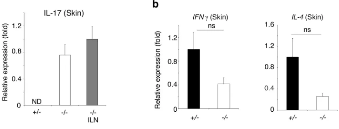
I investigated IL-17 production by lymphocytes in lymph nodes including ILNs and MLNs by intracellular cytokine staining. The population of IL-17-producing cells was increased in ILNs but not in MLNs in PLCδ1-/- mice (Fig. 13a). The number of IL-17-producing cells in the ILNs of PLCδ1-/- mice exhibited a 3.9-fold increase compared with that in PLCδ1+/- mice (Fig. 13b). IL-17 mRNA expression in ILNs analysed using real-time RT-PCR was increased in PLCδ1-/- mice (Fig. 13c). I then investigated the specific cellular source of IL-17 in ILNs of PLCδ1-/- mice. FACS analysis revealed that the main producers of IL-17 were the γδ-TCR-positive T cells (Fig. 13d). ILNs are skin-draining, whereas MLNs are not, suggesting that IL-17 was specifically upregulated in skin-draining lymph nodes. Indeed, another type of skin-draining lymph nodes, the axillary lymph nodes, also expressed high levels of IL-17 mRNA (Fig. 13e). These results suggest that the skin system plays a pivotal role in IL-17 upregulation. Importantly, IL-17 expression was detected in PLCδ1-/- skin but not in normal skin (Fig. 14a). In contrast to IL-17, the expression of
interferon (IFN) γ and IL-4 was not increased in skin of PLCδ1-/- mice (Fig. 14b). Thus,
Figure 11 Populations of CFU-GEMM, -GM, -M, -G, and BFU-E were unchanged in PLCδ1-/- bone marrow cells in vitro.
Populations of CFU-GEMM, -GM, -M, -G, and BFU-E in all colonies formed from
PLCδ1+/- and PLCδ1-/- BM cells are shown. The combined results from three independent
experiments are displayed. Percentages for population frequencies of total colonies are mean values for four mice in each group. Mice used in all experiments were 8–12 weeks old.
Figure 12 Loss of PLCδ1 elevated serum levels of IL-17.
(a) IL-17 concentration in the serum measured by ELISA. Mean ± SEM (n = 6). (b) Serum was
collected from four control and four PLCδ1-/- mice and pooled. Serum concentrations of IL-3, KC,
24
Figure 13 Loss of PLCδ1 increased IL-17 production in skin-draining lymphnodes.
(a) Representative FACS profiles of intracellular IL-17 in ILNs and mesenteric lymph nodes (MLNs). Cells were stimulated and intracellular IL-17 was detected (n = 6 for ILNs and n=3 for
MLNs). Three independent experiments were performed. (b) Absolute numbers of IL-17+ cells in
ILNs. Mean ± SEM (n = 6). The combined results from three independent experiments are displayed. (c) IL-17 mRNA expression in the ILNs determined by real-time RT-PCR. All values
are normalized to GAPDH. Results are displayed as arbitrary units (expression in PLCδ1+/- mice =
1). Mean ± SEM (n = 3). (d) Representative FACS profiles of γδ-TCR and intracellular IL-17 in ILNs (n = 3). Cells were stimulated and intracellular IL-17 was detected. Three independent experiments were performed. (e) IL-17 mRNA expression in the axillary lymph nodes (ALNs) determined by real-time RT-PCR. All values are normalized to GAPDH. Results are displayed as
arbitrary units (expression in PLCδ1+/- mice = 1). Mean ± SEM (n = 5). Mice used in all
Epidermal PLCδ1 is sufficient for normal IL-17 levels.
Among skin cells, keratinocytes express the highest levels of PLCδ1 (Nakamura et al., 2005). In addition, IL-17 was upregulated in skin and skin-draining lymph nodes. I therefore hypothesized that the PLCδ1 in keratinocytes primarily regulates IL-17 levels. To test this hypothesis, I investigated if reintroduction of PLCδ1 into keratinocytes of PLCδ1-/- mice could restore normal IL-17 levels. The expression of PLCδ1 in keratinocytes was mainly regulated by the transcription factor Foxn1 (Nakamura et al., 2008) and I therefore used a
Foxn1 promoter-driven PLCδ1 gene (Foxn1::PLCδ1) (Fig. 15a) to restore PLCδ1 expression
in PLCδ1-/- keratinocytes in a manner resembling that of endogenous PLCδ1. Mice carrying
Foxn1::PLCδ1 (Fig. 15b) appeared normal and did not exhibit any overt changes.
Intercrossing PLCδ1-/- mice and mice carrying Foxn1::PLCδ1 produced PLCδ1-/- mice
carrying Foxn1::PLCδ1 (Tg/KO mice). PLCδ1 expression was restored in the skin of Tg/KO mice, but not in other organs such as the lungs, brain, ILNs, spleen and bone marrow (Fig. 15c). PLCδ1 protein was weakly expressed in Tg/KO thymuses, but these thymuses did not show obvious phenotypes, such as a disturbed balance of T cell subtypes. Although the level
Figure 14 IL-17 mRNA was increased in PLCδ1-/- skin.
(a) IL-17 mRNA expression in skin determined by real-time RT-PCR. PLCδ1-/- ILN was used as
the positive control for IL-17 expression. All values are normalized to GAPDH. Results are
displayed as arbitrary units (expression in PLCδ1-/- ILN = 1). Mean ± SEM (n = 5).
(b) Levels of expression of IFN γ and IL-4 mRNA in the skin of 8–12-week-old control and
26
of expression of PLCδ1 protein was somewhat lower in Tg/KO than in wild-type and heterozygous skin (Fig. 15d), the immunofluorescence observation indicated that both endogenous and transgene-derived PLCδ1 protein was expressed in suprabasal epidermis (Fig. 15e). These expression patterns were consistent with enriched expression of PLCδ1 and Foxn1 in differentiated keratinocytes (Nakamura et al., 2008; Lee et al., 2008). At the histological level, the introduction of Foxn1::PLCδ1 gene rescued epidermal hyperplasia and immune cell infiltration that were observed in PLCδ1-/- mice (Ichinohe et al., 2007) (Fig. 16a,
b). Importantly, IL-17 expression was remarkably decreased in Tg/KO skin (Fig. 17a) compared with PLCδ1-/- skin. Residual expression of IL-17 mRNA in Tg/KO skin may be caused by a lower level of PLCδ1 protein expression in Tg/KO than in control skin. Tg/KO mice showed no lymphadenopathy of the ILNs (Fig. 17b and Table 4), and IL-17-producing cells were not increased in the ILNs in Tg/KO mice (Fig. 17c, d). Thus, reintroduction of
PLCδ1 into keratinocytes ameliorated local IL-17 upregulation. In addition, IL-17
Figure 15 Generation of Tg/KO mice.
(a) Structure of the Foxn1::PLCδ1 gene. Poly A: the bovine growth hormone polyadenylation
sequence. (b) PCR analysis of genomic DNA from the tails of wild-type and Foxn1::PLCδ1 Tg
mice. Products derived from Foxn1::PLCδ1 and endogenous PLCδ1. (c) Immunoblotting of
PLCδ1 and β-actin in tissues from control, PLCδ1-/- (KO), and Tg/KO mice. (d) β-actin was
included as a loading control. (e) Skin stained with antibody against PLCδ1 (red) and Hoechst stain (blue). Dotted lines denote the dermal-epidermal border. Scale bar = 50 µm. (e)
28
Figure 16 Tg/KO mice showed neither epidermal hyperplasia nor immune cell infiltration.
(a) Haematoxylin-eosin (HE) stained dorsal skin sections. Scale bar = 50 µm. Scale bar = 100 µm. (b) Skin stained with antibodies against CD3 (green), against F4/80 (green) or with Hoechst stain
(blue). Scale bar = 100 µm. 8–12-week-old mice were used. Heterozygotes with Foxn1::PLCδ1
Figure 17 PLCδ1 expression in keratinocytes was sufficient for normal granulocyte counts and IL-17 levels in PLCδ1-/- mice.
(a) IL-17 mRNA expression in the skin of control, PLCδ1-/- (KO), and Tg/KO mice determined by
real-time RT-PCR. All values are normalized to GAPDH. Results are displayed as arbitrary units (expression in the skin of KO mice = 1). Mean ± SEM (n = 5). (b) Macroscopic appearance of ILNs in control and Tg/KO mice. Scale bar = 1 mm. (i) Representative FACS profiles of intracellular IL-17 in ILNs. Cells were stimulated and intracellular IL-17 was detected (n = 4).
Three independent experiments were performed. (c) Absolute numbers of IL-17+ cells. Mean ±
SEM (n = 4). The combined results from three independent experiments are displayed. (d) IL-17 concentration in serum measured by ELISA. Mean ± SEM (n = 3). (e) Representative FACS
profiles of CD11b+ Gr-1+ granulocytes in the PBL, spleen, and BM of Tg/KO mice (n = 4).
8–12-week-old mice were used. Heterozygotes with Foxn1::PLCδ1 were used as controls.
30
Table 4 Weights of spleen, inguinal, and mesenteric lymph nodes in control and Tg/KO mice Spleen (mg) ILNs (mg) MLNs (mg) Control 55.3 ± 1.6 (n=4) 3.0 ± 0.4 (n=8) 15.3 ± 0.8 (n=7) Tg/KO 54.9 ± 1.4 (n=5) 4.0 ± 0.4 (n=8) 14.5 ± 0.7 (n=7)
ILNs = inguinal lymph nodes; MLNs = mesenteric lymph nodes.
Epidermal PLCδ1 regulates local and systemic IL-17 levels.
I examined if loss of PLCδ1 in keratinocytes caused IL-17 upregulation and granulocytosis by generating keratinocyte-specific conditional PLCδ1-knockout (cKO) mice with K14 promoter-driven Cre transgenic mice (Fig. 18a, b). I confirmed that PLCδ1 was deleted in the epidermis of the cKO mice and that its expression was not altered in other organs such as the lungs, brain, ILNs, spleen and bone marrow (Fig. 18c). Since the K14 promoter is active in the thymus, I examined the expression of PLCδ1 in the thymus, finding that PLCδ1 was downregulated in the thymus of cKO mice (Fig. 18c). However, I did not observe any obvious abnormalities in cKO thymus. IL-17 was upregulated in the skin of cKO mice (Fig. 19a), while IFN γ and IL-4 expression levels remained unchanged (Fig. 19b). Interestingly, IL-17 mRNA was upregulated more in the epidermis than in whole skin of cKO mice (Fig. 20a). CD3-positive T cells were major IL-17 producers in the cKO epidermis (Fig. 20b). The number of CD3-positive T cells was 1.7-fold higher in cKO than in control epidermis, suggesting that upregulation of IL-17 mRNA in cKO epidermis is due, at least in part, to an increase in T cells. Further analysis revealed that IL-17 was expressed by Vγ3-positive γδ T cells in cKO epidermis (Fig. 20c). I also found that the level of IL-17 mRNA was not significantly altered by depletion of Langerhans cells (Fig. 20d). cKO mice also showed increased ILN size (Fig. 21a , Table 5) and cell numbers (mean ± SEM cell numbers, 3.7 ± 0.9 × 10⁶ in controls vs 14 ± 2.4 × 10⁶ in cKO mice, both n = 6). The number of IL-17-producing cells in the ILNs of cKO mice was more than six times that in the control mice (Fig. 21b). The main IL-17 producing cells in cKO ILNs were γδ T cells (Fig. 21c).
IL-17 upregulation in ILNs of cKO mice was also confirmed by real-time RT-PCR (Fig. 21d).
Production of the granulopoietic cytokine G-CSF is induced by IL-17 in many cell types, and I therefore examined if CM derived from cKO skin-draining lymph nodes induced G-CSF expression. CM from cKO skin-draining lymph nodes induced higher G-CSF expression in fibroblasts compared with CM from control skin-draining lymph nodes (Fig. 22a). Importantly, CM from cKO skin-draining lymph nodes pretreated with anti-IL-17 neutralizing antibody did not cause G-CSF upregulation (Fig. 22a), strongly suggesting that cells in the
from skin and skin-draining lymph nodes causes serum IL-17 elevation, then serum IL-17 concentrations should also be increased in cKO mice. Indeed, serum IL-17 levels were high in
cKO mice (Fig. 22b), strongly suggesting that local IL-17 upregulation is linked to elevation
of serum IL-17 levels. In addition, serum G-CSF concentrations were significantly increased in cKO mice compared with control mice (Fig. 22c). I further investigated the granulocyte population in cKO mice and found that cKO mice showed granulocytosis (Fig. 22d), consistent with local and serum IL-17 upregulation. Reduced numbers of bone marrow B lymphocytes and erythrocytes were also observed in cKO mice (Fig. 23). Taken together, these results indicate that PLCδ1 in keratinocytes is required for the maintenance of normal IL-17 levels and granulocyte counts.
Figure 18 Generation of cKO mice.
(a) Genomic structure of the PLCδ1 gene (WT). Exons are indicated by filled boxes. The
structures of the targeting vector (TV) for disrupting the mouse PLCδ1, targeted allele (MT), allele after FLP recombination (After FLP), and allele after Cre recombination (After Cre) are displayed. (b) PCR genotyping of control and cKO mice. (c) Immunoblotting of PLCδ1 and β-actin in tissues
32
Figure 19 Epidermal loss of PLCδ1 increased IL-17 mRNA in skin.
(a) IL-17 mRNA expression in skin. KO ILN was used as positive control for IL-17 expression. Results are displayed as arbitrary units (expression in KO ILN = 1). Mean ± SEM (n = 3). (b)
Levels of expression of IFN γ and IL-4 mRNA in the skin of 8–12-week-old control and cKO
mice were determined by real-time RT-PCR. All results were normalized relative to expression of
GAPDH mRNA, with levels of expression in the skin of control mice set at 1. Results are
Figure 20 γδTCR+Vγ3+ cells mainly produced IL-17 in cKO epidermis. (a) IL-17 mRNA expression in epidermis (epi) and whole skin (skin). Results are displayed as arbitrary units (expression in cKO whole skin = 1). Mean ± SEM (n = 3). (b) Representative FACS profiles of IL-17 and CD3 of epidermis (n = 3). Three independent experiments were performed.
(c) γδTCR+Vγ3+ cells were isolated from 8–12-week-old cKO epidermal cell suspensions using
34
Table 5 Weights of spleen, inguinal, and mesenteric lymph nodes in control and cKO mice
Statistical significance was assessed using a Student's t-test. **P < 0.01. ILNs = inguinal lymph nodes; MLNs = mesenteric lymph nodes.
Spleen (mg) ILNs (mg) MLNs (mg) Control 69.3 ± 3.5 (n=5) 6.8 ± 1.4 (n=10) 12.2 ± 1.7 (n=7) cKO 100 ± 7.6** (n=5) 14.9 ± 1.6** (n=10) 13.5 ± 1.2 (n=7)
Figure 21 Epidermal loss of PLCδ1 resulted in lymphadenopathy and IL-17 overproduction in ILNs.
(a) Macroscopic appearance of ILNs. Scale bar = 2 mm. (b) Absolute numbers of IL-17+ cells in
Figure 22 Epidermal loss of PLCδ1 increased serum levels of IL-17 and G-CSF, leading to granulocytosis.
(a) G-CSF expression in Swiss 3T3 cells treated with skin-draining lymph-node CM in the presence of anti-IL-17 neutralizing antibody or isotype control. Results are displayed as arbitrary units (expression in cells treated with control skin-draining lymph-node CM and isotype control = 1). Mean ± SEM (n = 4). (b) IL-17 concentration in serum. Mean ± SEM (n = 10). c) G-CSF
concentration in the serum. Mean ± SEM (n = 6). (d) Representative FACS profiles of CD11b+
Gr-1+ granulocytes (n = 8). (a) normalized to GAPDH. 8–12-week-old mice were used. Statistical
36
Epidermal PLCδ1 regulates IL-23 expression in skin
I investigated the mechanisms of IL-17 upregulation in the epidermis by analysing the expression of the IL-17-inducing cytokine IL-23. IL-23 and IL-12 are functionally related as heterodimeric cytokines that share the IL-12/23p40 subunit (Oppmann et al., 2000). The mRNAs for the subunits of the IL-23 heterodimer (IL-12/23p40 and IL-23p19) were upregulated in cKO skin (Fig. 24a), while IL-12p35, which encodes the IL-12-specific subunit, was not upreglated (Fig. 24a). In contrast, a dramatic decrease in IL-23 expression was detected after reintroduction of PLCδ1 into keratinocytes (Fig. 24b). Since Tg/KO skin showed no drastic increase in IL-17 (Fig. 17a) while cKO skin showed remarkable IL-17 upregulation (Fig. 19a), the expression level of IL-23 was closely correlated with that of IL-17 in skin. Similar to IL-17, IL-23p19 mRNA and protein were upregulated in cKO epidermis (Fig. 24c, d). Immunofluorescence experiments showed faint IL-23p19 immunoreactivity in control epidermis, whereas keratinocytes in the basal layer of cKO epidermis showed strong IL-23p19 immunoreactivity (Fig. 24e). IL-23p19 expression was also assessed in primary
Figure 23 Loss of PLCδ1 in keratinocytes changed erythrocyte and B lymphocyte populations in bone marrow.
Representative FACS profiles of B220+, and Ter119+ cells in BM (n = 2–8 per genotype). Mice
keratinocyte cultures. cKO keratinocytes did not show increased expression of IL-23p19, regardless of their differentiation status (Fig. 24f). Thus, loss of PLCδ1 from keratinocytes did not upregulate IL-23 in this in vitro system, suggesting that interactions between PLCδ1-deficient keratinocytes and other epidermal cells may be required for IL-23 production. I then examined whether the epidermal increase in IL-23 was linked to IL-17 upregulation in the cKO epidermis. IL-23 was neutralized using its specific p19 subunit antibody and IL-17 expression was then examined in control and cKO epidermal sheets. IL-17 mRNA levels in the cKO epidermal sheet were clearly decreased in the presence of anti-IL-23p19 neutralizing antibody compared with levels in the presence of isotype control (Fig. 24g), indicating that IL-23 plays a critical role in IL-17 upregulation in cKO epidermis. I then investigated the mechanisms responsible for IL-23 upregulation by assessing activation of PLC and its downstream effector, PKC. I found that overall PLC activity was drastically decreased in cKO compared with control epidermis (Fig. 25a), indicating that, even in the presence of other PLC isoforms, loss of PLCδ1 impaired PLC activity in the epidermis. Consistent with the decrease in PLC activity, I found that the phosphorylation of PKC substrates was markedly decreased in cKO epidermis (Fig. 25b), indicating that loss of PLCδ1 from keratinocytes impaired the activation of the PLC downstream effector. I next determined if the PLC downstream signal affected IL-23 expression. PLC activation results in the generation of IP3 and DAG, leading to elevated concentrations of intracellular calcium ions and activation of PKC. I therefore treated epidermal sheets with the calcium ionophore, ionomycin, and PMA, a synthetic analogue of DAG and a PKC activator, to mimic PLC activation. IL-23p19 expression was upregulated in cKO epidermal sheets in the absence of ionomycin and PMA. Importantly, IL-23p19 upregulation was ameliorated in cKO epidermal sheets in the presence of ionomycin and PMA (Fig. 25c). Expression of the IL-12-specific subunit IL-12p35 was unchanged in the epidermis, regardless of the presence or absence of ionomycin and PMA (Fig. 25c). These results strongly suggest that IL-23 upregulation in the
38
Figure 24 The IL-17-inducing cytokine, IL-23 was upregulated in cKO epidermis.
(a) IL-12/23p40, IL-23p19, and IL-12p35 mRNA expression in the skin determined by real-time RT-PCR. All values are normalized to GAPDH. Results are displayed as arbitrary units (expression in control skin = 1). Mean ± SEM (n = 5). (b) IL-12/23p40, and IL-23p19 mRNA
expression in the skin of 8–12-week-old PLCδ1-/- (KO), and Tg/KO mice determined by real-time
RT-PCR. All values are normalized to GAPDH. Results are displayed as arbitrary units
Figure 24 continued
40
Figure 25 Impaired activation of PLC and its downstream effector caused
IL-23 upregulation in cKO epidermis.
cKO skin shares features of human psoriasis
42
Figure 26 Epidermal loss of PLCδ1 resulted in psoriasis-like dermatitis.
Figure 27 Psoriasis-related inflammatory genes were upregulated in cKO skin.
44
Figure 28 cKO skin shared features of human psoriasis.
Figure 29 PLCδ1 protein was decreased in epidermis of patients with psoriasis.
46
Loss of PLCδ1 in keratinocytes exacerbates contact hypersensitivity responses
DNFB-induced CHS of the skin in mice is commonly used as a model for studying the pathogenesis of allergic contact dermatitis (ACD), in which IL-17 plays a critical role (Nakae et al., 2002; He et al., 2009). I therefore assessed CHS responses in cKO mice. Mice were sensitized and challenged with DNFB, and the CHS response was assessed by measuring ear swelling. Upon challenge with DNFB, DNFB-sensitized control mice exhibited a CHS response with mild ear swelling, whereas cKO mice showed more prominent ear swelling with exaggerated edema and severe inflammatory cell infiltration (Fig. 30a, b). Interestingly, IL-17 neutralization resulted in abrogation of the exaggerated ear swelling in cKO mice almost to basal level at any time after challenge (Fig. 30c). These results indicate that the exacerbated CHS in cKO mice was IL-17-dependent.
Figure 30 Loss of PLCδ1 in keratinocytes exacerbated contact hypersensitivity responses.
(a) Time course of ear swelling after DNFB challenge. Ear swelling was measured at the indicated times. Mean ± SD (n = 4). (b) HE stains of ear after DNFB challenge. The ears of sensitized mice were painted with DNFB, collected 96 hr later and stained with HE. Lower panels are magnified views of the boxed regions in the upper panels. A scale bars in upper panel = 100 µm. A Scale bars in lower panel = 50 µm. (c) Sensitized mice were treated with anti-IL-17 neutralizing antibody or normal rat IgG before challenge and ear swelling was measured at the indicated times. Mean ± SD (n = 3). Mice used in all experiments were 8–12 weeks old. The data presented in (b) are representative of three mice per genotype. Statistical significance was assessed using a Student's
IV. Discussion
In all of mouse models used in this study, the level of expression of PLCδ1 in keratinocytes was inversely correlated with the levels of expression of IL-23 and IL-17 in skin and skin-draining LNs. Thus, loss of PLCδ1 in keratinocytes results in local activation of the IL-23/IL-17 axis (Fig. 31). Keratinocytes from lesional psoriatic skin express IL-23 (Johansen et al., 2009). In addition, human keratinocytes stimulated with nickel, a common hapten inducing CHS, produce IL-23 (Larsen et al., 2009). These results are consistent with my observation that IL-23p19 was upregulated in keratinocytes of cKO epidermis (Fig. 24e). Since IL-23p19 was upregulated mainly in basal layer of cKO interfollicular epidermis (Fig. 24e), essential and sufficient roles of suprabasal PLCδ1 in maintenance of normal IL-23p19 levels (Fig. 15e and Fig. 24b) are somewhat surprising. Interactions between suprabasal and basal keratinocytes might be important in regulation of IL-23p19 expression. I also found that γδ T cells in cKO epidermis expressed IL-17 (Fig. 20c). Interestingly, γδ T cells were recently reported to be major IL-17 producers in skin of IL-23-mediated psoriasiform dermatitis (Cai et al., 2011; Mabuchi et al., 2011; Macleod et al., 2013).
Aberrant activation of the IL-23/IL-17 axis in the skin is known to be involved in the development of inflammatory human skin diseases, especially psoriasis (Lee et al., 2004). Indeed, IL-23 injection into normal skin was sufficient for the development of psoriatic phenotypes in mice (Hedrick et al., 2009; Zheng et al., 2007), and a monoclonal antibody against IL-12/23p40 subunit, ustekinumab is efficacious for the treatment of patients with moderate-to-severe psoriasis (Leonardi et al., 2008). Although cKO skin shared some molecular features of psoriasis, it did not demonstrate the all histological characteristics of psoriatic skin. This may be because the expression of another key cytokine for the development of psoriasis, IL-22, was not upregulated in cKO skin. Nonetheless, as PLCδ1 expression was decreased in the epidermis of patients with psoriasis (Fig. 29) and in mouse IMQ-induced psoriasiform lesion (Fig. 26a), PLCδ1 may be involved in the pathogenesis of psoriasis. cKO mice also demonstrated increased sensitivity to hapten-induced CHS, a mouse model of human ACD. The fact that the exaggerated CHS response in cKO mice was inhibited by IL-17 neutralization demonstrated the involvement of PLCδ1 in IL-17-mediated ACD.
48
myeloproliferative disease characterized by increased granulocytes through elevated G-CSF production by keratinocytes (Meixner et al., 2008). As expression levels of JunB and G-CSF were unaltered in epidermis of cKO mice, PLCδ1 appears to cause granulocytosis by a different mechanism to that observed in keratinocyte-specific JunB-knockout mice.
The results of this study demonstrate that disruption of the PLCδ1 gene in keratinocytes disturbs not only local skin immune responses, but also the systemic homeostasis of haematopoietic cells, especially granulocytes. The proposed mechanism underlying the phenotypes seen in cKO mice is depicted in Fig. 31. These findings suggest that targeting body-surface-specific inflammatory pathways may prevent not only inflammatory skin diseases but systemic granulocytosis and related disorders.
Figure 31 Proposed model of local and systemic phenotypes induced by epidermal loss of PLCδ1.
V.
References
Aggarwal, S., Ghilardi, N., Xie, M.H., de Sauvage, F.J. & Gurney, A.L. Interleukin-23 promotes a distinct CD4 T cell activation state characterized by the production of interleukin-17. J. Biol. Chem. 278, 1910-1914 (2003)
Berridge, M.J. & Irvine, R.F. Inositol trisphosphate, a novel second messenger in cellular signal transduction. Nature. 312, 315-321 (1984)
Bleul, C.C. & Boehm, T. BMP signaling is required for normal thymus development. J.
Immunol. 175, 5213-5221 (2005)
Broome, A. M., Ryan, D. & Eckert, R. L. S100 protein subcellular localization during epidermal differentiation and psoriasis. J. Histochem. Cytochem. 51, 675-685 (2003) Cai, Y. et al. Pivotal Role of Dermal IL-17-Producing γδ T Cells in Skin Inflammation.
Immunity, 4, 596-610 (2011)
Creamer, D., Allen, M.H., Sousa, A., Poston, R. & Barke, J.N. Localization of endothelial proliferation and microvascular expansion in active plaque psoriasis. Br. J. Dermatol. 136, 859-865 (1997)
Dassule, H.R., Lewis, P., Bei, M., Maas, R. & McMahon, A.P. Sonic hedgehog regulates growth and morphogenesis of the tooth. Development. 127, 4775-4785 (2000)
Ettehadi, P. et al. Elevated tumour necrosis factor-alpha (TNF-alpha) biological activity in psoriatic skin lesions. Clin. Exp. Immunol. 96, 146-151 (1994)
Forlow, S.B. et al. Increased granulopoiesis through interleukin-17 and granulocyte colony-stimulating factor in leukocyte adhesion molecule-deficient mice. Blood. 98, 3309-3314 (2001)
Fried, L. et al. Inflammatory and prothrombotic markers and the progression of renal disease in elderly individuals. J. Am. Soc. Nephrol. 15, 3184-3191 (2004)
Fujino, S. et al. Increased expression of interleukin 17 in inflammatory bowel disease. Gut. 52, 65-70 (2003)
Fukami K. Structure, regulation, and function of phospholipase C isozymes. J. Biochem. 131, 293-299 (2002)
Goverman, J. Autoimmune T cell responses in the central nervous system. Nat. Rev. Immunol.
9, 393-407 (2009)
Grossman, R.M. et al. Interleukin 6 is expressed in high levels in psoriatic skin and stimulates proliferation of cultured human keratinocytes. Proc. Natl. Acad. Sci. USA. 86, 6367-6371 (1989)
Guo, Y., Rebecchi, M. & Scarlata, S. Phospholipase Cbeta2 binds to and inhibits phospholipase Cdelta1. J. Biol. Chem. 280, 1438-1447 (2005)
Happel, K.I. et al. Roles of Toll-like receptor 4 and IL-23 in IL-17 expression in response to Klebsiella pneumoniae infection. J. Immunol. 170, 4432-4436 (2003)
50
Hedrick, M.N. et al. CCR6 is required for IL-23–induced psoriasis-like inflammation in mice.
J. Clin. Invest. 119, 2317-2329 (2009)
Hern, S., Stanton, A.W., Mellor, R., Levick, J.R. & Mortimer, P.S. Control of cutaneous blood vessels in psoriatic plaques. J. Invest. Dermatol. 113, 127-132 (1999)
Homey, B. et al. Up-regulation of macrophage inflammatory protein-3 alpha/CCL20 and CC chemokine receptor 6 in psoriasis. J. Immunol.164, 6621-6632 (2000)
Ichinohe, M. et al. Lack of phospholipase C-delta1 induces skin inflammation. Biochem.
Biophys. Res. Commun. 356, 912-918 (2007)
Ishigame, H. et al. Differential roles of interleukin-17A and -17F in host defense against mucoepithelial bacterial infection and allergic responses. Immunity. 30, 108-119 (2009) Iwakura Y, Nakae S, Saijo S, Ishigame H. The roles of IL-17A in inflammatory immune
responses and host defense against pathogens. Immunol. Rev. 226, 57-79 (2008)
Johansen, C. et al. Characterization of the interleukin-17 isoforms and receptors in lesional psoriatic skin. Br. J. Dermatol. 160, 319-324 (2009)
Kouchi, Z. et al. Phospholipase Cdelta3 regulates RhoA/Rho kinase signaling and neurite outgrowth. J. Biol. Chem. 286, 8459-8471 (2011)
Larsen, J.M., Bonefeld, C.M., Poulsen, S.S., Geisler, C. & Skov, L. IL-23 and T(H)17-mediated inflammation in human allergic contact dermatitis. J. Allergy Clin.
Immunol.123, 486-492 (2009)
Lee, D., Prowse, D.M. & Brissette, J.L. Association between mouse nude gene expression and the initiation of epithelial terminal differentiation. Dev. Biol. 208, 362-374 (1999) Lee, E. et al. Increased expression of interleukin 23 p19 and p40 in lesional skin of patients
with psoriasis vulgaris. J. Exp. Med. 199, 125-130 (2004)
Leonardi, C. L. et al. Efficacy and safety of ustekinumab, a human interleukin-12/23 monoclonal antibody, in patients with psoriasis: 76-week results from a randomised, double-blind, placebo-controlled trial (PHOENIX 1). The Lancet. 371, 1665-1674 (2008) Lichti, U., Anders, J. & Yuspa, S.H. Isolation and short-term culture of primary keratinocytes,
hair follicle populations and dermal cells from newborn mice and keratinocytes from adult mice for in vitro analysis and for grafting to immunodeficient mice. Nature Protocols. 3, 799-810 (2008)
Lubberts, E. et al. Requirement of IL-17 receptor signaling in radiation-resistant cells in the joint for full progression of destructive synovitis. J. Immunol. 175, 3360-3368 (2005) Mabuchi, T., Takekoshi, T. & Hwang, S.T. Epidermal CCR6+ γδ T Cells Are Major
Producers of IL-22 and IL-17 in a Murine Model of Psoriasiform Dermatitis. J. Immunol.
187, 5026-5031 (2011)
Macleod, A. S. et al. Dendritic epidermal T cells regulate skin antimicrobial barrier function.
J. Clin. Invest. 123, 4364-4374 (2013)
Margolis, K.L. et al. Leukocyte count as a predictor of cardiovascular events and mortality in postmenopausal women: the Women’s Health Initiative Observational Study. Arch. Intern.
Med. 165, 500-508 (2005)
Micali, G., Lacarrubba, F., Musumeci, M.L., Massimino, D. & Nasca, M.R. Cutaneous vascular patterns in psoriasis. Int. J. Dermatol. 49, 249-256 (2010)
Murphy, M., Kerr, P. & Grant-Kels, J.M. The histopathologic spectrum of psoriasis. Clin.
Dermatol. 25, 524-528 (2007)
Nakae, S. et al. Antigen-specific T cell sensitization is impaired in IL-17-deficient mice, causing suppression of allergic cellular and humoral responses. Immunity. 17, 375-387 (2002).
Nakae, S., Nambu, A., Sudo, K. & Iwakura, Y. Suppression of immune induction of collagen-induced arthritis in IL-17-deficient mice. J. Immunol. 171, 6173-6177 (2003) Nakamura, Y. et al. Phospholipase C-δ1 and -δ3 are essential in the trophoblast for placental
development. Mol. Cell. Biol. 25, 10979-10988 (2005)
Nakamura, Y. et al. Phospholipase C-δ1 is an essential molecule downstream of Foxn1, the gene responsible for the nude mutation, in normal hair development. FASEB J. 22, 841-849 (2008)
Nakamura, Y. et al. Phospholipase Cdelta1 is required for skin stem cell lineage commitment.
EMBO J. 22, 2981-2991 (2003)
Nestle, F.O., Di Meglio, P., Qin, J.Z. & Nickoloff, B.J. Skin immune sentinels in health and disease. Nat. Rev. Immunol. 9, 679-691 (2009)
Nishizuka, Y. The molecular heterogeneity of protein kinase C and its implications for cellular regulation. Nature. 334, 661-665 (1988)
Ong, P. Y. et al. Endogenous antimicrobial peptides and skin infections in atopic dermatitis.
N. Engl. J. Med. 347, 1151-1160 (2002)
Oppmann, B. et al. Novel p19 protein engages IL-12p40 to form a cytokine, IL-23, with biological activities similar as well as distinct from IL-12. Immunity. 13, 715–725 (2000) Piskin, G., Sylva-Steenland, R.M., Bos, J.D. & Teunissen, M.B. In vitro and in situ
expression of IL-23 by keratinocytes in healthy skin and psoriasis lesions: enhanced expression in psoriatic skin. J. Immunol. 176, 1908-1915 (2006)
Ruggiero, C. et al. White blood cell count and mortality in the Baltimore Longitudinal Study of Aging. J. Am. Coll. Cardiol. 49, 1841-1850 (2007)
Sano, S. et al. Stat3 links activated keratinocytes and immunocytes required for development of psoriasis in a novel transgenic mouse model. Nat. Med. 11, 43-49 (2005)
Schwarzenberger, P. et al. IL-17 stimulates granulopoiesis in mice: use of an alternate, novel gene therapy-derived method for in vivo evaluation of cytokines. J. Immunol. 161, 6383-6389 (1998)
Schwarzenberger, P. et al. Requirement of endogenous stem cell factor and granulocyte-colony-stimulating factor for IL-17-mediated granulopoiesis. J. Immunol. 164, 4783-4789. (2000)
Shigeta. A. et al. An L-selectin ligand distinct from P-selectin glycoprotein ligand-1 is expressed on endothelial cells and promotes neutrophil rolling in inflammation. Blood. 13, 4915-4923 (2008)