岩手医科大学 審 査 学 位 論 文
(博 士)
67
I . Introduction
The regeneration of the liver from physical damage requires the involvement of various kinds of cells. A number of studies have reported that putative liver progenitor cells are one of the cell types that contribute to liver tissue repair
1 - 6 ). Recently, it has been reported that sox 9 -positive intrahepatic cholangiocytes are a major source of hepatocytes for liver tissue homeostasis
7 ).
However, it is difficult to apply the human equivalent of the oval cells (i.e., liver progenitor cells) during cell-based therapy in practice because their tissue of origin remains unknown
8).
On the other hand, it has been proposed that extrahepatic cells, as one of the hepatic lineages that respond to injury, contribute to liver regeneration
9). Earlier reports speculated regarding the involvement of extrahepatic
Potential involvement of extrahepatic cells in liver regeneration
Hirokatsu K atagiri
1), Satoshi N ishizuka
1), 2), Hiroyuki N itta
1)and Go W akabayashi
1)1)
Department of Surgery, School of Medicine,
Iwate Medical University, Morioka, Japan
2)
Institute for Biomedical Sciences, Iwate Medical University, Yahaba, Japan
(Received on January 21, 2014 & Accepted on January 27, 2014)
Extrahepatic cells have been considered a potential cellular resource for liver regeneration in response to liver damage. The genotyping of human living donor liver transplant (LDLT) recipients has reported that cells derived from those recipients of living donor liver transplantation differentiated into multiple types of liver component cells, suggesting that extrahepatic, as well as multipotent, cells contributed to liver regeneration. However, the consequences of the involvement of the extrahepatic cells in terms of clinically observed liver enlargement remain unknown.
In the present study, we suggest that cells that
might have been of extrahepatic origin contributed to the increase in liver volume, while the hypertrophy of the hepatocytes played an important role in the initial enlargement. Despite the rapid growth of the liver volume, cells in the mitotic phase were not evident. Taken together, these results suggest that: (i) Liver enlargement is achieved through some degree of extrahepatic cellular proliferation via liver progenitor cells, and (ii) cells from the recipient of living donor liver transplantation may contribute to the regeneration of the grafted liver.
Abstract
Key words : extrahepatic cells, liver regeneration, stem cells, transplantation
Hirokatsu K
atagiri, et al.
68
cells in liver regeneration based on the fact that cellular and genetic chimerism exists in transplanted livers
10 - 13 ). Investigators have also reported that cells from bone marrow are from an extrahepatic cellular source
14 - 20 ). In particular, some recent studies have shown that bone-marrow-derived mesenchymal stem cells (BM-MSCs) contribute to liver regeneration after liver damage
21-24). However, the consequences of the extrahepatic cells ’ involvement in liver regeneration are still unclear because it is difficult to keep track of human liver samples histologically. LDLT is one of the rare opportunities in which such pathological time-course samples are available, along with macroscopic images of liver regeneration.
To investigate the histological background against which extrahepatic cells contribute to liver regeneration, we counted the number of hepatocytes per area at three time points after LDLT. The number of hepatocytes indicates the volume of individual cells.
Thus, it would be an informative hallmark of
cellular hypertrophy or proliferation when it is compared to macroscopic liver volume. Here, we demonstrate how hepatocytes respond during the process of liver regeneration, in which extrahepatic cells may be involved, based on the data from human LDLT specimens.
II . Materials and methods 1. Human objects of LDLT
Ten pairs of recipients and donors who underwent LDLT between January 2007 and February 2011 at the Department of Surgery in Iwate Medical University Hospital were subjected to analysis. The subject LDLT pairs were restricted based on the availability of time-course samples. The specimens were 10 livers removed from recipients, 10 zero- point biopsies from donors, and 10 needle biopsies of the grafts. A zero-point biopsy is performed to test the quality of a donor liver before transplantation. The graft biopsies used in the present study were taken at various time points between 5 and 12 months after Table 1. Characteristics of donors and recipients of LDLT
1 2 3 4 5 6 7 8 9 10
R:recipient,D:donor, PBC:primary biliary cirrhosis, PSC:primary sclerosing cholangitis, TOB: Time of Biopsy, L: left lobe, C: caudate lobe, R: right lobe, ER: extended right lobe, LC:
liver cirrhosis, HCC: hepatocellular carcinoma, LF: liver failure, w: weeks, m: months.
Sex(R) F M F M F F M F F F
Age(R) 51 59 37 49 57 57 57 56 53 60
Sex(D) M M M F F F M M M F
Age(D) 24 27 37 55 33 32 30 30 25 32
Graft L + C
R L R L + C
R ER
R R R
Infection (-) HCV
(-) HCV HBV (-) HCV HCV HCV HCV
PBC LC,HCC PSC LC,HCC LC,HCC PBC LC,HCC LC,HCC LF LC
Case Primary disease TOB Early/Late
2w/6m
8w/14m
3w/6m
8w/16m
3w/6m
4w/6m
2w/7m
3w/10m
4w/6m
7w/8m
LDLT; they were retrospectively reviewed.
The study was reviewed and approved by the Ethics Committee of Iwate Medical University School of Medicine (HG H 23 - 13 ). Informed consent was obtained from all donors/
recipients according to the Institutional Review Board guidelines. The details of the LDLT set are shown in Table 1.
2. Macroscopic examinations of liver regeneration
In the process of following up after LDLT, CT volumetry (Toshiba Medical Systems, Tokyo) was performed in the portal phase (30 sec after the injection of the contrasting agent) at early and late time points. A regenerating liver image was constructed from a series of 1 - 2 mm slices from the DCOM file using the SYNAPSE VINCENT program (FUJIFILM, Tokyo) running on an Intel Xenon 2 . 93 GHz personal computer. The outline of the liver was first traced automatically and then fine- tuned manually for all slices. The volumes of
the inferior vena cava and extrahepatic portal veins were excluded.
3. Histological examinations
For the human LDLT biopsy specimens, samples were fixed with 10 % formalin for pathological examination. Three time-point specimens (i.e., the recipient, zero-point, and graft biopsies) were subjected to H&E staining for hepatocyte number counting (200
× 200 μm
2per view). Cells were counted in eight arbitrarily selected view fields, and the average number per field was calculated. The cell numbers at each time-point relative to the maximum number of each LDLT set were calculated.
III . Results
1. Macroscopic examinations of graft liver volume
The median weight and volume of the graft liver (before transplantation) were 594 g and 635cm
3, respectively. The median volumes of Fig. 1. CT and liver volume rendering. (A) Early and (B) late time points of Case 1; (C)
early and (D) late time-points of Case 7. The left and right panel in each box
indicate the CT and volume rendering of the regenerating liver, respectively.
the graft liver at early and late time points were 852 and 1149 cm
3, respectively. The median enlargement rate was 148 % at the early time point. Almost all cases showed the enlargement of liver volume, but one case ( 10 %) showed no volume gain, even at the late time point. The rate of enlargement is shown in Table 2 . Images of the CT and volume rendering of early and late time points for Cases 1 and 7 are shown (Fig. 1A-D). As shown, the liver volume gradually increased
and reached 285 % and 216 % enlargement in Cases 1 and 7, respectively. Median liver volume enlargement at the late time point for all cases was 177% (Table 2).
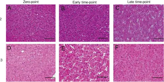
2. The number of hepatocytes per field The number of hepatocytes at the early time points was reduced to 79% of the median value, and it then recovered to almost that of the donor (median 98%, Table 3). Typical H&E images are shown in Figure 2 A-F.
Almost the entire LDLT dataset demonstrated
70 Hirokatsu K
atagiri, et al.
Table 2. Changes in graft volume after LDLT
1 2 3 4 5 6 7 8 9 10
517 667 429 603 405 560 1000 840 703 765 Case Initial volume(cm
3)
G
872 981 747 831 689 831 1029 1188 1305 832 Early (cm
3)
V1
Late(cm
3) V2 1475 1343 896 874 874 798 1400 1287 1712 1012
168.7 147.1 174.1 137.8 170.1 148.4 102.9 141.4 185.6 108.8 Initial → Early
Δ (%)*
Initial → Late Δ (%)**
285.3 201.3 208.9 144.9 215.8 142.5 140.0 153.2 243.5 132.3
* Δ ={(V1-G)/G} × 100%,** Δ ={(V2-G)/G} × 100%.
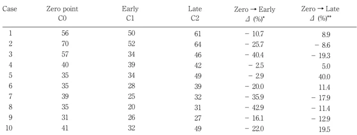
Table 3. Number of hepatocytes per unit view (cells/200 × 200 μm
2)
1 2 3 4 5 6 7 8 9 10
56 70 57 40 35 35 39 35 31 41 Case Zero point
C0
50 52 34 39 34 28 25 20 26 32 Early
C1
Late C2
61 64 46 42 49 39 32 31 27 49
− 10.7
− 25.7
− 40.4
− 2.5
− 2.9
− 20.0
− 35.9
− 42.9
− 16.1
− 22.0 Zero → Early
Δ (%)*
Zero → Late Δ (%)**
8.9
− 8.6
− 19.3 5.0 40.0 11.4
− 17.9
− 11.4
− 12.9 19.5
* Δ ={(C1-C0)/C0} × 100%,** Δ ={(C2-C0)/C0} × 100%.
the same “ v-shape ” pattern when plotted, indicating that cells were enlarged shortly after liver transplantation (Fig. 3). The liver volume at the late time point was significantly increased (median 179%), while the number of hepatocytes was not increased (median 98%), suggesting that mechanisms other than early hypertrophy may have been involved.
IV . Discussion
Here, we demonstrated that hepatocyte proliferation is more dominant in the late time point after LDLT, while hypertrophy is the first trigger of liver regeneration, possibly sharing the process with wound healing.
Importantly, the graft liver volume reached 179% during a 6-12-month time period, which should require a similar degree of cell division as is seen in rapidly growing neoplasms.
However, cells in the mitotic phase were not
evident in the graft samples, indicating that liver regeneration at the late stage requires an extrahepatic origin. Thus, cellular resources should be extrahepatic during the LDLT regeneration process.
Zero-point Early time-point Late time-point
Case 2
Case 3
Fig. 2. H&E staining of the liver in the process of regeneration. (A) Zero point, (B) early time point, and (C) late time point of case 2. (D) Zero point, (E) early time point, and (F) late time point of case 3. Scale bar: 20 μm.
×
0.5 Zero-point Early time-point Late time-point 0.6
0.7 0.8 0.9 1
* *
*
◆
◆
◆
●
●
●
◆
◆
◆
×
×
■+
+
+
■
■
■
■
■
■
■
■
Fig. 3. Changes in the number of hepatocytes.
The relative fraction of the maximum number of hepatocytes per area of each time point is plotted. The view area is 200
× 200 μm
2.
Zero-point Early time-point Late time-point
Hirokatsu K
atagiri, et al.
72
Supporting our hypothesis, there have been reports describing the fact that cells of recipient origin were detected in LDLT grafts
11 - 13 ). This observation immediately indicates the involvement of extrahepatic cells. Although the exact origin of the extrahepatic cells has not yet been clarified, some candidate stem cells of bone-marrow m e s e n c h y m a l s t e m c e l l o r i g i n a r e believed to contribute to liver regeneration, such as multilineage-differentiating stress- enduring (Muse) cells
25). In fact, it has been reported that human Muse cells voluntarily differentiate into AFP-positive cells in vitro
26). Both AFP and CK 19 have been considered to be markers for oval cells, which may differentiate into multiple types of liver tissue components
5). A specific type of bone- marrow mesenchymal stem cell should be an interesting target to investigate in terms of its application for selected cell-based therapy
22 ). It is important to clarify whether any types of liver progenitor cells are present in the process of tissue repair after injury
23 , 24 ). Although the tissue of origin of the liver progenitor cells remains unknown, the prevalence of a recipient signal in each tissue component may suggest that extrahepatic (i.e., recipient) cells that have multipotency,
at least partially, may have migrated and differentiated into liver component cells
15). In the case of male recipients and female liver donors, FISH analysis also revealed that liver component cells showed a Y chromosome signal, which indicates an extrahepatic cellular origin
11-13). In addition, extrahepatic cells that express the liver progenitor marker may be related to the cells composing hepatocytes and bile-duct-like structures in the periportal
area
14 ). Based on the present findings and
previous reports, we considered extrahepatic cells with differentiation potential to be one of the candidate cells for repairing liver damage.
Importantly, in the present study, we suggest that extrahepatic cells may be involved at the late time point after LDLT.
Currently, most liver regeneration studies have been performed using rodent models.
The present study, using human LDLD materials, should provide precious insights into developing possible alternative approaches to liver regeneration, such as cell-based therapy using bone marrow cells.
Conflict of interest: The authors have no conflict of interest to declare.
References 1) Brues AM and Marble BB: An Analysis of
Mitosis in Liver Restoration. J Exp Med 65, 15-27, 1937.
2) Alison M R, Golding M, Sarraf C E, et al.: Liver damage in the rat induces hepatocyte stem cells from biliary epithelial cells. Gastroenterology 110, 1182-1190, 1996.
3) Michalopoulos GK and DeFrances MC: Liver regeneration. Science 276, 60-66, 1997.
4) Theise ND, Saxena R, Portmann BC, et al.:
The canals of Hering and hepatic stem cells in humans. Hepatology 30, 1425-1433, 1999.
5) Wang X, Foster M, Al-Dhalimy M, et al.: The origin and liver repopulating capacity of murine oval cells. Proc Natl Acad Sci USA 100 (Suppl 1), 11881-11888, 2003.
6) Duncan AW, Dorrell C and Grompe M: Stem cells and liver regeneration. Gastroenterology 137, 466-481, 2009.
7) Furuyama K, Kawaguchi Y, Akiyama H, et al.:
Continuous cell supply from a Sox9-expressing progenitor zone in adult liver, exocrine pancreas and intestine. Nat Genet 43, 34-41, 2011.
8) Forbes SJ and Newsome PN: New horizons for stem cell therapy in liver disease. J Hepatol 56, 496-499, 2012.
9) Sell S: Heterogeneity and plasticity of hepatocyte lineage cells. Hepatology 33, 738-750, 2001.
10) Qian S, Demetris AJ, Murase N, et al.: Murine liver allograft transplantation: tolerance and donor cell chimerism. Hepatology 19, 916-924, 1994.
11) Kleeberger W, Rothamel T, Glockner S, et al.:
High frequency of epithelial chimerism in liver transplants demonstrated by microdissection and STR-analysis. Hepatology 35, 110-116, 2002.
12) Hove WR, van Hoek B, Bajema IM, et al.:
Extensive chimerism in liver transplants:
vascular endothelium, bile duct epithelium, and hepatocytes. Liver Transpl 9, 552-556, 2003.
13) Ng IO, Chan KL, Shek WH, et al. : High frequency of chimerism in transplanted livers.
Hepatology 38, 989-998, 2003.
14) Petersen BE, Bowen WC, Patrene KD, et al.:
Bone marrow as a potential source of hepatic oval cells. Science 284, 1168-1170, 1999.
15) Alison MR, Poulsom R, Jeffery R, et al.:
Hepatocytes from non-hepatic adult stem cells.
Nature 406, 257, 2000.
16) Lagasse E, Connors H, Al-Dhalimy M, et al.:
Purified hematopoietic stem cells can differentiate into hepatocytes in vivo. Nat Med 6, 1229-1234, 2000.
17) Theise ND, Nimmakayalu M, Gardner R, et al.:
Liver from bone marrow in humans. Hepatology 32, 11-16, 2000.
18) Schwartz RE, Reyes M, Koodie L, et al.:
Multipotent adult progenitor cells from bone
marrow differentiate into functional hepatocyte- like cells. J Clin Invest 109, 1291-1302, 2002.
19) Russo FP, Alison MR, Bigger BW, et al.: The bone marrow functionally contributes to liver fibrosis. Gastroenterology 130, 1807-1821, 2006.
20) Wang L, Wang X, Xie G, et al.: Liver sinusoidal endothelial cell progenitor cells promote liver regeneration in rats. J Clin Invest 122, 1567-1573, 2012.
21) Abdel Aziz MT, Atta HM, Mahfouz S, et al.:
Therapeutic potential of bone marrow-derived mesenchymal stem cells on experimental liver fibrosis. Clin Biochem 40, 893-899, 2007.
22) Kuo TK, Hung SP, Chuang CH, et al.: Stem cell therapy for liver disease: parameters governing the success of using bone marrow mesenchymal stem cells. Gastroenterology 134, 2111-2121, 2121 e2111-2113, 2008.
23) Kanazawa H, Fujimoto Y, Teratani T, et al.:
Bone marrow-derived mesenchymal stem cells ameliorate hepatic ischemia reperfusion injury in a rat model. PLoS One 6, e19195, 2011.
2 4 ) L i J a n d L i L : I m m e d i a t e i n t r a p o r t a l transplantation of human bone marrow mesenchymal stem cells prevents death from fulminant hepatic failure in pigs. Hepatology 2012.
25) Wakao S, Kitada M, Kuroda Y, et al.:
Multilineage-differentiating stress-enduring (Muse) cells are a primary source of induced pluripotent stem cells in human fibroblasts. Proc Natl Acad Sci USA 108, 9875-9880, 2011.
26) Kuroda Y, Kitada M, Wakao S, et al.: Unique
multipotent cells in adult human mesenchymal
cell populations. Proc Natl Acad Sci USA 107,
8639-8643, 2010.
74
肝再生における肝外細胞の関与の可能性
片桐弘勝
1),西塚 哲
1),2), 新田浩幸
1),若林 剛
1)1)
岩手医科大学医学部,外科学講座
2)