Plaque Assay Method for Adenovirus Type 5 with the Culture of HEK Cells Synchronously Infected with the Virus.
Yoshiaki UEDA, Mitsunobu AKASHI, and Kaoru HAYASHI
ABSTRACT: The mixture of antiserum and antigen at each adequate dilution was added the seed virus contained X × 104 PFU per ml. The mixture was diluted upto the concentration contained 50 to 100 plaques per ml per a dish and 2 ml of the last dilution was made. The last dilution of the mixture was added equal volume of HEK cell suspension contained 2 or 3×10⁶ cells per ml and shaked at 37℃ for 20 or 30 minutes. One ml of these mixture of antiserum, antigen and seed virus was plated into a dish, and 4 ml of maintainance medium contained 0.5% calf serum and 0.75% carboxymethylcellulose was added and spread over in a dish. After incubation for 5 or 6 days, the cell sheet was fixed with 10% formaldehyde saline solution and washed and stained with Gimsa solution. The plaques formed in this method were clear and easy to count. The activity of antigen A, C and P of adenovirus type 5 for the blocking antibody against purified adenovirus type 5 was studied with the application of this plaque assay method.
Department of Virology, Institute for Tropical Medicine, Nagasaki University
(Chief: Prof. Kaoru Hayashij
For the quanぬtive examination of animal viruses, the plaque assay method was
extremely estimated since Dulbecco (1952) had reported it. The plaque formation of animal viruses was usually performed with the agar overlay method. However, the advantaged
knoいrledge of the inhibitory effect of agar on the cell growth and the plaque formation
of viruses was shown by Takemoto and Liedhaver (1961). In contrast, the use of carboxymethjdcelluolse instead of agar in the overlay medium was shown to assure the uniform results for the plaque formation of certain viruses by Hotchin (1955) and Rapp et al. (1959).
Recently, the plaque formation o土dengue viruses and certain group B arboviruses under the carboxymethylcellulose overlay medium was performed and demonstrated excellent results by Schulze and Schlesinger (1963) and Makino and Mitune (1975).
For the plaque assay method, many workers usually used a monolayer of cells, not suspended cells, at the initial inoculation of the virus. It was, however, found that when the adequate number of suspended cells was infected with the virus and plated into
Coユtribution No. 734 from the Institute for Tropical Medicine, Nagasaki University
Received for publication, October 17, 1975
dishes, the plaque formation in the cell sheet was able to observe after the incubation for several days in this study. Furthermore, the block test was able to perform such as follows : the suspended cells were infected synchronously with the virus survived from the neutralization reaction with the excess antibody in the mixture of antiserum and antigen.
This method is useful for the application of block test of the antiserum with antigens・
In this paper, the results of the block activity of antigen A, C and P of adenovirus type 5 will be described.
MATERIAS AND METHODS
Seed virus: Adenovirus type 5, AD 75 strain, was propagated in HEK cellculture and the seed virus was partially purified by equilibrium density centrifugation in cesium chloride as described by Russell et al・ (1967)・
Cells : A line of human embryo kidney cells (HEK) cultured in Eagle′s medium containing
tryptose phosphate broth and lO% calf serum for the growth medium and o・5% calf
serum for the maintainance medium were used.
Antisera: Guinea pig antiserum perpared against the partially purified virus was kindly
gll en by Dr・ Pereira・
Antigen A, C and P : Soluble antigen obtained by the cesium chloride density gradient centrifugation was fractionated with DEAE Sephadex A50 column chromatography. The column was eluted with increasing molarities of sodium chloride in a step‑wise gradient as stated by Pereira (1967).
P antigen was prepared as described by Russell et al・ (1967)・ RK 13 cells infected with adenovirus type 5 in the presence of cytosine arabinoside at a concentration of 20 ug per ml was harvested at 12 to 14 hours after incubation・ Infected cellswere washed
with phosphate buffered saline and scrapped them林 The pellet of cells obtained by
centrifugation was disrupted by sonication at low temperature and centrifuged at 98,OOQg for one hour・ Such a supernatant fluid obtained from infected cells was the earlyextract called Iしantigen as described by Russell et al・ (1967).
Neutralization test : Serial dilutions of antiserum were added the equivalent virus solution contained l・5×10⁴ PFU per ml・ After the mixture was kept at room temperature for one
hour, it was diluted upto l‥ 100 and added the equivalent cell suspension consisted of
2 or 3×10⁶ cells per ml林 These mixture were also shaken at 37‑C for 30 minutes and
each 1 ml of mixtures was plated into dishes with 5 cm in diameter. Each dishes
added 4 ml of the overlay medium of Eagles solution contained o・5% calf serum and 0.75%
carboxymethylcellulose. After incubation for several days, the cell sheets were fixed with
lO% formaldehyde saline solution and stained with Gimsa solution lor the observation of
plaques・ The neutralization titer of the antiserum was determined by the ordinal method
of the calculation of plaque reduction. Table 1 shows the method of neutralization technique
using suspended cells for the initial inoculation of the virus・
Table l林 Method for neutralization test
l・ 0.2 ml of antiserum at the twofold dilution was added O林2 ml of xxlO4/ml of virus dilution・
2. 1.2 rnl of diluent without antiserum was added O・2 ml of xxlO4/ml of virus solution for the calc什 Iation of the original virus titer.
3. Mixtures of virus and antiserum or diluent were kept at room temperature for 60 minutes. They were diluted up to 1:100, and the last dilution consist of 2 ml in each tubes. In each the last dilution of mixtures, the seed virus will be contained 50 to 100 plaques per a dish.
4・ Preparation of the cells: Monolayer cells of HER was trypsinized, washed and prepared adeqい.tate
numbers of bijou in which 2 ml of cell suspension were contained 2.i XIO cells per ml.
5林Each 2 ml of HEK cell suspension were added 2 ml of diluted mixture of virus and antiserum or
diluent, and they were shaken at 370C for 30 minutes.
6林 One ml of mixtures consisted of cells, antiserum and virus was plated into a petri dish・ Two petri
dishes were used for each dilution.
7林Four ml of maintaince medium contained 0.5% calf serum and 0.75% carboxymethylcellulose (CMC)
was added and spread out in each dishes・
8. After incubation at 370C for 5 or 6 days, discarding the maintainance medium in each dishes,cells were fixed with lO% formaldehyde saline solution for 30 minutes/ After washing the cells, they were stained with Gimsa solution and the number of plaques was counted・
Block test: The excess antibody in the mixture of antiserum and antigen was determined
by the proportion of the plaque reduction o壬the seed virus・ Table 2 shows the method for the technique of the block test.
Table 2・ Method for block test
1. 0.2 ml of a certain dilution of antiserum was added 0.2 ml of antigen tested, and the mixture was kept at room temperature for 60 minutes.
2・ Mixtures of antiserum and antigen were added each 0.2ml of xxlO*/ml of virus solution and keptat
room temperature for 60 minutes・
3. For the calculation of the original virus titer, the diluent was added instead of the antigen in stage2.
4林 Further techniques were followed as similar as the method described in Table 1.
Adsorption test: Two ml of the seed virus of 1.5×10² PFU per ml was added 2 mlof suspended HEK cells of 2.29×10⁶ ce】Is per ml・ The mixture in the serial tubes was shaken at 37oC for various time・ The cells were washed three times with phosphate buffered saline by low speed centrifugation, and resuspended in 2 ml of Eagles medium
contained O・5% cal土serum林 Then, each 1 ml of cell suspension was spread over in two
dishes. After one hour incubation in the CO2 atmosphere, 4 ml of overlay medium consisted of Eagles solution contained 0.5% calf serum and O・5% carboxymethylcellulose was
added and incubated土or seaeral days. The observation of plaques was performed, after
the cell sheets were壬ixed and stained as described above林
RESULTS
l・ Number of cells and concentration of calf serum in the overlay medium contained 0. 75% carboxymethylcellulose
One ml of HEK cell suspension solution at 〜,arious concentrations was plated into
a dish with 5 cm diameter and covered with 4 ml of the overlay medium and kept at 37 C in the CO? atmosphere for several days. As seen in Table 3, the destructionof cells was observed in cases of the cell number counted 4×10⁶ cells per ml ormoreover.
The adequate number of cells士or the formation of cell sheets was found in cases of 1 to 3
× 106 cells per ml in the overlay medium contained o.5% calf serum and o.75%
carboxymethylcellulose ・
Table 3. Conditions of cell number and calf serum concentration for the growth of HEK cells in the overlay medium contained o. 75% carboxymethylcellul。se
serum concentration cell number
per ml
1. 0 condition of monolayer cells 1 × 10⁶
2 × 10⁶ 3 × 10⁶ 4 × 10⁶ 5 × 10⁶ 6 × 10⁶ 7 × 10⁶ 8 × 10⁶
good and thin good and thin good but thick some cells destructed most cells destructed most cells destructed most cells destructed most cells destructed
good and thin good and thin some cells destructed
most cells destructed most cells destructed most cells desctucted most cells obstructed most cells destructed
Overlay medium was used the miture of equivalent volume of 199 and Eagles basal medium
containing 0. 75% carboxymethylcellulose.
2. Serum concentration in the over一ay medium
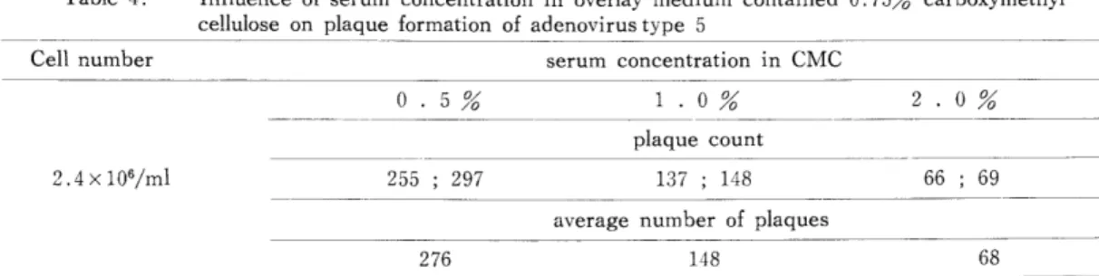
HEK cells infected synchronously with adenovirus type 5 as described in the method, and one ml of them was plated into a dish and covered with 4 ml of the overlay medium contained various concentrations of calf serum・ After the incubation for six days in the CO2
atmosphere, the cell sheets were士ixed and stained as described in the method. As seen in
Table 4, the adequate number and size of plaques was obtained in the overlay medium
contained o.5% calf serum・
3. Adsorbtion test
Eighteen ml of 2.29×10⁶ HEK cells per ml suspended with Eagles medium contained
o.5xJ calf serum was added 2 ml of adenovirus type 5 contained l.SxlO3 PFU per ml. The
mixt‑e was divided into 10 tubes and shaken in the water bath at 37C for various times・
Table 4林 Influence of serum concentration in overlay medium contained 0.75% carboxymethyl
cellulose on plaque formation of adenovirus type 5
Cell number serum concentration in CMC
2.4x10⁶/ml
0 . 5% 1 ・ 0%
plaque count 255 ; 297 137 ; 148
9n‑/ 」 u7o
66 : 69 average number of plaques
276
Remarks: The abbreviation of CMC means the overlay medium contained 0. 75/0 carboxymethylcellいilose・
At each time, the virus adsorbed onto HEK cells was examined as described in the method. As seen in Table 5, it was found that the virus was adsorbed onto the cells at a level o土90% or more during 20 minutes・
Table 5. Adsorption test of purified adenovirus type 5 on HEK cells
㍍㍍㍍㍍ J者、㍍ ㍍㍍㍍ ㍍ ㍍ ㍍
adsorption time at 370C (min・)
0 10 20 30 60 120
227
plaque number average number given in experiment 1
lO8 124
(0%) (76%)
121 106
percent of adsorption
(92%) (100%)
average number given in experiment lO6 116 percent of adsorption
116 119
(100%) (100%)
119 124
(0%) (87%) (91%) (100%)
Remarks:Seedviruswasusedatthetiterof1.5×103mlPFUandcellnumberwasgiven at2林29×106/mlineachexperimentasseenincaseofOmimnteinTable.
4・Neutralizationtest
AsseeninTable6,theneutralizationtiteroftheantiserumofguineapig
immunizedwithpurifiedadenovirustype5wasgivenatthelevelof1:320intheproportion o土83%intheplaquereductionusingthetechniquedescribedinthemethod.
Table6.NeutralizationtestofantiserumagainstpurifiedvirionofAdenovirustype5with homolgousantigen
anti dilu冒eru i。n㌢plaque number芸vueおage her
40 1 : 80 160 1 : 320 1 : 640 1280 2560 mamtamance
medium
1 , 1 2 , 3 2 , 2 8 , 12 43 47 54 57 56 62 59 54
1
√、
し)
1
10 45 55 59 57
Remarks: Seed virus was applied the titer of 1.5×10ソml PFU and cell number
was given at 3.0×lOVml in this experiment・
5. Block test of the antiserum with the antigen A (hexon)
From the result of neutralization test, the antiserum dilution contained 4 units (1 : 80)
of the neutralization titer was used for the block test・ As seen in Table ′ the activity of
the hexon antigen for the blocking antiserum was indicated at the level of 1 :160 in the
proportion of 81% in the plaque reduction・
Table 7・ Block test of the antiserum against purified adenovirus type 5 with the antigen A (hexon)
ami dilu… erumantigen i。ndiluti。n芸1ua關ue iber 80
・ク
'f
'/
・*
ク
・ク
mamtainance medium
20 40 80 1 : 160 320 640 1280
59 42 49 46 44 42 43 4 , 6 4 , 2 2 , 2 53
average number
5∩
48 43 46 5 Q U
2 51
Remarks: The seed virus was used at the titer of 1.8×10⁴ PFU per ml and the number of cells was given 2林8 ×10⁶ cells per ml in this experiment・
6.BlocktestoftheantiserumwiththeantigenC(fiber)
Thoughitseemstobeslightlyblockedtheantiserumwiththeoriginalconcentrationof theantigenC,itwasconsideredthattheantigenCdidnotcompletelyblocktheantiserum・
Table8・BlocktestofantiserumagainstpurifiedvirionofAdenovirustype5withantigenC antiserumantigenC 一者
diluti。ndiluti。n芸La良ueaverage bernumber 1 : 80
//
',
ノク
'/
'/
^
maintainai】ce medium
1 : 1 1 : 5 10 20 40 80 160
18 22 5 , 7 4 , 6 7 , 4 7 , 3 8 , 6 7 , 9 73 79
25 7 5 6 5 7 8 76
Remarks: Seed virus was applied at the titer of 2・4×10ソml PFU and cell number was given 2.3x 10⁶/rnl in this experiment.
7. Block test of the antiserum with P‑antigen
lt was obviously found that the P‑antigen has not the activity to block the antiserum
against the purified virus・
Table 9. Block test of antiserum against purified adenovirus type 5 with the P‑antigen induced in HEK cells infected with adenovirus type 5
antiserum dilution 1 : 80
/>
ク
(/fs
//
//
maintainance medium
antigen dilution
i! ≠ i!
1 : 5 10 20 1 : 40 80 160
plaque number
8 , 7 10 14 6 , 2 ,1 , O 3 , o 3 , 7 136 , 122
avarage number
8 9 12 4 5
・1 5 129
Remarks: Seed virus was used at the titer of 3.9×10* PFU per ml and the number of cells
was given 2.4×10⁶ per ml in this experiment林
DISCUSSION AND SuMMARY
since Takemoto and Liebharber (1961) had reported the inhibitory effect of ager on the cell growth and the plaque formation of certain animal viruses, the carboxyme‑
thylcellulose overlay medium was frequently used for the plaque assay method・ In fact, the plaque formation of dengue viruses under the carboxmethylcellulose overlaj, medium was observed without the decrease in number and size as described by Makino and Mifune in our laboratory・ However, it had been demonstrated by Russell et al. (1967) that the adsorption of the virus onto the suspended cells was earlier and efficiently advanced than that on monolayer cells. When the suspended HEK cells infected with adenovirus type 5 was plated
into dishes, the plaque formation in the cell sheets was observed under the constant condition・
It was demonstrated by Wilcox and Ginsberg (1963) and Kjellen and Pereira (1968) that the production of adenovirus type 5 neutralizing antibody is mainly induced by the antigen of hexon・ It was also found that the antigen of hexon was able to completely block the antiserum against purified virus with using the block test in this study. In contrast, though it seemed to block slightly the antiserum with the antigen C, it was considered that the antigen C may have not enough capacity for the neutralization of the antiserum against purified virus. These findings were supported from the experiments carried out by Kjellen and Pereira・ It was also demonstrated in this study that the P‑antigen induced in HEK cells infected with adenovirus type 5 could not block the antiserum against purified virus・
AKNOWLEDEMENT
The authors are indebted to Dr・ Pereira who generously supplied the antiserum・ we would like to thank Dr. Pereira and Dr. Russell for his encouragement during the adenovirus
study.
同時感染したHEK細胞を用いたアデアウイルス5型のプラーク法 上田芳秋,明石光伸,林薫(長崎大学熱帯医学研究所ウイルス学部門)
適宜に階段稀釈した抗血清とX×104 PFUのウイルスを混じ室温1時間放置した後,直ちに1:100ま で稀釈する.この稀釈液と等量の維持に浮游させた2〜3×10⁶/mlのHEK細胞とを混和し,37℃
30分間軽く振盪する.径5cmのシヤレーに1mlずつ入れ,細胞をガラス面に拡げ,0.5%仔牛血清, 0.75%カルボオキシメチールセルローズを含んだ維持液を加え蔽う.5日ないし6日目に細胞をギム ザ液で染色し,プラークを算える.以上が抗血清の中和抗体の測定法である.抗血清と抗原(分画そ の他)を加え充分反応させた後,更らにX×104PFUのウイルスを追加し室温60分放置し,以後の 手順は上記の方法に従う,以上が抗原による抗血清中の抗体のブロック能を知る方法である.以上の 方法を用いて,アデノウイルス5型の抗血清に対するA抗原,C抗原及びP抗原のブロック能を検査 した.抗血清が完全にブロックされたのはA抗原によってのみであり, C抗原及びP抗原は抗血清の ブロック能を有しないことを知った.
熱帯医学 第17巻 第3号151‑158頁, 1975年12月 REFERENCES
1) Bonifas, V, & Schlesinger, R. W. (1959)‑ Nutritional requirements for plaque production by abenovirus. Fed. Proc., 18, 560.
2) Dulbecco, R. (1952). ‥ Production of plaques in monolayer tissue cultures by single particles 。f an
animal virus. Proc― Natl. Acad. Sci., 32, 747.
3) Hayashi, K & Russell, W. C. (1968). ‥ A study of the development of adenovirus antigens by
the immunofluorescent technigne. Virol. , 34, 470‑480.
4) Hotchin, J. E・ (1955)‑ Use of methylcellulose gel as a substitute for agar in tissue culture overlays'
Nature, 175, 352―
5) Kjellen, L. (1961). : A study of adenovirus‑host cell system by the plaque technique. Virol., 14, 234‑239.
6) Kjellen, L. & Pereira, H. G. (1968). : Role of adenovirus antigens in the induction 。f virus neutralizing antibody. J. Gen. Virol., 2, 177‑185.
7) Makino, Y. & Mifune, K. (1975). : Sensitivity of rapid plaque assay method of denguevirus.
Trop. Med., 16, 163―170.